. 2023 Nov 28;120(48):e2309995120.
doi: 10.1073/pnas.2309995120. Epub 2023 Nov 20.

Mapping the configurational landscape and aggregation phase behavior of the tau protein fragment PHF6

Affiliations

Mapping the configurational landscape and aggregation phase behavior of the tau protein fragment PHF6

Evan Pretti et al. Proc Natl Acad Sci U S A. .

Abstract

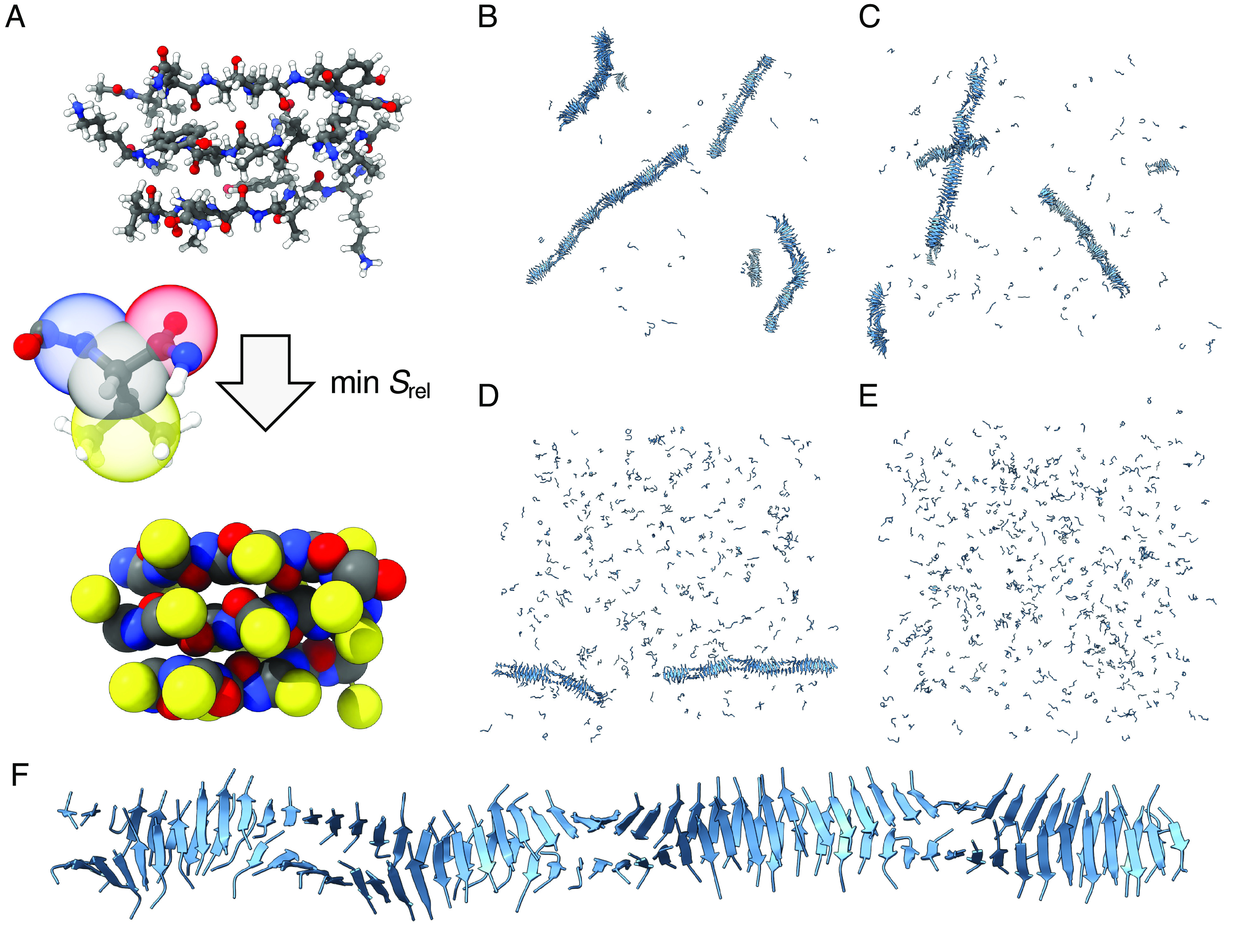
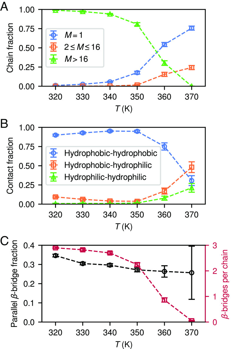
The PHF6 (Val-Gln-Ile-Val-Tyr-Lys) motif, found in all isoforms of the microtubule-associated protein tau, forms an integral part of ordered cores of amyloid fibrils formed in tauopathies and is thought to play a fundamental role in tau aggregation. Because PHF6 as an isolated hexapeptide assembles into ordered fibrils on its own, it is investigated as a minimal model for insight into the initial stages of aggregation of larger tau fragments. Even for this small peptide, however, the large length and time scales associated with fibrillization pose challenges for simulation studies of its dynamic assembly, equilibrium configurational landscape, and phase behavior. Here, we develop an accurate, bottom-up coarse-grained model of PHF6 for large-scale simulations of its aggregation, which we use to uncover molecular interactions and thermodynamic driving forces governing its assembly. The model, not trained on any explicit information about fibrillar structure, predicts coexistence of formed fibrils with monomers in solution, and we calculate a putative equilibrium phase diagram in concentration-temperature space. We also characterize the configurational and free energetic landscape of PHF6 oligomers. Importantly, we demonstrate with a model of heparin that this widely studied cofactor enhances the aggregation propensity of PHF6 by ordering monomers during nucleation and remaining associated with growing fibrils, consistent with experimentally characterized heparin-tau interactions. Overall, this effort provides detailed molecular insight into PHF6 aggregation thermodynamics and pathways and, furthermore, demonstrates the potential of modern multiscale modeling techniques to produce predictive models of amyloidogenic peptides simultaneously capturing sequence-specific effects and emergent aggregate structures.

Keywords: amyloid aggregation; multiscale modeling; tau protein.


Conflict of interest statement

Competing interests statement:The authors declare no competing interest.

Figures

Fig. 1.
Fig. 1.
Illustration of the CG model and representative snapshots of simulated aggregates. (A) Representative configuration of the reference system containing 3 PHF6, with an illustration of the mapping operation to the corresponding CG configuration. (BE) Systems of 512 PHF6 with ρ=10mM and T=340, 350, 360, 370K, respectively, after 10μs of MD simulation. (F) A representative fibril containing 106 PHF6 from a simulation at 350K.
Fig. 2.
Fig. 2.
Temperature dependence of properties of aggregating systems. (A) Fractions of isolated chains (M=1) and chains in clusters of sizes 2M16 and M>16, from the last 5μs of 10μs MD simulations of 512 PHF6 with ρ=10mM. (B) Likewise, fractions of non-hydrogen-bonded contacting chains for which the closest pair of sidechains consisted of residues from the hydrophobic and hydrophilic sides of PHF6. (C) Fractions of β-bridge-participating residues with parallel (vs. antiparallel) orientations, and total numbers of β-bridges per chain. Error bars show SDs of the measured properties.
Fig. 3.
Fig. 3.
Oligomer structures and thermodynamic properties. (AC) A representative tetramer, octamer, and 16-mer, respectively, from replica exchange simulations of PHF6 at T=300K and ρ=10mM. (D) Fractions of non-hydrogen-bonded contacting chains for which the closest pair of sidechains consists of residues from the hydrophobic and hydrophilic sides of PHF6, for a system with N=16, ρ=10mM, and T=350K. (E) Free energies of oligomers of size M in a ρ=10mM solution. (F) Critical oligomer sizes estimated from free energies (Upper Panel) and corresponding free energy barrier heights (Lower Panel: Curves are truncated when the most probable value of M reaches the largest system size N=16). Error bars and bands show 95% CIs of mean values, or in the case of M, median values.
Fig. 4.
Fig. 4.
Fibril seeding and fibril–solution coexistence. (A) Initial state and (B) state after 1μs, of a fibrillar seed of size 32 (orange) in a solution of 32 PHF6 (blue) in a volume corresponding to a total concentration ρ=9mM, and T=350K. (C) Fibril size (solid) and chains never having detached from the fibril (dashed) from 5 replicates each, T=350K. Means and SDs over 200 ns intervals are shown. (D) Theoretical coexistence curve (red – –) of fibrillar PHF6 with solution, fit to observed solution concentrations (green ) in equilibrium with fibrils. Blue ° and orange × symbols indicate the presence or absence, respectively, of persistent aggregates in 25μs simulations of 64 PHF6. Error bars show 95% CIs.
Fig. 5.
Fig. 5.
Heparin-induced aggregation of PHF6. (A) Initial stages of heparin-induced aggregation at T=350K and ρ=2.5mM from a system of 64 PHF6 containing a heparin chain of 60 saccharide units. (B) Fully grown fibril under these conditions. (C) Fractions of chains in clusters of different sizes from the last 2μs of 5 replicates each of 10μs simulations of 64 PHF6 with a heparin chain of 60 saccharide units at T=350K. (D) Likewise, fractions of β-bridge-participating residues with parallel (vs. antiparallel) orientations, and total numbers of β-bridges per chain. (E) Radius of gyration of heparin chains and total electrostatic interaction energy between heparin and lysine sidechain CG sites. Error bars show SDs of the measured properties.

References

    1. Ballatore C., Lee V. M. Y., Trojanowski J. Q., Tau-mediated neurodegeneration in Alzheimer’s disease and related disorders. Nat. Rev. Neurosci. 8, 663–672 (2007). - PubMed
    1. Goedert M., Eisenberg D. S., Crowther R. A., Propagation of tau aggregates and neurodegeneration. Annu. Rev. Neurosci. 40, 189–210 (2017). - PubMed
    1. Sanders D., et al. , Distinct tau prion strains propagate in cells and mice and define different tauopathies. Neuron 82, 1271–1288 (2014). - PMC - PubMed
    1. Vaquer-Alicea J., Diamond M. I., Joachimiak L. A., Tau strains shape disease. Acta Neuropathol. 142, 57–71 (2021). - PMC - PubMed
    1. Fitzpatrick A. W. P., et al. , Cryo-EM structures of tau filaments from Alzheimer’s disease. Nature 547, 185–190 (2017). - PMC - PubMed

LinkOut - more resources