. 2023 Dec;10(36):e2303457.
doi: 10.1002/advs.202303457. Epub 2023 Nov 20.

Lactobacillus Intestinalis Primes Epithelial Cells to Suppress Colitis-Related Th17 Response by Host-Microbe Retinoic Acid Biosynthesis

Affiliations

Lactobacillus Intestinalis Primes Epithelial Cells to Suppress Colitis-Related Th17 Response by Host-Microbe Retinoic Acid Biosynthesis

Qi-Wen Wang et al. Adv Sci (Weinh). 2023 Dec.

Abstract

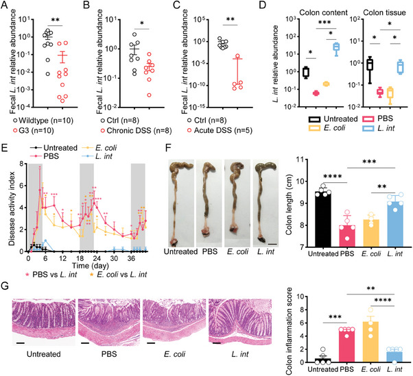
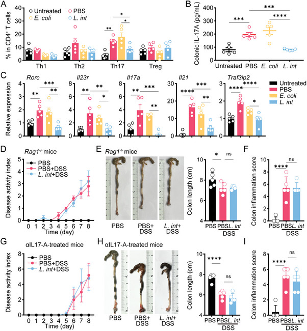
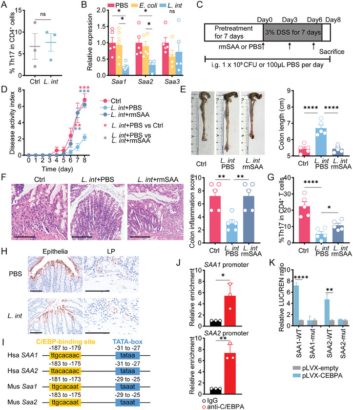
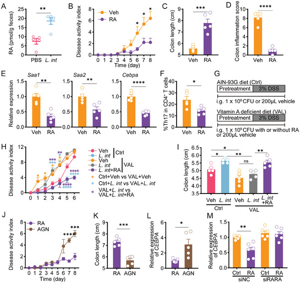
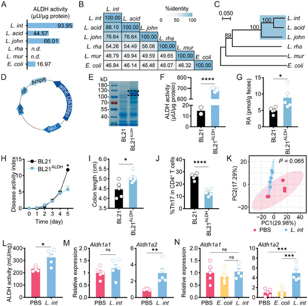
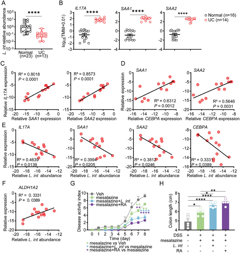
Gut microbiome is integral to the pathogenesis of ulcerative colitis. A novel probiotic Lactobacillus intestinalis (L. intestinalis) exerts a protective effect against dextran sodium sulfate-induced colitis in mice. Based on flow cytometry, colitis-associated Th17 cells are the target of L. intestinalis, which is supported by the lack of protective effects of L. intestinalis in T cell-null Rag1-/- mice or upon anti-IL-17-A antibody-treated mice. Although L. intestinalis exerts no direct effect on T cell differentiation, it decreases C/EBPA-driven gut epithelial SAA1 and SAA2 production, which in turn impairs Th17 cell differentiation. Cometabolism of L. intestinalis ALDH and host ALDH1A2 contributed to elevated biosynthesis of retinoic acid (RA), which accounts for the anti-colitis effect in RAR-α -mediated way. In a cohort of ulcerative colitis patients, it is observed that fecal abundance of L. intestinalis is negatively associated with the C/EBPA-SAA1/2-Th17 axis. Finally, L. intestinalis has a synergistic effect with mesalazine in alleviating murine colitis. In conclusion, L. intestinalis and associated metabolites, RA, have potential therapeutic effects for suppressing colonic inflammation by modulating the crosstalk between intestinal epithelia and immunity.

Keywords: Th17; colitis; microbes; retinoic acid.


Conflict of interest statement

The authors declare no conflict of interest.

Figures

Figure 1
Figure 1
L. intestinalis relieved DSS‐induced chronic colitis. A) Fecal abundance of L. intestinalis was measured in wild‐type and G3 mice. B,C) Fecal abundance of L. intestinalis was measured in a control group, and murine DSS‐induced chronic colitis (B) or acute colitis (C). D) Fecal abundance of L. intestinalis was compared among mice treated without (Untreated), or with DSS accompanied by PBS, E. coli, or L. intestinalis (L. int) gavage respectively (n = 5). E–G) The pathology of colitis was evaluated among the control group (Untreated) and the three chronic colitis groups with PBS, E. coli, or L. intestinalis (L. int) gavage respectively by disease activity index (E), colon length (scale bar, 1 cm) (F) and histological score (scale bar, 100 µm) (G) (n = 5). Error bars indicate mean ± SEM. * p < 0.05; ** p < 0.01; *** p < 0.001; **** p < 0.0001. p values were based on Mann–Whitney test and one‐way ANOVA with post‐hoc test.
Figure 2
Figure 2
L. intestinalis relieved colitis in a Th17‐dependent way. A–C) Frequencies of four CD4+ T subsets were analyzed by multicolor flow cytometry in colon LP of untreated mice and chronic DSS‐treated mice with gavage of PBS, E. coli, or L. intestinalis (L. int) (A). Th17 response in colon tissue was analyzed by ELISA (B) and expression of Th17‐related genes (C). D–F) The pathology of colitis was evaluated among Rag1 −/− mice, which were treated by acute DSS with gavage of PBS or L. intestinalis (L. int) or only treated by PBS gavage, by disease activity index (D), colon length (E), and pathological scores (F). G‐I) The pathology of colitis and systemic inflammation was evaluated among αIL17‐A‐treated mice, which were treated by acute DSS with gavage of PBS or L. intestinalis (L. int) or only treated by PBS gavage, by disease activity index (G), colon length (H), and pathological scores (I). n = 5. Error bars indicate mean ± SEM. * p < 0.05; ** p < 0.01; *** p < 0.001; **** p < 0.0001. p values were based on one‐way ANOVA with post‐hoc test.
Figure 3
Figure 3
L. intestinalis suppressed Th17 cell differentiation by targeting epithelial SAA production. A) In vitro Th17 differentiation assay was performed on naïve CD4+ T cells isolated from mouse spleen and treated with PBS (Ctrl) or L. intestinalis (L. int) (n = 3). B) Expression of Saa1/2/3 was evaluated in bulk colon samples from chronic DSS‐treated mice with gavage of PBS, E. coli, or L. intestinalis (L. int) (n = 5). C) Pretreatment with gavage of PBS or L. intestinalis (L. int) was started on the 7th day before DSS treatment and until the end of the experiment. On days zero, three, and six of DSS course, all mice were intraperitoneally injected (i.p.) with recombinant mouse SAA proteins (rmSAA) or PBS. D–G) The pathology of colitis was evaluated among PBS‐i.p. mice with PBS(PBS+PBS) or L. intestinalis (L. int+PBS) gavage, as well as rmSAA‐i.p. mice with L. intestinalis (L. int+rmSAA) gavage, by disease activity index (D), colon length (E), pathological scores (scale bar, 200 µm) (F), and frequencies of Th17 cells in colon LP (G) (n = 5). H) Representative fluorescent immunohistochemistry images indicated the expression of Saa1/2 in colon epithelia and LP from chronic DSS‐treated mice with gavage of PBS and L. intestinalis (L. int). I) The C/EBP‐binding motifs were conservative among the sequences of promoter regions from human (Hsa) SAA1/2 genes or mouse (Mus) Saa1/2 genes. J) The enrichment at SAA1 and SAA2 promoter was detected by ChIP‐qPCR in C/EBPA‐overexpressing HEK‐293T cells using anti‐C/EBPA or control IgG. K) The firefly luciferase (LUC) was driven by wildtype (WT) promoter or mutate (mut) regions of SAA1 and SAA2. The trans‐activation ability of C/EBPA was measured by the LUC / REN ratio in HEK‐293T cells co‐transfected with pLVX‐empty or pLVX‐CEBPA. Error bars indicate mean ± SEM. ns, no significance; * p < 0.05; ** p < 0.01; **** p < 0.0001. p values were based on Student's t‐test and one‐way ANOVA with post‐hoc test.
Figure 4
Figure 4
L. intestinalis‐associated RA reduced C/EBPA‐driven SAA production in RAR‐α‐mediated manner. A) Quantification of RA was performed in the feces of acute DSS‐treated mice with PBS or L. intestinalis (L. int) gavage (n = 5). B–F), The pathology of acute colitis and related gene expressions were evaluated between DSS‐treated mice with vehicle (Veh) or RA gavage, by disease activity index (B), colon length (C), pathological scores (D), colonic expression of Saa1, Saa2, and Cebpa (E), and frequencies of Th17 cells (F) (n = 5). G) 2 groups were fed with AIN‐93G as a control diet (Ctrl). Three groups were fed with a vitamin A‐deficient diet (VAL). Pretreatment with gavage of PBS, L. int, or L. int+RA was started on the 7th day before DSS treatment and until the end of the experiment. All mice received a 6‐day 3% DSS treatment. H,I) The pathology of acute colitis was evaluated among Ctrl or VAL‐fed mice with respective gavage, by disease activity index (H) and colon length (I) (n = 5). J–L), The pathology of acute colitis and related gene expressions were evaluated between DSS‐treated mice with vehicle RA or RA+AGN193109 (AGN) administration, by disease activity index (J), colon length (K), and Cebpa expression (L) (n = 5). M) In HT29 cells treated by siRARA, the effect of RA on the expression of C/EBPA was analyzed. Error bars indicate mean ± SEM. ns, no significance; * p < 0.05; ** p < 0.01; *** p < 0.001; **** p < 0.0001. p values were based on Student's t‐test and one‐way ANOVA with post‐hoc test.
Figure 5
Figure 5
L. intestinalis promoted RA synthesis through its own ALDH and by enhancing host ALDH. A) The ALDH activity was detected in six different species. B) Percentage identity analysis was performed on ALDH protein sequences of six different species. C) Phylogenetic tree analysis of ALDH protein sequences of six different species was constructed. Bootstrap analysis values for 1000 replicates were shown. D–F), Plasmid containg L. intestinalis adhE (D) was transduced into bacterial expression systems BL21ALDH. The ALDH expression and activity were detected (E–F). G–J), Quantification of RA was performed in the feces of acute DSS‐treated mice with BL21 or BL21ALDH gavage (G). The pathology of acute colitis was evaluated between DSS‐treated mice with BL21 or BL21ALDH gavage, by disease activity index (H) and colon length (I), and frequencies of Th17 cells (J) (n = 5). K) β‐diversity of the fecal was compared between chronic DSS‐treated mice with PBS gavage and with L. intestinalis (L. int) gavage (n = 5). L,M) The ALDH activity (L) and related gene expression (M) were detected between colon tissues of DSS‐induced acute colitis mice with PBS, and L. intestinalis (L. int) gavage (n = 5). N) Expression levels of Aldh1a1 and Aldh1a2 were evaluated in bulk colon samples from chronic DSS‐treated mice with gavage of PBS, E. coli, and L. intestinalis (L. int) (n = 5). Error bars indicate mean ± SEM. ns, no significance; * p < 0.05; ** p < 0.01; *** p < 0.001. p values were based on Student's t‐test, PERMANOVA test, and one‐way ANOVA with post‐hoc test.
Figure 6
Figure 6
L. intestinalis suppressed the C/EBPA‐SAA1/2‐Th17 axis in UC patients and exerted therapeutic effect on DSS‐induced colitis. A) Relative abundance of L. intestinalis was compared between normal colon samples and UC patient inflammatory samples. B) Colonic expression of IL17A, SAA1, and SAA2 was compared between UC patients and healthy people from GEO data (GSE128682). C–F) The correlation analysis was performed in colon samples from UC patients to determine the relationship between the expression of IL17A and SAA1/2 (C), between expression of SAA1/2 and CEBPA (D), and between expression of genes (IL17A, SAA1/2, CEBPA, ALDH1A2) and L. intestinalis abundance (L. int) (E–F) (n = 13). G,H) The pathology of acute colitis was evaluated among DSS‐treated mice treated with vehicle (Veh), or mesalazine alone or in combination with L. intestinalis (L. int) or RA, by disease activity index (G) and colon length (H) (n = 5). Error bars indicate mean ± SEM. * p < 0.05; ** p < 0.01; *** p < 0.001; **** p < 0.0001. p values were based on Mann–Whitney test, Student's t‐test, one‐way ANOVA with post‐hoc test, and Pearson correlation test.

References

    1. Leblanc J.‐F., Segal J. P., De Campos Braz L. M., Hart A. L., Nutrients 2021, 13, 1780. - PMC - PubMed
    1. Round J. L., Mazmanian S. K., Proc Natl Acad Sci U S A. 2010, 107, 12204. - PMC - PubMed
    1. Fan L., Qi Y., Qu S., Chen X., Li A., Hendi M., Xu C., Wang L., Hou T., Si J., Chen S., Gut Microbes 2021, 13, e1826746. - PMC - PubMed
    1. Zhao J., Lu Q., Liu Y., Shi Z., Hu L., Zeng Z., Tu Y., Xiao Z., Xu Q., J Immunol Res 2021, 8816041. - PMC - PubMed
    1. Hang S., Paik D., Yao L., Kim E., Trinath J., Lu J., Ha S., Nelson B. N., Kelly S. P., Wu L., Zheng Y., Longman R. S., Rastinejad F., Devlin A. S., Krout M. R., Fischbach M. A., Littman D. R., Huh J. R., Nature 2019, 576, 143. - PMC - PubMed

Publication types

Supplementary concepts