. 2023 Dec;37(12):e23316.
doi: 10.1096/fj.202301722R.

Lactate produced by alveolar type II cells suppresses inflammatory alveolar macrophages in acute lung injury

Affiliations

Lactate produced by alveolar type II cells suppresses inflammatory alveolar macrophages in acute lung injury

René M Roy et al. FASEB J. 2023 Dec.

Abstract

Alveolar inflammation is a hallmark of acute lung injury (ALI), and its clinical correlate is acute respiratory distress syndrome-and it is as a result of interactions between alveolar type II cells (ATII) and alveolar macrophages (AM). In the setting of acute injury, the microenvironment of the intra-alveolar space is determined in part by metabolites and cytokines and is known to shape the AM phenotype. In response to ALI, increased glycolysis is observed in AT II cells, mediated by the transcription factor hypoxia-inducible factor (HIF) 1α, which has been shown to decrease inflammation. We hypothesized that in acute lung injury, lactate, the end product of glycolysis, produced by ATII cells shifts AMs toward an anti-inflammatory phenotype, thus mitigating ALI. We found that local intratracheal delivery of lactate improved ALI in two different mouse models. Lactate shifted cytokine expression of murine AMs toward increased IL-10, while decreasing IL-1 and IL-6 expression. Mice with ATII-specific deletion of Hif1a and mice treated with an inhibitor of lactate dehydrogenase displayed exacerbated ALI and increased inflammation with decreased levels of lactate in the bronchoalveolar lavage fluid; however, all those parameters improved with intratracheal lactate. When exposed to LPS (to recapitulate an inflammatory stimulus as it occurs in ALI), human primary AMs co-cultured with alveolar epithelial cells had reduced inflammatory responses. Taken together, these studies reveal an innate protective pathway, in which lactate produced by ATII cells shifts AMs toward an anti-inflammatory phenotype and dampens excessive inflammation in ALI.

Keywords: acute lung injury; acute respiratory distress syndrome; alveolar epithelium; alveolar macrophage; glycolysis; hypoxia-inducible factor; lactate.


Conflict of interest statement

DISCLOSURES

The authors declare no conflicts of interest.

Figures
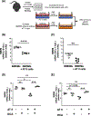
FIGURE 1
FIGURE 1
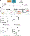
AT II cells suppress cytokine release in macrophages. Schematic of macrophages–AT II co-culture system (A): AT I cells and BMDMs were isolated from the same animal and incubated in a non-contact insert co-culture system for 48 h prior to stimulation with 50 ng/mL LPS. Twenty-four hours after LPS stimulation, BMDMs were harvested. mRNA expression of IL-6 (B) and CXCL1 (C) was determined with qPCR in BMDMs cultured with and without ATIIs (Controls) after 24 h. BMDM cells were cultured either alone or co-cultured with AT II cells with respective controls and incubated with 2.5 mM of glycolysis inhibitor dichloroacetic acid (DCA) and stimulated with LPS. After 24 h, cytokine mRNA expression was determined with qPCR (D, E). n = −5, data expressed as mean ± SD, *p < .05, **p < .01, ***p < .001, ****p < .0001. Data were analyzed with two-tailed, unpaired, Student’s t-test (B, C) or one-way ANOVA with Tukey’s correction for multiple comparisons (D, E).
FIGURE 2
FIGURE 2
Intratracheal L-lactate shifts resident macrophages pro-resolving phenotype. Schematic of mouse experiments: Acute lung injury was induced by either 0.125 M HCl (Acid) or 5 μ/g intratracheal (i.t.) LPS. Six hours after the induction of lung injury, mice received 50 μL 25 mM L-lactate. The injury control group received 50 μL pH-controlled PBS. Age-, sex-, and weight-matched C57/B6 mice were used. The experiment was terminated after 24 h (A). Flow cytometry sorting strategy. Monocytes, neutrophils, B cells, and T cells were excluded. CD45+ population was then segregated into resident AMs (Dump, CD45+, CD64+, SigF+, CD11b) and recruited AMs (CD45+, CD64+, SigF, CD11b+) (B). Arg-1 expression (C, E), respectively, IL-10 expression (G, F) in resident airway macrophages were determined by flow cytometry and expressed as mean fluorescence intensity (MFI). n = 5–7, data expressed as mean ± SD, **p < .01, ***p < .001. Data were analyzed with two-tailed, unpaired, Student’s t-test.
FIGURE 3
FIGURE 3
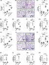
Intratracheal L-lactate attenuates acute lung injury. Acute lung injury was induced in age-, sex-, and weight-matched C57/B6 mice by 5 μ/g intratracheal (i.t.) LPS (A–G) or 0.125 M HCl i.t. (H–N) 6 h after the induction of lung injury mice received 50 μL 25 mM L-lactate (LPS+ lactate). The injury control group received 50 μL pH-controlled PBS. Lactate was measured in bronchoalveolar lavage fluid (BALF) at the end of the experiment (A, H). Representative images of H&E-stained lungs (B, I). A semi-quantitative cumulative lung injury score was performed, which is a combined score of cellular infiltrates, interstitial congestion, hyaline membrane formation, and hemorrhage (C, J). mRNA expression in whole lung tissue was determined by qPCR (D–G and K–N). n = 4–6/group. Data are represented as mean ± SD, ns = not significant, **p < .01, ****p < .0001. Data were analyzed with one-way ANOVA with Tukey’s correction for multiple comparisons.
FIGURE 4
FIGURE 4
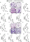
Intra-tracheal L-lactate reconstitutes Hif1aloxp/loxp SPC-ER-Cre+ animals. Acute lung injury was induced in age-, sex-, and weight-matched alveolar epithelial cell-specific conditional knockout mice (Hif1aloxp/loxp SPC-ER-Cre+ or control animals (SPC-ER-Cre+) by 5 μ/g intratracheal (i.t.) LPS (A–G) or 0.125 M HCl (H–N) i.t. 6 h after the induction of lung injury mice received 50 μL 25 mM L-lactate (LPS+ lactate). The injury control group received 50 μL pH-controlled PBS. Lactate was measured in bronchoalveolar lavage fluid (BALF) at the end of the experiment (A, H). Representative images of H&E-stained lungs (B, I). A semi-quantitative cumulative lung injury score was performed, which is a combined score of cellular infiltrates, interstitial congestion, hyaline membrane formation, and hemorrhage (C, J). mRNA expression in whole lung tissue was determined by qPCR (D–G and K–N). n = 4–6/group. Data are represented as mean ± SD, ns = not significant, *p < .05, **p < .01, ***p < .001, ****p < .0001. Data were analyzed with one-way ANOVA with Tukey’s correction for multiple comparisons.
FIGURE 5
FIGURE 5
Mice with alveolar type 2 specific suppression of Hif1a are unable to shift resident macrophages’ pro-resolving phenotype in response to ALI. Acute lung injury was induced by intratracheal LPS (A–E) or HCl (G–J) in Hif1aloxp/loxp surfactant Cre+ (Hif1aloxp/loxp SPC-ER-Cre+) mice in age-, sex-, and weight-matched controls (SPC-ER-Cre+). Six hours after the induction of lung injury, mice received 50 μL 25 mM L-lactate with control group receiving 50 μL pH-controlled PBS. Twenty-four hours after induction of ALI, the AMs were harvested by bronchoalveolar lavage. mRNA expression in airway macrophages was determined by qPCR. n = 4–5/group. Data are represented as mean ± SD, ns = not significant, *p < .05, **p < .01, ***p < .001, ****p < .0001. Data were analyzed with one-way ANOVA with Tukey’s correction for multiple comparisons.
FIGURE 6
FIGURE 6
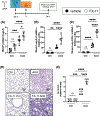
Pharmacological inhibition of LDHA exacerbates acid aspiration-induced acute lung injury. Eight- to 10-week-old C57BL/6 mice received LDHA inhibitor FX-11 i.p 24 h prior to induction of ALI with acid instillation with i.t. HCl. Control groups received vehicle. After 24 h, the lungs were removed. Protein concentration was measured in BALF with Bradford-assay (A). mRNA expression in whole lung tissue was determined by qPCR (B, C). Representative images of H&E-stained lungs (D). A semi-quantitative cumulative lung injury score was performed, which is a combined score of cellular infiltrates, interstitial congestion, hyaline membrane formation, and hemorrhage (E) n = 4–5/group. Data are represented as mean ± SD, ns = not significant, *p < .05, **p < .01, ***p < .001, ****p < .0001. Data were analyzed with one-way ANOVA with Tukey’s correction for multiple comparisons.
FIGURE 7
FIGURE 7
Lactate produced by alveolar epithelial cells shifts primary human airway macrophage phenotype. Schematic of experiments (A). A549 cells (with stable knockdown of LDHA (A549LDHA−/− or scrambled controls (A549scr)) and primary human airway macrophages (MΦ) isolated from lungs rejected for donation were incubated in a non-contact insert co-culture system for 48 h prior to stimulation with 50 ng/mL LPS. Twenty-four hours after LPS stimulation, primary human airway macrophages were harvested and cytokines were measured with qPCR (A–F). n = 5/group. Data are represented as mean ± SD, ns = not significant, *p < .05, **p < .01, ***p < .001, ****p < .0001. Data were analyzed with one-way ANOVA with Tukey’s correction for multiple comparisons.

References

    1. Elks PM, van Eeden FJ, Dixon G, et al. Activation of hypoxia-inducible factor-1alpha (Hif-1alpha) delays inflammation resolution by reducing neutrophil apoptosis and reverse migration in a zebrafish inflammation model. Blood 2011;118:712–722. - PubMed
    1. Eckle T, Brodsky K, Bonney M, et al. HIF1A reduces acute lung injury by optimizing carbohydrate metabolism in the alveolar epithelium. PLoS Biol 2013;11:e1001665. - PMC - PubMed
    1. Vohwinkel CU, Burns N, Coit E, et al. HIF1A-dependent induction of alveolar epithelial PFKFB3 dampens acute lung injury. JCI Insight 2022;7:e157855. doi:10.1172/jci.insight.157855 - DOI - PMC - PubMed
    1. Vohwinkel CU, Coit EJ, Burns N, et al. Targeting alveolar-specific succinate dehydrogenase a attenuates pulmonary inflammation during acute lung injury. FASEB J 2021;35:e21468. - PMC - PubMed
    1. Bain CC, MacDonald AS. The impact of the lung environment on macrophage development, activation and function: diversity in the face of adversity. Mucosal Immunol 2022;15:223–234. - PMC - PubMed

Publication types

LinkOut - more resources