Review
. 2024 Feb;11(8):e2305837.
doi: 10.1002/advs.202305837. Epub 2023 Nov 20.

Synthetic Cells Revisited: Artificial Cells Construction Using Polymeric Building Blocks

Affiliations
Review

Synthetic Cells Revisited: Artificial Cells Construction Using Polymeric Building Blocks

Viviana Maffeis et al. Adv Sci (Weinh). 2024 Feb.

Abstract

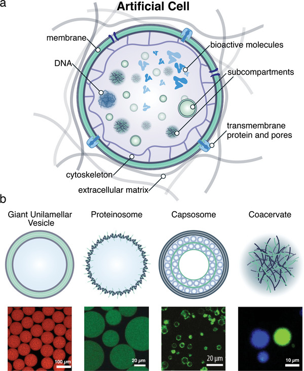
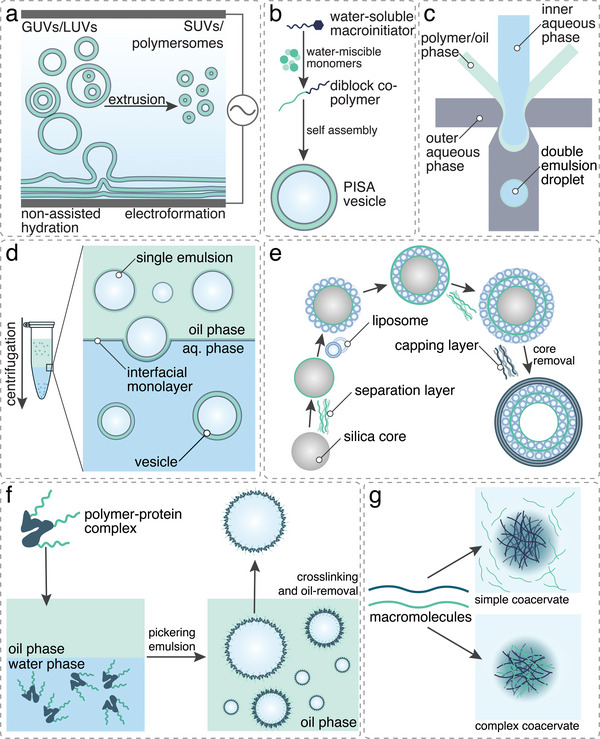
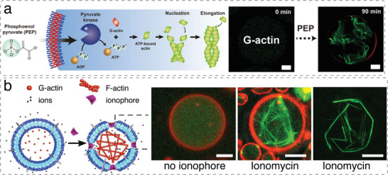
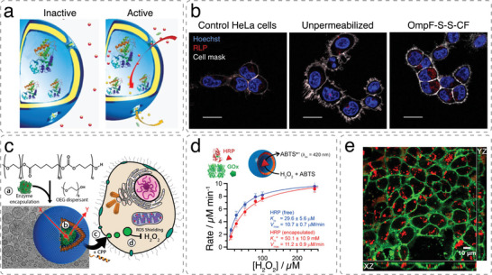
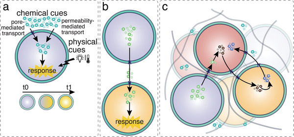
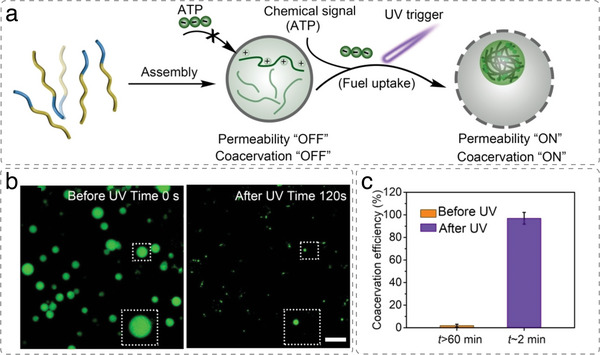
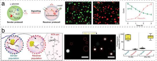
The exponential growth of research on artificial cells and organelles underscores their potential as tools to advance the understanding of fundamental biological processes. The bottom-up construction from a variety of building blocks at the micro- and nanoscale, in combination with biomolecules is key to developing artificial cells. In this review, artificial cells are focused upon based on compartments where polymers are the main constituent of the assembly. Polymers are of particular interest due to their incredible chemical variety and the advantage of tuning the properties and functionality of their assemblies. First, the architectures of micro- and nanoscale polymer assemblies are introduced and then their usage as building blocks is elaborated upon. Different membrane-bound and membrane-less compartments and supramolecular structures and how they combine into advanced synthetic cells are presented. Then, the functional aspects are explored, addressing how artificial organelles in giant compartments mimic cellular processes. Finally, how artificial cells communicate with their surrounding and each other such as to adapt to an ever-changing environment and achieve collective behavior as a steppingstone toward artificial tissues, is taken a look at. Engineering artificial cells with highly controllable and programmable features open new avenues for the development of sophisticated multifunctional systems.

Keywords: artificial cells; artificial organelles; artificial signaling; bottom-up; collective behavior; communicative networks; polymers.


Conflict of interest statement

The authors declare no conflict of interest.

Figures

Figure 1
Figure 1
The artificial cell possesses the capacity to emulate the structure of a biological cell in a simplified manner. Its hierarchical multi‐compartmentalization enables active compounds within distinct nanocompartments to engage in straightforward cascade reactions and communication when organized within cell consortia.
Figure 2
Figure 2
a) Schematic illustration of a large outer micro‐compartment with incorporation of hierarchical systems and their organization in functional membrane bound or membrane less compartments. b) Schematic overview of polymeric based architecture used for cell mimics. Reproduced with permission.[ 72 , 77 , 79 , 80 ] Copyright 2019, Wiley‐VCH. Copyright 2016, American Chemical Society. Copyright 2019, American Chemical Society. Reproduced with permission under terms of the CC‐BY‐NC‐ND license.[ 72 ] Copyright 2017, American Chemical Society.
Figure 3
Figure 3
Schematic representation of selected production methods for artificial cells and organelles: a) film rehydration, electroformation, and subsequent extrusion of block copolymers, b) polymerization‐induced self‐assembly (PISA), c) double emulsions microfluidic formation of vesicles, d) emulsion centrifugation, e) layer‐by‐layer assembly of capsosomes, f) pickering emulsion self‐assembly of proteinosomes, and g) self‐assembly of coacervates.
Figure 4
Figure 4
Triggering actin filament formation inside GUVs. a) Actin polymerization inside p‐GUVs induced by phosphoenol pyruvate (PEP). Monomeric (G‐) actin is polymerized in the presence of ATP, resulting in filamentous (F‐) actin. Actin polymerization (green) inside GUV (red) over 90 min upon PEP addition. Scale bars, 20 µm. Reproduced under terms of the CC‐BY license.[ 42 ] Copyright 2022, Springer Nature. b) Actin polymerization inside p‐GUVs induced by the influx of MgCl2. p‐GUVs encapsulating G‐actin and the actin crosslinker filamin were incubated with 150 mm MgCl2. In the absence of the ionophore ionomycin, Mg2+ is not transported across the p‐GUV membrane (red) and confined actin (green) remains monomeric. When ionomycin was added to the surrounding solution, it reconstituted into the membrane of the p‐GUV and facilitated the entry of Mg2+ into the GUV cavity where it triggered actin filament formation and bundling. Projections of actin filaments and filament bundles in p‐GUVs recorded with super‐resolution 3D structured illumination microscopy. Scale bars, 5 µm. Reproduced under terms of the CC‐BY license.[ 34 ] Copyright 2020, Wiley‐VCH.
Figure 5
Figure 5
Catalytic nanocompartments encapsulating one type of enzyme demonstrate their activity inside cells. Permeability toward substrates and products is achieved through a,b) membrane proteins or c–e) by using intrinsically permeable polymer membranes. a) Schematic representation of modified OmpF acting as a gate in catalytic polymersome nanocompartments. b) Cellular uptake and intracellular activation of fluorescently labelled HRP‐loaded polymersomes producing resorufin‐like product (RLP; red). Scale bar 20 µm. Reproduced with permission.[ 179 ] Copyright 2018, Springer Nature. c) Preparation of biodegradable enzyme‐loaded PEG‐b‐PCLgTMC polymersomes via self‐assembly and model of function as artificial organelles inside cells. Semipermeable enzyme‐loaded nanocompartments were decorated with a cell‐penetrating peptide (CPP) to promote cellular integration as antioxidant organelles. d) Michaelis–Menten kinetics plot of HRP‐loaded artificial organelles and free enzyme. Encapsulation within polymersomes leads to an increase in K M due to the diffusion barrier. e) Uptake of polymersomes in HEK293T cells. Confocal microscopy images showing the subcellular distribution of TAT‐polymersomes encapsulating AF647BSA (red) after 24 h of incubation at a concentration of 0.4 mg mL−1. Plasma membranes are stained with CellMask green. Reproduced with permission under terms of the CC‐BY‐NC‐ND license.[ 83 ] Copyright 2018, American Chemical Society.
Figure 6
Figure 6
Cascade reactions between enzymes co‐encapsulated within the same compartment and enzymes in clustered compartments. a) Schematic illustrating artificial peroxisome based on enzymatic cascade for the detoxification of superoxide radicals and related H2O2 inside cells (upper panel). The artificial organelle is based on the simultaneous encapsulation of a set of antioxidant enzymes in a polymersome, with a membrane equipped with channel proteins. Flow cytometry analysis demonstrates the protective effect of artificial peroxisomes (PQ‐Aps) when cells (viability 100%) are treated with paraquat (PQ) (down). Reproduced with permission.[ 82 ] Copyright 2013, American Chemical Society. b) Schematic representation of the AMG–GOx–LPO cascade between two clustered polymersomes, tethered via complementary ssDNA (upper panel). Enzymatic activity of AMG(GOx)–LPO clusters (blue), unclustered compartments (red), (GOx)–LPO clusters with AMG in the solution (magenta) and Amplex Red autoxidation (black) (down). Reproduced with permission under terms of the CC‐BY‐NC license.[ 81 ] Copyright 2021, Royal Society of Chemistry.
Figure 7
Figure 7
Cascade reactions in multicompartment systems based on p‐GUVs. a) Different enzymes were encapsulated within PS‐b‐PIAT polymersomes, mixed with cytosolic enzyme and substrates and encapsulated in PB‐b‐PEO p‐GUVs to fabricate polymersome‐in‐p‐GUV artificial cells hosting multicompartment catalysis. Different cascade reactions of increased complexity were investigated and monitored with fluorescent spectroscopy. Reproduced with permission.[ 45 ] Copyright 2014, Wiley‐VCH. b) Enzyme‐loaded silica nanocapsules were used as organelles to construct a catalytic p‐GUV multicompartment system. The GOx‐HRP cascade in the microreactor was followed using confocal laser scanning microscopy to measure the generation of fluorescent resorufin product. Reproduced with permission.[ 211 ] Copyright 2022, Wiley‐VCH.
Figure 8
Figure 8
Intracellular activity of capsosomes containing enzyme‐loaded liposomes. a) β‐D‐Glucose can permeate through the liposomes membrane and be converted by GOx into d‐gluconolactone and H2O2. H2O2 reacts with Amplex Red reagent in the presence of HRP to generate the fluorescent resorufin. b) Fluorescence intensity of the enzymatic cascade conversion monitored by fluorescence spectroscopy at 4 and 24 h. Macrophages had been incubated with the capsosomes prior to the addition of substrates for 4 h to allow cell internalization (n  = 3, ***p < 0.001). c) Differential interference contrast (DIC, left) and confocal laser scanning (CLSM, right) microscopy images showing the enzymatic cascade conversion of β‐d‐glucose into the red fluorescent product resorufin by capsosomes loaded with GOx and HRP after and 24 h. Control images, in which the substrates were incubated with the macrophages in the absence of capsosomes, are shown at the bottom. Reprinted with permission.[ 161 ] Copyright 2017, American Chemical Society.
Figure 9
Figure 9
Coacervate‐in‐proteinosome artificial cells hosting GOx‐HRP cascade. HRP is integrated in the proteinosome membrane (green circle) while GOx is associated with the ATP/PDDA coacervate phase. a) Electrostatically induced spatial positioning and relocation of entrapped coacervate and GOx from arrangement I (thin coacervate shell located directly on the HRP‐active membrane) to arrangement II (entrapped dispersion of coacervate microdroplets. b) Confocal fluorescence microscopy images of a single proteinosome showing i) green fluorescent HRP membrane with PDDA and GOx encapsulated inside, j) coacervate phase forming a thin layer under the proteinosome membrane after the addition of ATP, recorded as blue‐filtered image, and l) relocation of the GOx/coacervate phase into droplets away from the HRP‐containing membrane after the addition of NaCl. c) Increase in [ABTSox] over time for the GOx‐HRP cascade reactions in coacervate‐in‐proteinosome artificial cells organized in arrangements I or II. Reproduced with permission.[ 77 ] Copyright 2019, Wiley‐VCH.
Figure 10
Figure 10
Signaling pathways in artificial cells. a) Outside‐in signaling. Chemical and physical signals from the environment trigger activity inside the artificial cell. b) Cell–cell communication through diffusive transmission and reception of chemical cues. c) Intercellular signaling networks for coordinated activity in ensembles of artificial cells.
Figure 11
Figure 11
Light‐triggered formation of sub‐compartments inside SP‐GPs. a) Cartoon illustration of the light‐triggered formation of coacervate‐in‐polymersome. The external chemical signal (ATP) cannot initially cross the vesicle membrane. Upon UV irradiation, the enhanced SP‐GP permeability enables the crossing of ATP into the aqueous lumen of the vesicles, inducing the condensation of poly‐l‐lysine (PLL) and the formation of internal coacervate droplets. b) Confocal microscopy images of fluorescently labeled PLL present inside SP‐GPs. The external solution contained 1 mg mL−1 ATP. Images show PLL before UV and PLL coacervation 120 s after UV irradiation. Scale bar: 50 µm. c) Relative coacervation efficiency with and without UV irradiation (20 s UV irradiation). Reproduced with permission under terms of the CC‐BY‐NC license.[ 225 ] Copyright 2022, Wiley‐VCH.
Figure 12
Figure 12
Self‐division in polymersome during cooling from 39 to 28 °C observed by confocal microscopy. PDMA30b‐PNIPAM200‐BODIPY:PDMA30b‐PNIPAM200 (1 : 100) (90 mg mL−1) and membrane PBD51b‐PEO27. Reproduced with permission under terms of the CC‐BY‐NC license.[ 224 ] Copyright 2022, Wiley‐VCH.
Figure 13
Figure 13
Cell–Cell communication between two artificial cells. a) Fluorescence‐activated signal communication behavior between two populations of synthetic protocells, based on low pH‐mediated dimerization of transmembrane DNA receptors. Left: Cartoon of coacervate microdroplets encapsulated with GOx/CAT enzyme that serve as sender protocells capable of H+ release in the presence of glucose. L‐GUVs decorated with pH‐responsive artificial receptors serve as receiver protocells capable of H+‐mediated signal transduction and fluorescence signal amplification inside the l‐GUVs through an intravesicular peroxidase‐like cascade reaction. Middle: Fluorescent confocal imaging of binary populations shows the signal communications between sender (green domain) and receiver (red domain) protocells, upon the addition of glucose for t = 0 and 180 min. Scale bar: 30 µm. Right: Mean fluorescence intensity plots of a mixed protocell population interacting for 180 min, showing the decrease of HPTS fluorescence (green) for sender protocells and the correlated increase of resorufin fluorescence (red) from Amplex Red oxidation in receiver protocells. Reproduced with permission.[ 232 ] Copyright 2023, Wiley‐VCH. b) Left: Schematic showing protein shuttling between two different coacervate‐based artificial cells. The sender population was loaded with eYFP and UPT strand, the receiver population was equipped with NTA‐amylose (NTA‐am) and Ni2+. Upon addition of REL strands, His‐tagged eYFP was released from the sender population and incorporated by the receiver population. Middle: Confocal laser scanning microscopy images showing the release and subsequent uptake of eYFP (100 nm) from the sender population (blue) to the receiver population (red). After adding the REL strand, the solution was incubated for 60 min at room temperature. The experiment was performed in PBS containing 5 mm MgCl2 and 3.75 µm NiCl2, pH 7.4, I = 185 mm. Right: Box plots displaying the quantified fluorescence intensity of eYFP inside the sender and receiver artificial cell population before and after addition of the REL strand. Fluorescence intensity was determined inside the core of ≥15 coacervates. ▪ represents the mean, ♦ represents outliers. Scale bars represent 20 µm. Reproduced under the terms of the CC‐BY license.[ 271 ] Copyright 2022, Wiley‐VCH.
Figure 14
Figure 14
Enzyme‐mediated amplitude modulation within thermoresponsive prototissue spheroids. a) Scheme representing the coupling of contractile behavior to an AGL/GOx cascade reaction within a prototissue spheroid (blue dashed circle) consisting of bio‐orthogonally linked AGL‐containing RITC‐labelled azide‐functionalized proteinosomes (red circles) and GOx‐containing FITC‐labelled alkyne‐functionalized proteinosomes (green circles). The prototissue reversibly contracts/relaxes in the presence of peptide Fmoc‐Ala‐Ala‐OH at pH 8.5 (left). Addition of maltose (Mal) triggers the enzyme cascade and reduces the pH below 5, initiating peptide hydrogelation, which hinders re‐expansion (middle). Removal of Mal and Fmoc‐Ala‐Ala‐OH, and restoration of an alkaline pH disassemble the hydrogel and re‐establishes reversible contractile behavior (right). b) Graph showing amplitude modulation of contractile behavior of uncaged prototissue spheroids for the conditions described in a for repeated thermal cycling between 25 and 47 °C (blue plot). Corresponding cycles in temperature are shown in red. c) Time‐dependent fluorescence microscopy images acquired at 25 °C of a single prototissue spheroid subjected to the conditions described in a and b, showing switching off and on of reversible contractile behavior. Red fluorescence, AGL‐containing RITC‐labelled azide‐functionalized proteinosomes. Green fluorescence, GOx‐containing alkyne‐functionalized proteinosomes. The beat number is indicated at the top left of each image, and corresponds to the graph in (b). Scale bar, 50 µm. Reproduced with permission.[ 58 ] Copyright 2018, Springer Nature.

Similar articles

Cited by

References

    1. Adir O., Albalak M. R., Abel R., Weiss L. E., Chen G., Gruber A., Staufer O., Kurman Y., Kaminer I., Shklover J., Shainsky‐Roitman J., Platzman I., Gepstein L., Shechtman Y., Horwitz B. A., Schroeder A., Nat. Commun. 2022, 13, 2328. - PMC - PubMed
    1. Chen G., Levin R., Landau S., Kaduri M., Adir O., Ianovici I., Krinsky N., Doppelt‐Flikshtain O., Shklover J., Shainsky‐Roitman J., Levenberg S., Schroeder A., Proc. Natl. Acad. Sci. U. S. A. 2022, 119, e2207525119. - PMC - PubMed
    1. Dos Santos E. C., Angelini A., Hürlimann D., Meier W., Palivan C. G., Chemistry 2020, 2, 470.
    1. Mukwaya V., Mann S., Dou H., Commun. Chem. 2021, 4, 161. - PMC - PubMed
    1. Toparlak Ö. D, Zasso J., Bridi S., Serra M. D., Macchi P., Conti L., Baudet M.‐L., Mansy S. S., Sci. Adv. 2020, 6, eabb4920. - PMC - PubMed

Publication types

LinkOut - more resources