. 2023 Dec;26(6):525-543.
doi: 10.4048/jbc.2023.26.e47. Epub 2023 Nov 17.

Characterization of Expression and Function of the Formins FHOD1, INF2, and DAAM1 in HER2-Positive Breast Cancer

Affiliations

Characterization of Expression and Function of the Formins FHOD1, INF2, and DAAM1 in HER2-Positive Breast Cancer

Minna Peippo et al. J Breast Cancer. 2023 Dec.

Abstract

Purpose: Human epidermal growth factor receptor 2 (HER2)-targeted therapies, such as trastuzumab, benefit patients with HER2-positive metastatic breast cancer; however, owing to traditional pathway activation or alternative signaling, resistance persists. Given the crucial role of the formin family in shaping the actin cytoskeleton during cancer progression, these proteins may function downstream of the HER2 signaling pathway. Our aim was to uncover the potential correlations between formins and HER2 expression using a combination of public databases, immunohistochemistry, and functional in vitro assays.

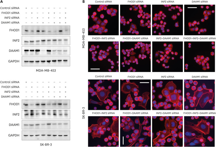
Methods: Using online databases, we identified a negative prognostic correlation between specific formins mRNA expression in HER2-positive cancers. To validate these findings at the protein level, immunohistochemistry was performed on HER2 subtype breast cancer tumors to establish the links between staining patterns and clinical characteristics. We then knocked down individual or combined formins in MDA-MB-453 and SK-BR-3 cells and investigated their effects on wound healing, transwell migration, and proliferation. Furthermore, we investigated the effects of erb-b2 receptor tyrosine kinase 2 (ERBB2)/HER2 small interfering RNA (siRNA)-mediated knockdown on the PI3K/Akt and MEK/ERK1 pathways as well as on selected formins.

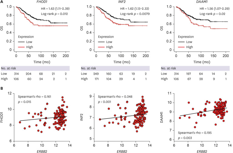
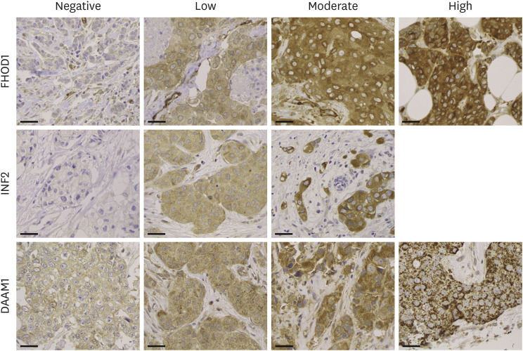
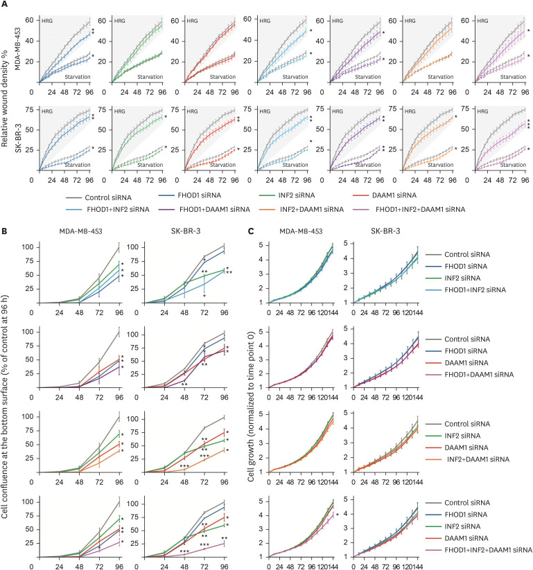
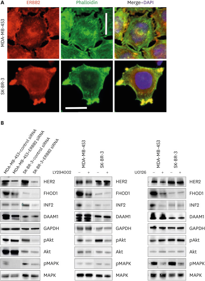
Results: Our results revealed that correlations between INF2, FHOD1, and DAAM1 mRNA expression and ERBB2 in HER2-subtype breast cancer were associated with worse outcomes. Using immunohistochemistry, we found that high FHOD1 protein expression was linked to higher histological grades and was negatively correlated with estrogen and progesterone receptor positivity. Upon formins knockdown, we observed effects on wound healing and transwell migration, with a minimal impact on proliferation, which was evident through single and combined knockdowns in both cell lines. Notably, siRNA-mediated knockdown of HER2 affected FHOD1 and INF2 expression, along with the phosphorylated Akt/MAPK states.

Conclusion: Our study highlights the roles of FHOD1 and INF2 as downstream effectors of the HER2/Akt and HER2/MAPK pathways, suggesting that they are potential therapeutic targets in HER2-positive breast cancer.

Keywords: Breast Neoplasms; Cytoskeletal Proteins; Formins; Receptor, ErbB-2; Signal Transduction.


Conflict of interest statement

The authors declare that they have no competing interests.

Figures

Figure 1
Figure 1. Kaplan–Meier overall survival and correlations between ERBB2 and formin mRNA expression. (A) Kaplan–Meier OS analyses of HER2-positive breast cancer samples reveal significantly poorer prognosis associated with FHOD1, INF2, and DAAM1 mRNA expression. The number of cases is denoted at the bottom of the figures. (B) ERBB2 mRNA expression in HER2-positive subtype breast tumors (n = 230) exhibits a significant positive correlation with FHOD1, INF2, and DAAM1. Kaplan–Meier analyses and correlations for all formins, excluding GRIDIP, are depicted in Supplementary Figures 1 and 2. ERBB2 = erb-b2 receptor tyrosine kinase 2; HER2 = human epidermal growth factor receptor 2; OS = overall survival; HR = hazard ratio.
Figure 2
Figure 2. Immunohistochemistry. Tissue microarrays were sectioned at 3.5 μm and immunostained using FHOD1, INF2, or DAAM1 antibodies, following the streptavidin-peroxidase technique. The samples were evaluated based on the predominant staining intensity and categorized into four levels: negative, low, moderate, or strong. No instances of strong INF2 intensity were detected. Magnification: 400×. Scale bars = 50 µm.
Figure 3
Figure 3. Localization of FHOD1, INF2, and DAAM1 in MDA-MB-453 and SK-BR-3 breast cancer cell lines. Upper panel: FHOD1 (red) is predominantly seen as cytoplasmic dots. Middle panel: INF2 (red) is distributed in the cytoplasm colocalized with phalloidin (green) and lamellipodia. Lower panel: DAAM1 (red) appears as cytoplasmic dots with co-localization with actin filaments. Magnification: 400×. Scale bars = 20 µm.
Figure 4
Figure 4. Small interfering RNA-mediated knockdown of FHOD1, INF2, DAAM1, or their combinations. (A) Knockdown effectiveness was assessed through immunoblotting 48-hour post-transfection, with GAPDH as a loading control. (B) Representative images of transfected cells with control, FHOD1, INF2, DAAM1, or combined siRNAs. Alexa Fluor 488-conjugated phalloidin and DAPI were used to visualize actin filaments and nuclei, respectively. Magnification: 400×. Scale bars = 50 µm. GAPDH = glyceraldehyde 3-phosphate dehydrogenase; siRNA = small interfering RNA; DAPI = 4′,6-diamidino-2-phenylindole.
Figure 5
Figure 5. Effect of knockdown of FHOD1, INF2, DAAM1 or their combination in wound healing, transwell migration, and proliferation. (A) Scratch wounds were performed 48-hour post siRNA transfection, and the medium subsequently replaced by starvation medium or starvation medium supplemented with 50 ng/mL HRG to speed migration. The wound relative densities were monitored over 96 hours. (B) Forty-eight hours after transfection, cells were seeded on the upper surface of the transwell migration chamber, while the lower chamber contained medium supplemented with 10% serum as a chemoattractant. Confluence of cells on the bottom surface of the membrane was monitored for 96 h and normalized based on the initial top surface cell area for each well. (C) For proliferation assessment, cells were plated to cover 5%–10% of the area 48-hour post-transfection, and the growth of confluence measured over 144 hours. Comparisons between cells treated with control siRNA and those subjected to FHOD1, INF2, DAAM1, and their combinations were analyzed using Student’s t-test, where *p ≤ 0.05, **p ≤ 0.01, and ***p ≤ 0.001 were considered statistically significant.
siRNA = small interfering RNA; HRG = heregulin.
Figure 6
Figure 6. HER2/ERBB2 knockdown alters FHOD1 and INF2 expression through Akt and MAPK pathways. (A) Subcellular localization of HER2 (in red) in MDA-MB-453 and SK-BR-3 cells, with ERBB2 concentrated in actin-rich cellular protrusions, observed through phalloidin staining (green). DAPI (blue) indicates nuclei. Magnification: 400×. Scale bars = 20 µm. (B) Cells were treated with ERBB2 siRNA for 48 hours, and the knockdown efficacy validated by western blotting in both MDA-MB-453 and SK-BR-3 cells. In MDA-MB-453 cells, HER2/ERBB2 knockdown decreased FHOD1, INF2, and DAAM1 levels, concomitant with a reduction in pMAPK. In SK-BR-3 cells, ERBB2 knockdown reduced FHOD1 and INF2 expression, accompanied by lowered Akt/pAkt and MAPK phosphorylation levels. Both cell lines were treated with MEK1/2 inhibitor UO126, PI3K inhibitor LY294002, or the control vehicle DMSO. Effective pathway inhibition was verified by western blotting of Akt and MAPK phosphorylation states. Treatment with PI3K or MAPK inhibitors in MDA-MB-453 cells emulated the effects of HER2/ERBB2 knockdown, decreasing FHOD1 and INF2 expression. In contrast, inhibiting PI3K or MAPK alone did not induce alterations in FHOD1 and INF2 levels in SK-BR-3 cells. Notably, DAAM1 levels remained unaffected by PI3K or MAPK inhibitor treatments in both cell lines. HER2 = human epidermal growth factor receptor 2; ERBB2 = erb-b2 receptor tyrosine kinase 2; DAPI = 4′,6-diamidino-2-phenylindole; siRNA = small interfering RNA.

Similar articles

Cited by

References

    1. Sung H, Ferlay J, Siegel RL, Laversanne M, Soerjomataram I, Jemal A, et al. Global cancer statistics 2020: GLOBOCAN estimates of incidence and mortality worldwide for 36 cancers in 185 countries. CA Cancer J Clin. 2021;71:209–249. - PubMed
    1. WHO. Breast Tumours. WHO Classification of Tumours. 5th ed. Geneva: WHO Classification Editorial Board; 2019.
    1. Holbro T, Beerli RR, Maurer F, Koziczak M, Barbas CF, 3rd, Hynes NE. The ErbB2/ErbB3 heterodimer functions as an oncogenic unit: ErbB2 requires ErbB3 to drive breast tumor cell proliferation. Proc Natl Acad Sci U S A. 2003;100:8933–8938. - PMC - PubMed
    1. Sirkisoon SR, Carpenter RL, Rimkus T, Miller L, Metheny-Barlow L, Lo HW. EGFR and HER2 signaling in breast cancer brain metastasis. Front Biosci (Elite Ed) 2016;8:245–263. - PMC - PubMed
    1. Tiwari RK, Borgen PI, Wong GY, Cordon-Cardo C, Osborne MP. HER-2/neu amplification and overexpression in primary human breast cancer is associated with early metastasis. Anticancer Res. 1992;12:419–425. - PubMed