Review
. 2024 Apr;20(4):206-217.
doi: 10.1038/s41581-023-00786-x. Epub 2023 Nov 20.

The immunoregulatory roles of non-haematopoietic cells in the kidney

Affiliations
Review

The immunoregulatory roles of non-haematopoietic cells in the kidney

George C Tsokos et al. Nat Rev Nephrol. 2024 Apr.

Erratum in

Abstract

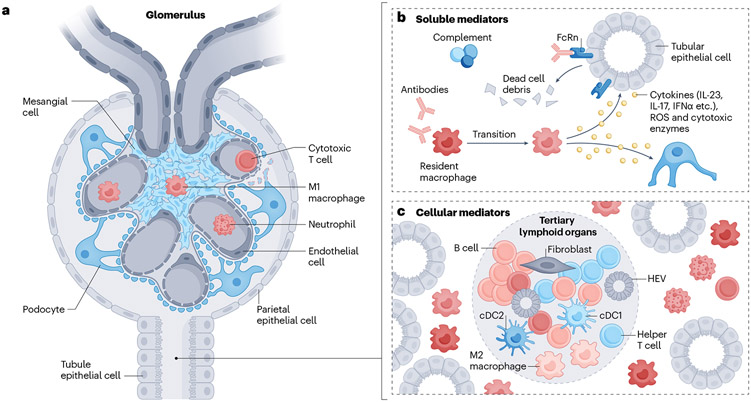
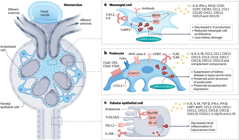
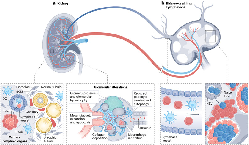
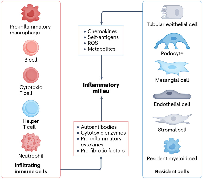
The deposition of immune complexes, activation of complement and infiltration of the kidney by cells of the adaptive and innate immune systems have long been considered responsible for the induction of kidney damage in autoimmune, alloimmune and other inflammatory kidney diseases. However, emerging findings have highlighted the contribution of resident immune cells and of immune molecules expressed by kidney-resident parenchymal cells to disease processes. Several types of kidney parenchymal cells seem to express a variety of immune molecules with a distinct topographic distribution, which may reflect the exposure of these cells to different pathogenic threats or microenvironments. A growing body of literature suggests that these cells can stimulate the infiltration of immune cells that provide protection against infections or contribute to inflammation - a process that is also regulated by draining kidney lymph nodes. Moreover, components of the immune system, such as autoantibodies, cytokines and immune cells, can influence the metabolic profile of kidney parenchymal cells in the kidney, highlighting the importance of crosstalk in pathogenic processes. The development of targeted nanomedicine approaches that modulate the immune response or control inflammation and damage directly within the kidney has the potential to eliminate the need for systemically acting drugs.


Figures

Fig. 1 ∣
Fig. 1 ∣. Soluble and cellular mediators of inflammation in the kidney.
a, immune cells, including macrophages and dendritic cells, may enter the glomerulus to instigate damage; cytotoxic T cells might also enter and cause podocyte apoptosis when the basement membrane is injured. b, Activated components of the complement system and/or autoantibodies can attack kidney parenchymal cells (KPCs), such as tubular epithelial cells, directly, to cause tissue damage. Alternatively, they can recruit and/or activate myeloid cells to induce the release of cytokines, reactive oxygen species (ROS) and cytotoxic enzymes. c, Different immune cell clusters composed of different types of lymphocytes can be observed in the interstitial region. Some clusters display features typical of secondary lymphoid organs, including the presence of T cell and B cell zones aligned with the formation of high endothelial venules (HEVs) and new lymphatic vessels; these clusters are termed tertiary lymphoid organs.
Fig. 2 ∣
Fig. 2 ∣. Immune features of kidney parenchymal cells.
Kidney parenchymal cells, including podocytes, tubular epithelial cells and mesangial cells, express molecules that are typical of the innate and adaptive immune systems. These molecules have important roles in the recruitment of immune cells and in the development of inflammatory responses. a, Mesangial cells have long been known to express cytokines and chemokines in response to circulating inflammatory signals. For example, IL-6 can independently cause mesangial proliferation. Signalling molecules, such as CaMK4, may control the production of cytokines, and may therefore represent a treatment target. b, Podocytes produce and express molecules typically produced by cells of the immune system, including HLA, costimulatory molecules, the neonatal Fc receptor (FcRn), TLRs, and chemokines and their receptors, along with complement components and receptors. The transcription factor TWIST1 may suppress the production of CCL2 in healthy glomeruli. Podocyte injury may lead to the altered expression of molecules such as CaMK4, which can directly compromise the expression of molecules involved in their function (e.g. nephrin) or structure (e.g. synaptopodin). c, Similarly, TECs may produce cytokines and chemokines and their receptors, which may promote the recruitment of inflammatory cells. Such cells may, through the further production of cytokines, contribute to the damage of TECs. Again, alterations in the expression of molecules such as CaMK4 may contribute to the accumulation of immune cells and may therefore representa therapeutic target. FcRn, neonatal Fc receptor; TLR, Toll-like receptor; PDL1, programmed death ligand 1; IRAK, IL-1 receptor-associated kinase; TRAF6, TNF receptor-associated factor 6; BAFF, B cell–activating factor; LMP7, low–molecular mass polypeptide-7; CAMK4, calcium/calmodulin dependent protein kinase IV.
Fig. 3 ∣
Fig. 3 ∣. Lymphoid organs in kidney disease.
a, Aged and chronically diseased kidneys can contain atrophic tubules, sclerotic glomeruli, and fibrosis, as well as tertiary lymphoid organs, which are lymphoid aggregates of T cells and B cells, high endothelial venules (HEVs) and lymphatic vessels. b, Kidney-resident dendritic cells (DCs) and T cells can circulate via lymphatic vessels from the kidney cortex to the kidney-draining lymph node (KLN), where they interact with T cells in the paracortex with the assistance of fibroblastic reticular cells. The fibroblastic reticular cells produce conduits composed of extracellular matrix (ECM), which provide structural support for the proliferation and expansion of HEVs, through which naïve T cells enter the KLN from the systemic circulation. Activation of T cells in the KLN is critical to the pathogenesis of glomerulonephritis, ischaemia–reperfusion injury and unilateral ureteral obstruction in mice.
Fig. 4 ∣
Fig. 4 ∣. Intercellular crosstalk within the kidneys between resident and infiltrating cells.
Immune cells attack tissue resident cells by producing various pathogenic factors such as antibodies, pro-inflammatory cytokines, pro-fibrotic factors and cytotoxic enzymes. Conversely, injured tissue resident cells, including resident immune cells and kidney parenchymal cells, respond by altering the local inflammatory milieu, which contributes further to the progression of renal inflammation. ROS, reactive oxygen species.

References

    1. Pisetsky DS Pathogenesis of autoimmune disease. Nat. Rev. Nephrol 19, 509–524 (2023). - PMC - PubMed
    1. Mohan C, Zhang T & Putterman C Pathogenic cellular and molecular mediators in lupus nephritis. Nat. Rev. Nephrol 19, 491–508 (2023). - PubMed
    1. Maeda K. et al. CaMK4 compromises podocyte function in autoimmune and nonautoimmune kidney disease. J. Clin. Invest 128, 3445–3459 (2018). - PMC - PubMed
    1. Li H. et al. IL-23 reshapes kidney resident cell metabolism and promotes local kidney inflammation. J. Clin. Invest 131, e142428 (2021). - PMC - PubMed
    1. Tsokos GC Systemic lupus erythematosus. N. Engl. J. Med 365, 2110–2121 (2011). - PubMed

Publication types

LinkOut - more resources