. 2023 Nov 29;137(22):1721-1751.
doi: 10.1042/CS20230319.

Hallmarks of ageing in human skeletal muscle and implications for understanding the pathophysiology of sarcopenia in women and men

Affiliations

Hallmarks of ageing in human skeletal muscle and implications for understanding the pathophysiology of sarcopenia in women and men

Antoneta Granic et al. Clin Sci (Lond). .

Abstract

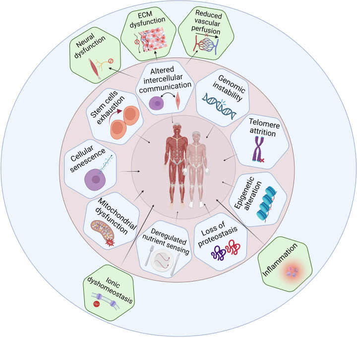
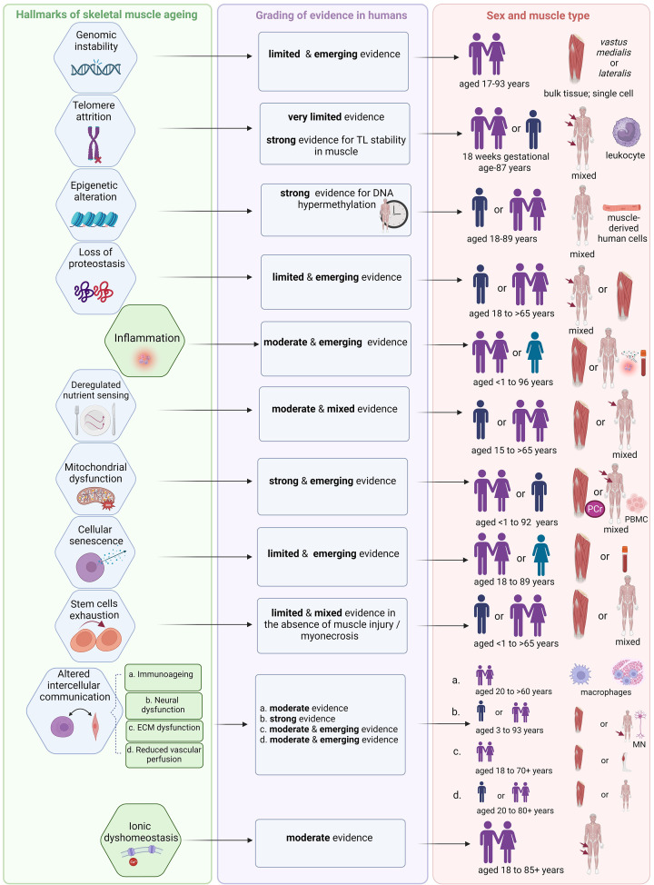
Ageing is a complex biological process associated with increased morbidity and mortality. Nine classic, interdependent hallmarks of ageing have been proposed involving genetic and biochemical pathways that collectively influence ageing trajectories and susceptibility to pathology in humans. Ageing skeletal muscle undergoes profound morphological and physiological changes associated with loss of strength, mass, and function, a condition known as sarcopenia. The aetiology of sarcopenia is complex and whilst research in this area is growing rapidly, there is a relative paucity of human studies, particularly in older women. Here, we evaluate how the nine classic hallmarks of ageing: genomic instability, telomere attrition, epigenetic alterations, loss of proteostasis, deregulated nutrient sensing, mitochondrial dysfunction, cellular senescence, stem cell exhaustion, and altered intercellular communication contribute to skeletal muscle ageing and the pathophysiology of sarcopenia. We also highlight five novel hallmarks of particular significance to skeletal muscle ageing: inflammation, neural dysfunction, extracellular matrix dysfunction, reduced vascular perfusion, and ionic dyshomeostasis, and discuss how the classic and novel hallmarks are interconnected. Their clinical relevance and translational potential are also considered.

Keywords: hallmarks of ageing; sarcopenia; skeletal muscle.


Conflict of interest statement

A.G., K.S., M.G., and A.A.S. declare no competing interests associated with the manuscript. T.S. is an employee of Regeneron and holds stock in Regeneron.

Figures

Figure 1
Figure 1. Schematic diagram of skeletal muscle tissue and of a single muscle fibre organisation
Panels (A,B) present schematic diagrams of the skeletal muscle tissue and muscle extracellular matrix (ECM)-tendon organisation. The muscle ECM is categorised into epimysium (a layer surrounding the entire muscle), perimysium (a layer surrounding muscle fascicles or the muscle fibre bundles, which are further grouped together to form the muscle tissue), and endomysium (a basal lamina that is in intimate contact with individual myofibres). In the longitudinal section of the muscle (panel B), the endomysium is enclosed within the fascicles, whereas the perimysium is presented continuously with the tendon. Panel (C) represents a single myofibre comprised of the myofibrils organised into sarcomeres enclosed in the sarcolemma. The sarcoplasmic reticulum entangles the fibrils, and the transverse (T) tubules intersect them. Mitochondria are secured near the T-tubules to sarcoplasmic reticulum junction and (or) are found in subsarcolemmal regions. Along the length of the myofibres are nerves (attached at the NMJ) and capillaries (often near satellite cells). Panels A and B from Gillies, A.R. and Lieber, R.L. (2011) Structure and function of the skeletal muscle extracellular matrix. Muscle Nerve 44, 318–331. Panel C was adapted from [15] and used with the permission of Wiley.
Figure 2
Figure 2. Comparison of skeletal muscle morphology and architecture in young and old adults
Panel (A) depicts representative cross-sections of the vastus lateralis (VL) muscle biopsies from young (left; aged ∼24 years) and old (right; aged ∼70 years) healthy, active men, immuno-stained with antibodies specific to adult myosin heavy chain (MYH) isoforms: blue (anti-MYH7, slow type 1 myofibres), green (anti-MYH2, fast type 2A myofibres), and red (anti-MYH1, fast type 2X myofibres) (scale bar: 100 μm). Compared with the relatively uniform size of myofibres in young muscles (left; Panel A), the main features of old muscle are a wider range (variability) of size and shapes especially in type 2 myofibres (right; Panel (A), white arrow). Panel (B) shows a magnetic resonance imaging (MRI) of human thigh with area of quadriceps muscle (grey) and surrounding fatty tissue (white) in young (left) compared with loss of muscle mass in older men (right). Panel (A) from [38] was used with the permission from Cell Press. Panel (B) from Herrmann, M., Engelke, K., Ebert, R., Müller-Deubert, S., Rudert, M., Ziouti, F. et al. (2020) Interactions between Muscle and Bone-Where Physics Meets Biology. Biomolecules10, 432, with permission from MDPI.
Figure 3
Figure 3. Hallmarks of ageing as applied to human skeletal muscle
Hallmarks of ageing were evaluated for their involvement in skeletal muscle ageing and sarcopenia. The nine classic hallmarks [1] are depicted in the inner (pink) circle. The outer (blue) circle highlights potential new hallmarks of skeletal muscle ageing and sarcopenia. Created with BioRender.com.
Figure 4
Figure 4. Key components of motor unit and neuromuscular junction
Schematic diagram of the motorneuron-skeletal muscle connection that is critical for the transfer of electrical signals for myofibre contraction. The motorneuron cell body in the spinal cord, sends an electrical signal down the motorneuron axon to the NMJ on the surface of an individual myofibre. The pre-synaptic nerve terminal release vesicles containing the neurotransmitter acetylcholine, this binds to receptors on the post-synaptic folds on the myofibre surface (sarcolemma). This signal is transferred along the myofibre surface and then into the T-tubules to initiate contraction. In mature human NMJs, the post-synaptic junction on each myofibre forms complex folds, a typical form of mature NMJ (in red; bottom right). The NMJ is innervated by a single motor axon (in purple; bottom right). Created with BioRender.com. ACh receptor, acetylcholine receptor.
Figure 5
Figure 5. Summary of evidence from human studies for each hallmark of ageing applied to skeletal muscle
For each of the nine classic (blue) and five novel hallmarks of ageing (green), the strength of evidence for human skeletal muscle ageing is graded in the middle column and depicted in the end column according to the sex (with age range) and type of muscle (vastus lateralis or mixed) most used in these studies. While specific data are available and emerging for some of these hallmarks in the context of sarcopenia (see individual Hallmark sections) these data are limited. Because of the relative paucity of studies for women and for investigating sex-specific differences across the lifespan for hallmarks of ageing, possible sex-related differences could not be determined at this time. For example, out of six individual studies included for Hallmark: Cellular senescence, only one feasibility pilot study explored senescence markers in men and women separately [195]. Moreover, consensus on clinical diagnosis of sarcopenia is still emerging: consequently, earlier studies likely included sarcopenic individuals who were not diagnosed as such. Therefore, at this stage, it is appropriate to evaluate the hallmarks based on evidence for normal ageing human muscle. This provides the background for progression of sarcopenia (as an adverse manifestation of normal ageing) [10,11] and, in limited cases, provides direct evidence of involvement of a hallmark in sarcopenia. Created with BioRender.com. ECM, extracellular matrix; MN, motor neuron; PBMC, peripheral blood mononuclear cells; PCr, phosphocreatine by 31P magnetic resonance spectroscopy; TL, telomere length.

References

    1. López-Otín C., Blasco M.A., Partridge L., Serrano M. and Kroemer G. (2013) The hallmarks of aging. Cell 153, 1194–1217 10.1016/j.cell.2013.05.039 - DOI - PMC - PubMed
    1. López-Otín C., Blasco M.A., Partridge L., Serrano M. and Kroemer G. (2023) Hallmarks of aging: An expanding universe. Cell 186, 243–278 10.1016/j.cell.2022.11.001 - DOI - PubMed
    1. Schmauck-Medina T., Molière A., Lautrup S., Zhang J., Chlopicki S., Madsen H.B.et al. . (2022) New hallmarks of ageing: a 2022 Copenhagen ageing meeting summary. Aging 14, 6829–6839 10.18632/aging.204248 - DOI - PMC - PubMed
    1. Fraser H.C., Kuan V., Johnen R., Zwierzyna M., Hingorani A.D., Beyer A.et al. . (2022) Biological mechanisms of aging predict age-related disease co-occurrence in patients. Aging Cell 21, e13524 10.1111/acel.13524 - DOI - PMC - PubMed
    1. Hanahan D. and Weinberg R.A. (2011) Hallmarks of cancer: the next generation. Cell 144, 646–674 10.1016/j.cell.2011.02.013 - DOI - PubMed