[Preprint]. 2024 Jan 30:2023.11.08.566169.
doi: 10.1101/2023.11.08.566169.

Aberrant hippocampal Ca2+ micro-waves following synapsin-dependent adeno-associated viral expression of Ca2+ indicators

Affiliations

Aberrant hippocampal Ca2+ micro-waves following synapsin-dependent adeno-associated viral expression of Ca2+ indicators

Nicola Masala et al. bioRxiv. .

Update in

Abstract

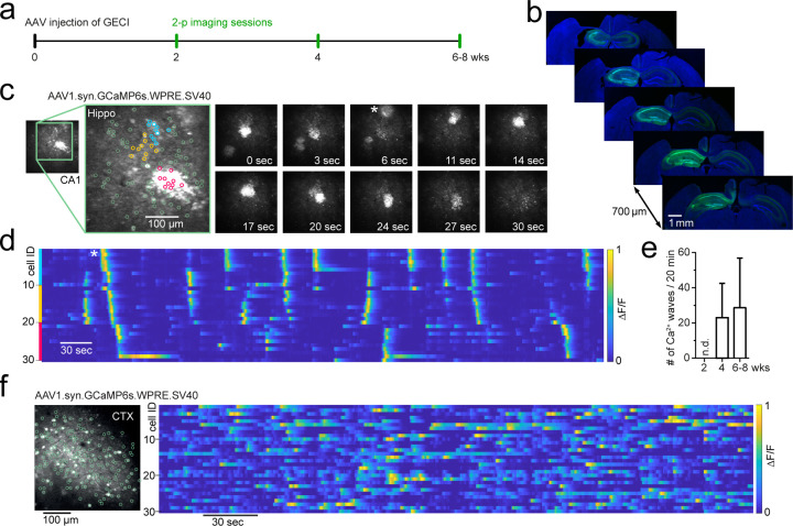
Genetically encoded calcium indicators (GECIs) such as GCaMP are invaluable tools in neuroscience to monitor neuronal activity using optical imaging. The viral transduction of GECIs is commonly used to target expression to specific brain regions, can be conveniently used with any mouse strain of interest without the need for prior crossing with a GECI mouse line and avoids potential hazards due to the chronic expression of GECIs during development. A key requirement for monitoring neuronal activity with an indicator is that the indicator itself minimally affects activity. Here, using common adeno-associated viral (AAV) transduction procedures, we describe spatially confined aberrant Ca2+ micro-waves slowly travelling through the hippocampus following expression of GCaMP6, GCaMP7 or R-CaMP1.07 driven by the synapsin promoter with AAV-dependent gene transfer, in a titre-dependent fashion. Ca2+ micro-waves developed in hippocampal CA1 and CA3, but not dentate gyrus (DG) nor neocortex, were typically first observed at 4 weeks after viral transduction, and persisted up to at least 8 weeks. The phenomenon was robust, observed across laboratories with various experimenters and setups. Our results indicate that aberrant hippocampal Ca2+ micro-waves depend on the promoter and viral titre of the GECI, density of expression as well as the targeted brain region. We used an alternative viral transduction method of GCaMP which avoids this artifact. The results show that commonly used Ca2+-indicator AAV transduction procedures can produce artefactual Ca2+ responses. Our aim is to raise awareness in the field of these artefactual transduction-induced Ca2+ micro-waves and we provide a potential solution.


Conflict of interest statement

Conflict of Interest None of the authors have a conflict of interest.

Figures

Figure 1:
Figure 1:. Development of Ca2+ micro-waves travelling through hippocampus following GCaMP transduction.
a: Experimental protocol to examine CA1 neuronal activity using two-photon imaging following AAV transduction of genetically encoded Ca2+ indicators. b: Immunohistochemical sections following last imaging session. GCaMP6s (AAV1.syn.GCaMP6s.SV40, addgene #100843) expression throughout the ipsilateral hippocampus and projection pathways in the contralateral hippocampus. c: Two-photon Ca2+ imaging of FOV in CA1 at 4wks p.i. showing aberrant Ca2+ micro-waves (see also Supplementary video 1). Magnified inset shows 3 colored neuronal subgroups (blue, orange, magenta) based on their spatial vicinity from a total population of 100 identified neurons (green). right: time series of two-photon Ca2+ imaging FOVs showing two Ca2+ micro-waves, the first at 0 sec, the second appearing at 6 sec (asterisk). The second wave progresses through FOV over dozens of seconds. d: Raster-plot of individual neuronal Ca2+ activity (ΔF/F, 1min moving window, traces max-normalized per neuron) from neighboring subgroups (colors correspond to c). Asterisk (same as in c): a Ca2+ micro-wave advances through neighboring neuronal subgroups. e: Occurrence rate of aberrant Ca2+ micro-waves with increasing expression time, following viral transduction of AAV1.syn.GCaMP6s.SV40 in mature C57BL6 wild-type animals. n.d. = none detected. f: Two-photon Ca2+ imaging FOV in the visual cortex at 6wks p.i. (left) with normal sparse spontaneous Ca2+ activity and no detected Ca2+ micro-waves (right; raster plot of ΔF/F, 1min moving window, traces max-normalized per neuron).
Fig. 2
Fig. 2. Aberrant Ca2+ micro-waves are consistent across laboratories and GECI variant.
a: Plot of the occurrence rate of aberrant Ca2+ micro-waves in CA1 at the different institutes at 6–8wks after injection of GCaMP6s or GCaMP6m. b: Ca2+ micro-wave diameters (left) and progression speed (right) in CA1 from each animal recorded across institutes. Inset: Histogram of fluorescent intensity taken across each Ca2+-wave within an animal. Green line is the average, areas outside dashed lines mark 10% lowest fluorescence values, which were excluded from analysis. c: Plot of the occurrence rate of aberrant Ca2+ micro-waves in CA1 following injection with commonly used GECIs (see table 1). d: Two-photon Ca2+ imaging FOV (left) in hippocampal CA1 following dual injection approach for conditional GCaMP6s expression (6 wks p.i.) with normal sparse spontaneous Ca2+ activity and no detection of Ca2+ micro-waves (right; raster plot of ΔF/F, 1min moving window, traces max-normalized per neuron).

References

    1. Application Specialist Team. (2023). “Storm in the hippocampus.” Retrieved from https://twitter.com/4Specialists/status/1639248903039057920
    1. Cai DJ, Aharoni D, Shuman T, … Silva AJ. (2016). A shared neural ensemble links distinct contextual memories encoded close in time. Nature, 534(7605), 115–118. - PMC - PubMed
    1. Chen T-W, Wardill TJ, Sun Y, … Kim DS. (2013). Ultrasensitive fluorescent proteins for imaging neuronal activity. Nature, 499(7458), 295–300. - PMC - PubMed
    1. Dana H, Sun Y, Mohar B, … Kim DS. (2019). High-performance calcium sensors for imaging activity in neuronal populations and microcompartments. Nature Methods 2019 16:7, 16(7), 649–657. - PubMed
    1. Dombeck DA, Harvey CD, Tian L, Looger LL, & Tank DW. (2010). Functional imaging of hippocampal place cells at cellular resolution during virtual navigation. Nature Neuroscience, 13(11), 1433–1440. - PMC - PubMed

Publication types