[Preprint]. 2023 Oct 8:2023.10.08.561411.
doi: 10.1101/2023.10.08.561411.

Semi-synthetic nanobody-ligand conjugates exhibit tunable signaling properties and enhanced transcriptional outputs at neurokinin receptor-1

Affiliations

Semi-synthetic nanobody-ligand conjugates exhibit tunable signaling properties and enhanced transcriptional outputs at neurokinin receptor-1

Nayara Braga Emidio et al. bioRxiv. .

Update in

Abstract

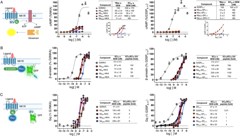
Antibodies have proven highly valuable for therapeutic development; however, they are typically poor candidates for applications that require activation of G protein-coupled receptors (GPCRs), the largest collection of targets for clinically approved drugs. Nanobodies (Nbs), the smallest antibody fragments retaining full antigen-binding capacity, have emerged as promising tools for pharmacologic applications, including GPCR modulation. Past work has shown that conjugation of Nbs with ligands can provide GPCR agonists that exhibit improved activity and selectivity compared to their parent ligands. The neurokinin-1 receptor (NK1R), a GPCR targeted for the treatment of pain, is activated by peptide agonists such as Substance P (SP) and neurokinin A (NKA), which induce signaling through multiple pathways (Gs, Gq and β-arrestin). In this study, we investigated whether conjugating NK1R ligands with Nbs that bind to a separate location on the receptor would provide chimeric compounds with distinctive signaling properties. We employed sortase A-mediated ligation to generate several conjugates consisting of Nbs linked to NK1R ligands. Many of these conjugates exhibited divergent and unexpected signaling properties and transcriptional outputs. For example, some Nb-NKA conjugates showed enhanced receptor binding capacity, high potency partial agonism, prolonged cAMP production, and an increase in transcriptional output associated with Gs signaling; whereas other conjugates were virtually inactive. Nanobody conjugation caused only minor alterations in ligand-induced upstream Gq signaling with unexpected enhancements in transcriptional (downstream) responses. Our findings underscore the potential of nanobody conjugation for providing compounds with advantageous properties such as biased agonism, prolonged duration of action, and enhanced transcriptional responses. These compounds hold promise not only for facilitating fundamental research on GPCR signal transduction mechanisms but also for the development of more potent and enduring therapeutics.


Conflict of interest statement

Declaration of interests: The authors declare no competing interests.

Figures

Figure 1.
Figure 1.. Receptor constructs and synthesis of Nb-peptide conjugates.
(A) NK1R signals through Gq, Gs (cAMP production) and β-arrestin. Epitope tags (alfa, 6e and BC2) were inserted into the N-terminus to enable Nb recognition. (B) Nanobodies and peptides were conjugated via Sortase A (SrtA)-mediated ligation. (C) Substance P (SP) activation of epitope-tagged NK1R. Receptor activation was evaluated by signal generated from a stably expressed cAMP-responsive luciferase construct. Data points (mean ± SD) correspond to technical replicates from a single representative experiment. Data from independent replicates are shown in Figure S4. (D) Biotin-labeled nanobodies, except NbGFP, bound to epitope-tagged NK1R, while no binding was observed with NK1R wild-type. Dose-response curves were generated from application of a sigmoidal concentration-response 3 parameter model. Data from independent replicates are shown in Figure S5.
Figure 2.
Figure 2.. Effects of peptides and conjugates on NK1R signaling.
(A) HEK293 cells stably transfected with Glosensor cAMP reporter (Promega Corp.) and epitope-tagged were treated with varying concentrations (1 pM – 10 μM) of the indicated peptides or conjugates. Activation was assessed by cAMP production after 6 min as described in Methods. Insets of concentration-responses without G3-peptides included are shown (under table) to allow better visualization of the activity of the weaker compounds. *Max activity values were calculated by normalizing the response at 1 μM for conjugates to that of G3NKA or G3SP6–11 at 10 μM (n=3). EC50 for conjugates with maximal responses lower than 30% were not determined (ND). (B) HEK293 cells stably expressing epitope-tagged NK1R were transfected with Gq TRUPATH Ga/b/g biosensor plasmid and treated with varying concentrations (0.1 pM – 1 μM) of the indicated peptides or conjugates and BRET signals were measured. (C) HEK293 cells stably expressing epitope-tagged NK1R were transfected with β-arrestin2-RlucII/rGFP-CAAX plasmids and treated with varying concentrations (0.1 pM – 1 μM). Representative concentration-response curves are shown, where data points correspond to mean ± SD from technical replicates. EC50 values were calculated from the fitting of a sigmoidal concentration-response model to data from n ≥ 3 independent experiments.
Figure 3.
Figure 3.. Impact of Nb conjugation on ligand competition binding and duration of cAMP activity.
(A, B) A competition binding assay was performed using flow cytometry to assess the ability of 100 nM (A) NKA and Nb-NKA or (B) SP6–11 and Nb-SP6–11 to outcompete the binding of fluorescently labeled SP to epitope-tagged NK1R. Data represent mean ± SEM from n ≥ 3 independent experiments. (C, D) Representative concentration-response curves or cAMP production kinetics (mean ± SD) for washout assays. 12 minutes after ligand addition, excess/unbound ligand was removed, fresh media was added to cells expressing (C) epitope-tagged or (D) wild-type NK1R, and cAMP responses were measured for an additional 30 min (washout) (n ≥ 3) according to Methods. Washout assay data were quantified as the area under the curve (AUC). Curves result from the fitting of a sigmoidal concentration-response model to data. Differences were evaluated for statistical significance with a one-way ANOVA followed by Dunnett correction. *p < 0.03, **p < 0.002, ***p < 0.0002, **** p < 0.0001
Figure 4.
Figure 4.. Evaluation of peptide and conjugate performance on transcription.
Transcriptional responses were evaluated through transfection of cells expressing epitope tagged NK1R with a luciferase reporter plasmid reporting on Gs or Gq signaling. Cells were incubated with ~35 nM of the indicated peptides or conjugates for 17 hours and transcription was measured as described in Methods. Data represent mean ± SEM from n ≥ 3 independent experiments. One-way ANOVA followed by Dunnett correction was performed to assess differences between free peptides and conjugates. *p < 0.03, **** p < 0.0001.
Figure 5.
Figure 5.. Model of epitope-tagged NK1R structures generated using AlphaFold 2 and summary of conjugate signaling activities.
(A) In the model in the left, the epitope tags are highlighted as follows: alfa orange, 6e maroon and BC2 purple. Compound bound to the receptor is NKA shown as orange sticks with receptor in gray. In the model in the right Nb6e is represented in blue and red spheres mark the C-terminus, which serves as the site of peptide conjugation. (B) Heat map summarizing ligand potencies (relative EC50 values) and efficacies (normalized maximal response values) for cAMP signaling and ligand potencies for Gq and β-arrestin2 signaling. Pink squares with X represent correspond to compounds for which activity was too weak to accurately calculate an EC50. *Max activity values were calculated by normalizing the response at 1 μM for conjugates to that of G3NKA or G3SP6–11 at 10 μM

References

    1. Cheloha RW, Harmand TJ, Wijne C, Schwartz TU, Ploegh HL. Exploring cellular biochemistry with nanobodies. J Biol Chem. 2020;295(45):15307–27. Epub 20200831. doi: 10.1074/jbc.REV120.012960. - DOI - PMC - PubMed
    1. Muyldermans S. Applications of Nanobodies. Annual Review of Animal Biosciences. 2021;9(1):401–21. doi: 10.1146/annurev-animal-021419-083831. - DOI - PubMed
    1. Sriram K, Insel PA. G Protein-Coupled Receptors as Targets for Approved Drugs: How Many Targets and How Many Drugs? Mol Pharmacol. 2018;93(4):251–8. Epub 20180103. doi: 10.1124/mol.117.111062. - DOI - PMC - PubMed
    1. Manglik A, Kobilka BK, Steyaert J. Nanobodies to Study G Protein–Coupled Receptor Structure and Function. Annual Review of Pharmacology and Toxicology. 2017;57(1):19–37. doi: 10.1146/annurev-pharmtox-010716-104710. - DOI - PMC - PubMed
    1. Heukers R, De Groof TWM, Smit MJ. Nanobodies detecting and modulating GPCRs outside in and inside out. Current Opinion in Cell Biology. 2019;57:115–22. doi: 10.1016/j.ceb.2019.01.003. - DOI - PubMed

Publication types