[Preprint]. 2023 Nov 9:2023.11.08.566350.
doi: 10.1101/2023.11.08.566350.

The HIV-1 capsid serves as a nanoscale reaction vessel for reverse transcription

Affiliations

The HIV-1 capsid serves as a nanoscale reaction vessel for reverse transcription

Jordan Jennings et al. bioRxiv. .

Update in

Abstract

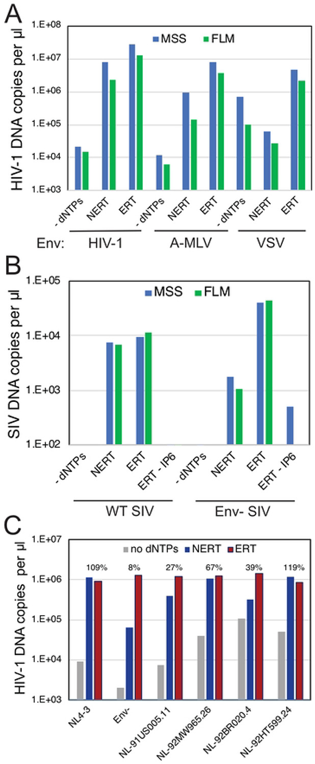
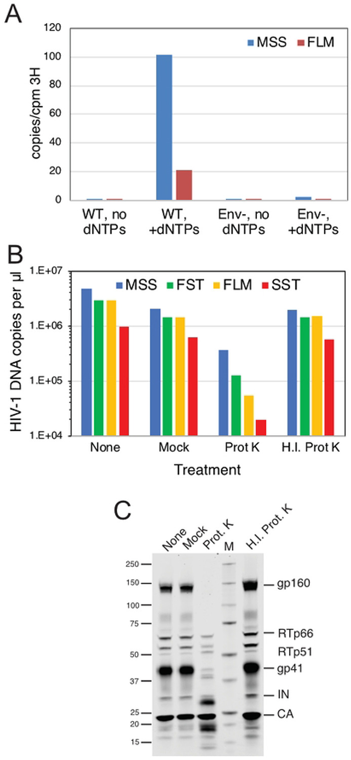
The viral capsid performs critical functions during HIV-1 infection and is a validated target for antiviral therapy. Previous studies have established that the proper structure and stability of the capsid are required for efficient HIV-1 reverse transcription in target cells. Moreover, it has recently been demonstrated that permeabilized virions and purified HIV-1 cores undergo efficient reverse transcription in vitro when the capsid is stabilized by addition of the host cell metabolite inositol hexakisphosphate (IP6). However, the molecular mechanism by which the capsid promotes reverse transcription is undefined. Here we show that wild type HIV-1 particles can undergo efficient reverse transcription in vitro in the absence of a membrane-permeabilizing agent. This activity, originally termed "natural endogenous reverse transcription" (NERT), depends on expression of the viral envelope glycoprotein during virus assembly and its incorporation into virions. Truncation of the gp41 cytoplasmic tail markedly reduced NERT activity, indicating that gp41 permits the entry of nucleotides into virions. Protease treatment of virions markedly reduced NERT suggesting the presence of a proteinaceous membrane channel. By contrast to reverse transcription in permeabilized virions, NERT required neither the addition of IP6 nor a mature capsid, indicating that an intact viral membrane can substitute for the function of the viral capsid during reverse transcription in vitro. Collectively, these results demonstrate that the viral capsid functions as a nanoscale container for reverse transcription during HIV-1 infection.


Figures

Fig 1.
Fig 1.. Hypothetical models for the role of the HIV-1 capsid in reverse transcription.
A. The container model posits that the capsid serves to maintain the concentrations of proteins and nucleic acids required for reverse transcription and predicts that the capsid must remain fully intact during the reaction. B. In the scaffold model, the inner face of the assembled capsid functions as a platform on which the reaction takes place, suggesting that the capsid may be semi-intact.
Fig 2.
Fig 2.. IP6-dependent ERT reactions are resistant to degradation by DNase I.
ERT reactions were performed with HIV-1 particles in the presence of the indicated concentrations of IP6, with or without added DNase I. Reactions were incubated for 4h. In reactions 10-12, PF74 was added to a concentration of 20 μM and DNAse I to 20 μg/ml, and the reactions were incubated for an additional 60 min. Products were purified and quantified by qPCR using primers specific for minus strand strong stop (MSS) and full length minus strand (FLM) amplicons. Results shown are representative of two independent experiments.
Fig 3.
Fig 3.. Reverse transcription in nonpermeabilized HIV-1 particles does not require addition of IP6 and is resistant to PF74.
Reactions were incubated for 14h. A. Reactions with wild type HIV-1 particles were performed in the presence or absence of dNTPs (dATP, dCTP, dGTP, and TTP; 0.1 mM each), 0.1% Triton X-100, and 0.1 mM IP6. B. Reactions were performed in the absence or presence of detergent and IP6 (NERT and ERT, respectively) with the indicated inhibitors: 10 μM azidothymidine triphosphate (AZTTP); 1 μM efavirenz (EFV); 10 μM stavudine triphosphate (d4TTP); 1 mM aldrithiol (AT-2); 10 μM PF-3450074 (PF74). The reaction designated “complete” contained dNTPs but no inhibitor. Results shown are representative of two independent experiments.
Fig 4.
Fig 4.. Reverse transcription in nonpermeabilized HIV-1 particles requires the gp41 CT.
A. Reactions containing wild type (WT), Env-deficient (Env), and the indicated gp41 C-terminal truncation mutants were performed in the absence (NERT) and presence (ERT) of detergent and IP6. Reactions were incubated for 14h and the early (MSS) and late (FLM) products quantified by qPCR. Numerical values in the graph represent the NERT to ERT ratio of FLM product levels in each reaction relative to that measured for wild type virions. B. Pelleted virions used in the reactions shown in A were analyzed by immunoblotting using a monoclonal antibody recognizing a membrane-proximal epitope in the gp41 CT. C. Ratio of band intensities of gp41 and CA shown in B. Error bars represent the range of values from two independent experiments. Results shown in each panel are representative of two independent experiments.
Fig 5.
Fig 5.. NERT activity depends on the viral Env protein.
NERT and ERT reactions were performed with the indicated viruses. A. Analysis of wild type HIV-1 particles (HIV-1), A-MLV pseudotyped HIV-1 particles (A-MLV), and VSV-G-pseudotyped HIV-1 particles (VSV). B. Analysis of wild type and Env- SIVmac239 particles. C. Analysis of wild type (NL4-3), Env-, and NL4-3 chimerae encoding Env proteins from HIV-1 primary isolates. Shown are the levels of late product synthesis detected in each NERT and ERT reaction. Results are representative of two independent experiments.
Fig 6.
Fig 6.. NERT efficiency in HIV-1 particles varies with the cellular source of the virions.
Wild type and Env- HIV-1 particles were harvested from the indicated T cell lines, concentrated, and assayed for NERT and ERT activity. Shown are the levels of early (MSS) and late (FLM) products. The numerical values above each sample represent the ratio of late product levels in the corresponding NERT and ERT reactions. Results shown are representative of two independent experiments.
Fig 7.
Fig 7.. Treatment of intact HIV-1 particles with protease reduces NERT activity.
A. NERT reactions in minimally processed wild type (WT) and Env- particles. Shown are the levels of early and late product synthesis in reactions containing and lacking dNTPs. B. NERT activity in concentrated virions after treatment for 1h with proteinase K (Prot K) or heat-inactivated proteinase K (H.I. Prot K). Reactions containing virions that were not incubated (None) and virions incubated with no proteinase K (Mock) served as controls. C. Immunoblot analysis of virions used in the reactions shown in B. The blot was probed with an antibody to the gp41 CT and with HIV-Ig, which recognizes RT, IN, and CA. Results shown are from one of two independent experiments.
Fig 8.
Fig 8.. HIV-1 particles with cleavable gp41 CT proteins exhibit reduced NERT activity.
A. Reactions were performed with the indicated wild type, Env-, and CT point mutant virions. Shown are the early (MSS and FST) and late (FLM and SST) products from the corresponding NERT and ERT reactions. B. Ratios of the DNA levels detected in the NERT and ERT reactions shown in A. C. Immunoblot analysis of the concentrated particles used in this experiment. Results shown are from one of two independent experiments.
Fig 9.
Fig 9.. NERT activity occurs in particles lacking a mature capsid.
A. NERT and ERT assays of the indicated wild type (NL4-3) and Gag cleavage mutants. CA5 particles contain uncleaved CA-SP1 protein; CA6 particles contain uncleaved CA-SP1-NC; MA-CA contains uncleaved MA-CA protein; MA-p2: uncleaved MA-CA-SP1; MA- NC: uncleaved MA-CA-SP1-NC; and MA-p6: uncleaved MA-CA-SP1-NC-SP1-p6. Shown are the early and late product DNA levels (FST and FLM, respectively). B. HIV-1 mutants bearing large deletions in CA are competent for NERT. Two mutants lacking nearly the entire N-terminal domain of CA were assayed for NERT and ERT with wild type and Env- particles. Numerical values shown represent the relative efficiency of ERT (FLM products normalized by exogenous RT activity levels in the viruses). The results shown are from one of two independent experiments.

References

    1. Campbell EM, Hope TJ. 2015. HIV-1 capsid: the multifaceted key player in HIV-1 infection. Nat Rev Microbiol 13:471–83. - PMC - PubMed
    1. Zila V, Muller TG, Muller B, Krausslich HG. 2021. HIV-1 capsid is the key orchestrator of early viral replication. PLoS Pathog 17:e1010109. - PMC - PubMed
    1. Tailor MW, Chahine EB, Koren D, Sherman EM. 2023. Lenacapavir: A Novel Long-Acting Capsid Inhibitor for HIV. Ann Pharmacother doi:10.1177/10600280231171375:10600280231171375. - DOI - PubMed
    1. Forshey BM, von Schwedler U, Sundquist WI, Aiken C. 2002. Formation of a human immunodeficiency virus type 1 core of optimal stability is crucial for viral replication. J Virol 76:5667–77. - PMC - PubMed
    1. Tang S, Murakami T, Agresta BE, Campbell S, Freed EO, Levin JG. 2001. Human immunodeficiency virus type 1 N-terminal capsid mutants that exhibit aberrant core morphology and are blocked in initiation of reverse transcription in infected cells. J Virol 75:9357–66. - PMC - PubMed

Publication types