[Preprint]. 2023 Nov 6:2023.11.06.565865.
doi: 10.1101/2023.11.06.565865.

Tissue-resident alveolar macrophages reduce O3-induced inflammation via MerTK mediated efferocytosis

Affiliations

Tissue-resident alveolar macrophages reduce O3-induced inflammation via MerTK mediated efferocytosis

M A Guttenberg et al. bioRxiv. .

Update in

Abstract

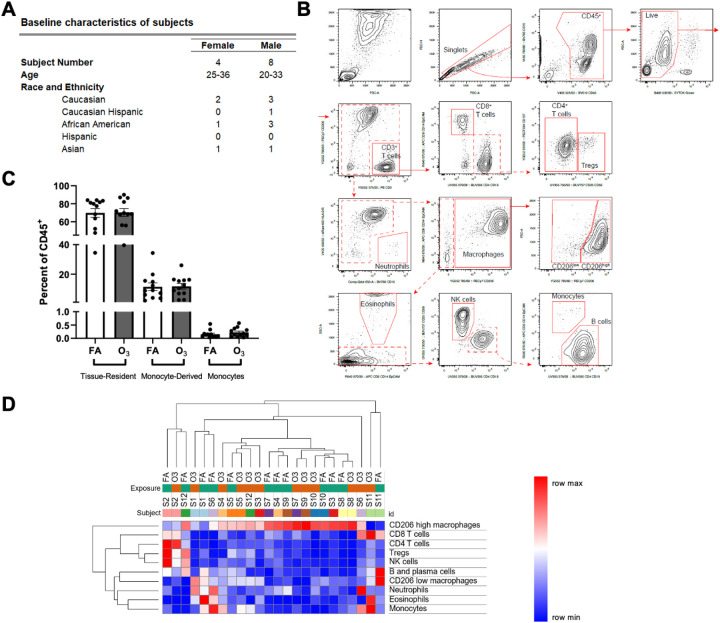
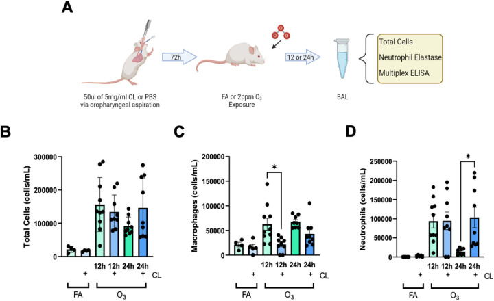
Lung inflammation, caused by acute exposure to ozone (O3) - one of the six criteria air pollutants - is a significant source of morbidity in susceptible individuals. Alveolar macrophages (AMØs) are the most abundant immune cells in the normal lung and their number increases following O3 exposure. However, the role of AMØs in promoting or limiting O3-induced lung inflammation has not been clearly defined. Here, we used a mouse model of acute O3 exposure, lineage tracing, genetic knockouts, and data from O3-exposed human volunteers to define the role and ontogeny of AMØs during acute O3 exposure. Lineage tracing experiments showed that 12, 24, and 72 h after exposure to O3 (2 ppm) for 3h all AMØs were tissue-resident origin. Similarly, in humans exposed to FA and O3 (200 ppb) for 135 minutes, we did not observe ~21h post-exposure an increase in monocyte-derived AMØs by flow cytometry. Highlighting a role for tissue-resident AMØs, we demonstrate that depletion of tissue-resident AMØs with clodronate-loaded liposomes led to persistence of neutrophils in the alveolar space after O3 exposure, suggesting that impaired neutrophil clearance (i.e., efferocytosis) leads to prolonged lung inflammation. Moreover, depletion of tissue-resident AMØ demonstrated reduced clearance of intratracheally instilled apoptotic Jurkat cells, consistent with reduced efferocytosis. Genetic ablation of MerTK - a key receptor involved in efferocytosis - also resulted in impaired clearance of apoptotic neutrophils followed O3 exposure. Overall, these findings underscore the pivotal role of tissue-resident AMØs in resolving O3-induced inflammation via MerTK-mediated efferocytosis.


Figures

Figure 1.
Figure 1.. Monocyte-derived alveolar macrophages (AMØ) are not recruited following acute O3 exposure.
A. Cx3cr1ERCre x zsGreen mouse model was used. Circulating blood monocytes and monocyte-derived AMØ express Cx3cr1, and after tamoxifen induction are GFP+, while tissue-resident AMØ remain unlabeled. Lineage label was induced with tamoxifen 24h prior to exposure with filtered air or O3 (2 ppm) for 3h. 12, 24, or 72h-post exposure, whole lungs were processed and analyzed by flow cytometry. Schematic made using BioRender.com B. Bar plots represent individual mouse GFP (tissue-resident AMØ, grey) and GFP+ (monocyte-derived AMØ, white) populations. ANOVA analysis was conducted using Tukey’s Honestly Significant Difference Test post-hoc. Data points represented with SEM; showing significant increase of tissue-resident AMØ from FA to 12 and 72h post O3 exposure. *p<0.05. C. Acute O3 exposure does not induce recruitment/differentiation of monocyte-derived AMØ. Representative flow cytometry plots of AMØs were negative for zsGreen/GFP positive cells, supporting no evidence of monocyte-derived AMØ recruitment. The red boxes indicate where GFP+ cells (monocyte-derived AMØ) would be observed if they were present in these samples. Percent GFP+ indicated within the respective box in red. Full flow cytometry gating strategy and antibodies can be found in the supplements (Supplemental Figure 1). Flow plots are representative of n=3–6 mice per timepoint and replicated x1. Mice used include 4 females at 12h, with all other mice being male.
Figure 2.
Figure 2.. Monocyte-derived alveolar macrophages (AMØ) are not recruited following acute O3 exposure in humans.
A. Human subjects were exposed to FA and O3 (2ppm) for 3h in separate visits. They then underwent a bronchoscopy in which BAL fluid was collected and processed and analyzed by flow cytometry. Demographic information is listed for all human subjects including sex, age, and ethnicity. B. Representative flow cytometry plots of the gating strategy used to identify alveolar macrophages were generated demonstrating populations of CD206Lo, monocyte-derived AMØ, and CD206Hi, tissue-resident AMØ, populations. The CD206Lo population did not increase following acute O3 exposure, supporting no evidence of monocyte-derived AMØ function in this context. The red boxes indicate where GFP+ monocyte-derived AMØ would be observed if they were present in these samples C. Acute O3 exposure does not induce recruitment of monocyte-derived AMØ. Bar plots represent human cellular response to FA (white) and O3 (grey) populations. Paired t-test analysis was conducted and data points are represented with SEM. No significant difference seen in tissue-resident AMØ, monocyte-derived AMØ, and monocyte populations post O3 exposure. D. Hierarchical clustering of the flow cytometry data from BAL samples. Column headers are color-coded by the exposure type (FA or O3) and subject. Samples were clustered by Euclidean distance using average linkage method. *p<0.05 Flow plots are representative of n=12 human subjects.
Figure 3.
Figure 3.. Airspace administration of clodronate (CL) depletes tissue-resident alveolar macrophages (AMØ).
A. C57Bl/6J female mice were dosed with clodronate or PBS (control) were harvested 72h post-dosing. The whole lung tissue samples were perfused, digested, and stained to identify the individual cells via flow cytometry. Bars shaded green indicate PBS/vehicle control while blue indicates CL administration. Data is from n=4 mice per group, p value is as identified. B. Cx3cr1ERCre x zsGreen mice were dosed with CL 72h before harvest then underwent lineage labeling with tamoxifen 48h post-dosing. They were then harvested and processed for flow cytometry. The lack of GPF+ AMØ (red box) suggests a lack of Cx3cr1ERCre x GFP+ monocyte-derived AMØs as a result of CL depletion of tissue-resident AMØ. Percent GFP+ indicated within the respective box in red. Flow plot is a representative sample of n=4. C. Bar plot represents individual mouse GFP (tissue-resident AMØ, grey) and GFP+ (monocyte-derived AMØ, white) populations, indicating no recruitment of monocyte-derived AMØ following CL depletion. Full flow cytometry gating strategy and antibodies can be found in the supplements (Supplemental Figure 1).
Figure 4.
Figure 4.. Tissue-Resident AMØ depletion leads to persistent O3-induced BAL neutrophilia.
A. Male mice were dosed with 50uL of 5mg/ml clodronate liposomes via oropharyngeal aspiration. 72h following clodronate depletion of tissue-resident AMØ, mice were exposed to FA or O3 (2 ppm) for 3h. Mice were then harvested 12/24h following exposure and the samples were processed for cell differentials and other measures of inflammation. Schematic made with BioRender.com. B. BAL total cells, (C.) macrophages, and (D.) neutrophils were enumerated following PBS or Clodronate (CL) administration and FA or O3 exposure. Bars shaded green indicate PBS/vehicle control while blue indicates CL administration. By total cell numbers, CL exposed mice exhibited a reduction of macrophages at 12h and increased neutrophils at 24h. n=4–10 mice per group/exposure/timepoint *p<0.05. ANOVA analysis was conducted using Tukey’s Honestly Significant Difference Test post-hoc.
Figure 5.
Figure 5.. Increased BAL cytokines associated with neutrophil recruitment in O3-exposed tissue-resident AMØ depleted mice.
BAL fluid from PBS or Clodronate and FA or O3 (2ppm) exposed male mice was assessed for cytokine expression by multiplex ELISA for inflammatory cytokines and known neutrophilic chemotactic factors. Bars shaded green indicate PBS/vehicle control while blue indicates CL administration. n=4–10 mice per group/exposure/timepoint *p<0.05. ANOVA analysis was conducted using Tukey’s Honestly Significant Difference Test post-hoc. ND indicates samples below the assay limit of detection.
Figure 6.
Figure 6.. Tissue-resident alveolar macrophage (AMØ) depletion decreased efferocytosis.
A. C57BL/6J male mice were dosed with CL or PBS. The mice were then instilled with apoptotic immortalized T cells, Jurkat cells, via oropharyngeal aspiration 22.5h after exposure for 1.5h immediately prior to the harvest. During the harvest, 24h post-exposure, BAL was collected and prepared for use in flow cytometry. Schematic made with BioRender.com. B. Apoptotic cell clearance was defined using Calcein AM, positive cells indicating live or early apoptotic cells. Healthy cells would not be engulfed by AMØs, thus decreased Calcein AM positive cells indicate clearance of apoptotic cells (efferocytosis). The graphs depict the ratio of counted recovered vs. instilled cells from the collected BAL. Bars shaded green indicate PBS/vehicle control while blue indicates CL administration. An unpaired T-test was conducted with n=10 mice per group/exposure/timepoint *p<0.05.
Figure 7.
Figure 7.. MerTK−/− mice have increased O3-induced airspace inflammation.
A. C57BL/6J male mice were exposed to O3 (2 ppm) for 3h and harvested 6h, 12h, 24h, 48h, 72h, and 7days later. mRNA expression of MerTK was assessed using the collected BAL cells. n=5 mice per time point. B. MerTK−/− mice and C57BL/6J mice (wildtype) were exposed to FA or O3 (2 ppm) for 3h. The mice were harvested 24h post-exposure and the BAL was collected and processed for total cell counts and differentials. Green bars indicate the C57BL/6J mice while the orange indicates the MerTK−/− genotype. n=9–19 mice per group. *p<0.05. ANOVA analysis was conducted using Tukey’s Honestly Significant Difference Test post-hoc. ND indicates samples below the limit of assay detection.
Figure 8.
Figure 8.. Increased BAL cytokines associated with neutrophil recruitment in O3-exposed MerTK−/− mice.
BAL fluid from C57BL/6J and MerTK−/− mice FA or O3 (2ppm) exposed mice was assessed for cytokine expression by multiplex ELISA for inflammatory cytokines and known neutrophilic chemotactic factors. Green bars indicate the C57BL/6J mice while the orange indicates the MerTK−/− genotype. n=8–19 mice per group/exposure/timepoint. *p<0.05. ANOVA analysis was conducted using Tukey’s Honestly Significant Difference Test post-hoc. ND indicates samples below the limit of assay detection.

References

    1. Fuller R. et al. Pollution and health: a progress update. Lancet Planet. Heal. 6, e535–e547 (2022). - PubMed
    1. Liao H., Chen W. T. & Seinfeld J. H. Role of climate change in global predictions of future tropospheric ozone and aerosols. J. Geophys. Res. Atmos. 111, (2006).
    1. Guttenberg M. A., Vose A. T. & Tighe R. M. Role of Innate Immune System in Environmental Lung Diseases. Curr. Allergy Asthma Rep. 21, 34 (2021). - PMC - PubMed
    1. Lim C. C. et al. Long-term exposure to ozone and cause-specific mortality risk in the United States. Am. J. Respir. Crit. Care Med. 200, 1022–1031 (2019). - PMC - PubMed
    1. Samet J. M. et al. The National Morbidity, Mortality, and Air Pollution Study. Part II: Morbidity and mortality from air pollution in the United States - PubMed. https://pubmed.ncbi.nlm.nih.gov/11354823/ (2000). - PubMed

Publication types