[Preprint]. 2023 Nov 9:2023.11.06.564847.
doi: 10.1101/2023.11.06.564847.

A vasopressin circuit that modulates sex-specific social interest and anxiety-like behavior in mice

Affiliations

A vasopressin circuit that modulates sex-specific social interest and anxiety-like behavior in mice

Nicole Rigney et al. bioRxiv. .

Update in

Abstract

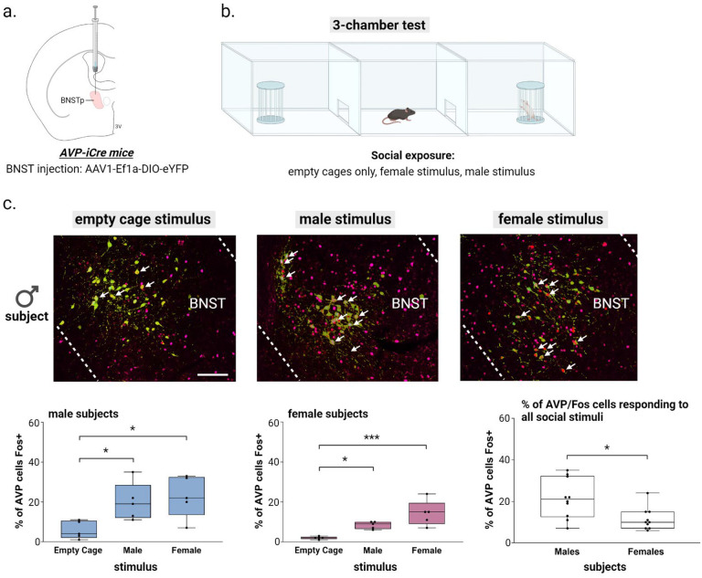
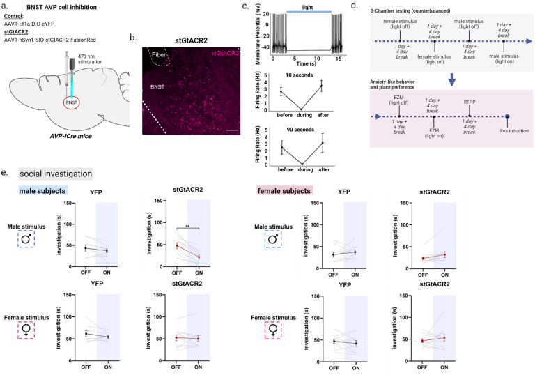
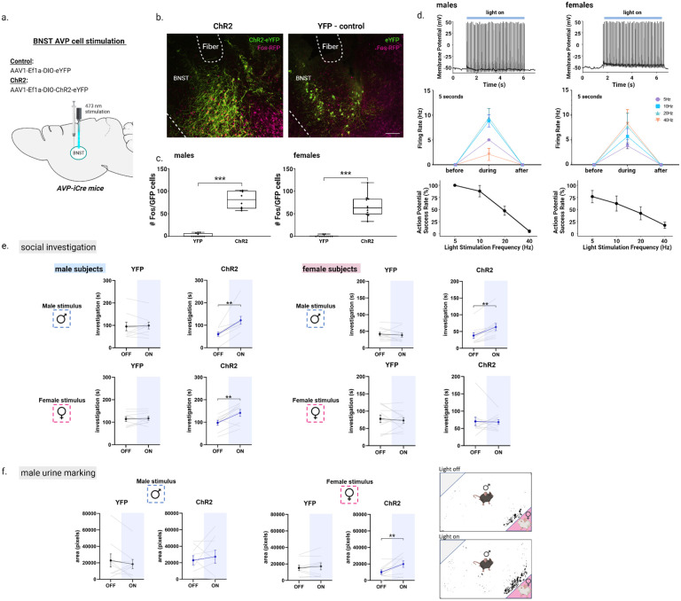
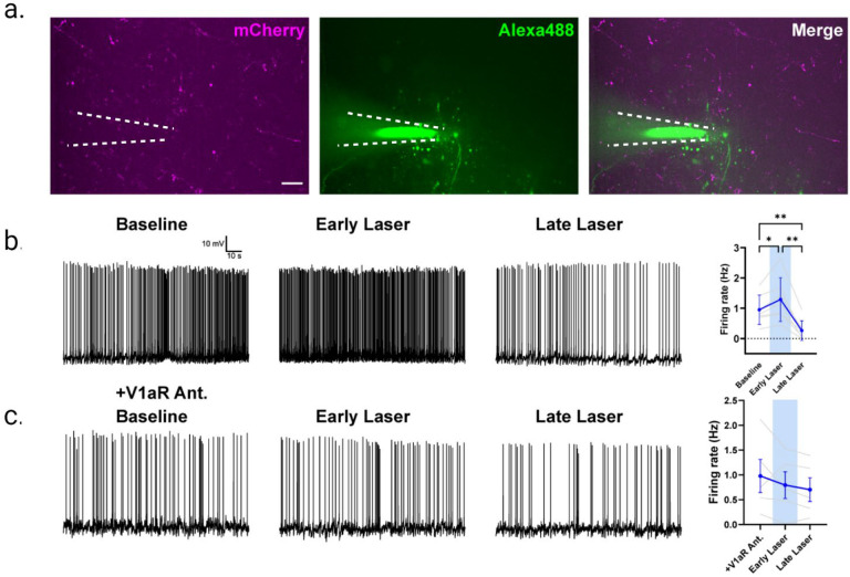
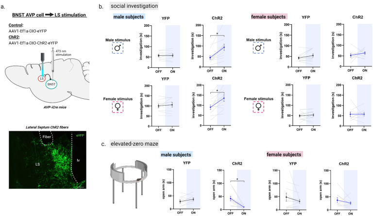
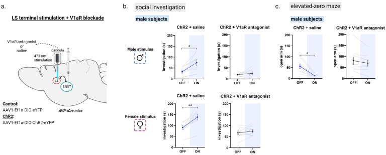
One of the largest sex differences in brain neurochemistry is the male-biased expression of the neuropeptide arginine vasopressin (AVP) within the vertebrate social brain. Despite the long-standing implication of AVP in social and anxiety-like behavior, the precise circuitry and anatomical substrate underlying its control are still poorly understood. By employing optogenetic manipulation of AVP cells within the bed nucleus of the stria terminalis (BNST), we have unveiled a central role for these cells in promoting social investigation, with a more pronounced role in males relative to females. These cells facilitate male social investigation and anxiety-like behavior through their projections to the lateral septum (LS), an area with the highest density of sexually-dimorphic AVP fibers. Blocking the vasopressin 1a receptor (V1aR) in the LS eliminated stimulation-mediated increases in these behaviors. Together, these findings establish a distinct BNST AVP → LS V1aR circuit that modulates sex-specific social interest and anxiety-like behavior.


Figures

Figure 1 –
Figure 1 –
BNST AVP cell and Fos colocalization during social exposure in the 3-chamber test (a) BNST injection site and virus. (b) Three-chamber social testing. (c) (top) Example images of merged BNST-AVP cells (green) and Fos+ cells (magenta). (bottom) Boxplots of the percentage of AVP cells colocalized with Fos in each 3-chamber stimulus condition (empty cage, male, female) for male and female subjects. A Two-way ANOVA revealed a significant effect when comparing activated BNST AVP cells with the type of stimulus received (F(1,24) = 12.89, p = 0.00016, η2 = 0.8), a significant effect when comparing the sex of the subjects (F(1,24) = 10.69, p = 0.003, η2 = 0.62), yet there was no interaction between sex and stimulus type. A post hoc ANOVA followed by Bonferroni-corrected pairwise tests showed that male mice exposed to male or female conspecifics had increased BNST AVP-Fos colocalization compared to males kept in an empty three-chamber apparatus containing empty stimulus cages (F(1,12) = 5.8, p = 0.017, η2 = 0.4; male vs. empty cage: p = 0.05, female vs. empty cage: p = 0.025). Similarly, female mice also showed Fos activation to social cues in BNST AVP cells (F(1,12) = 13.23, p = 0.0009, η2 = 0.69; male vs. empty cage: p = 0.05, female vs. empty cage: p = 0.0007). Mean ± SEM data represented. Dots indicate individual data points. White arrows indicate Fos and AVP cell colocalization. Scale bar = 25 μm. *p < 0.05, ** p < 0.01, ***p < 0.001.
Figure 2 –
Figure 2 –
Optogenetic inhibition of AVP-BNST cells decreases male-male social investigation. (a) Bilateral BNST injection and fiber implantation site; coordinates: DV: −4.4, AP: +0.15, ML: ±0.8; modified from Paxinos and Franklin (2012). (b) Example image of BNST AVP cells infected with inhibitory stGtACR adeno-associated virus (FusionRed) (c) representative trace from whole-cell current-clamp recording of stGTACR2-expressing BNST cell silenced by light application at 10Hz for 10 seconds and firing rate of stGTACR2-expressing BNST cell suppressed by the blue light for 10 seconds (n=8 cells) and 90 seconds (n=9 cells). (d) Experimental timeline. All subjects were tested within the 3-chamber apparatus on 4 separate days (4-day break in between) with all stimuli and light conditions counterbalanced. The same subjects were further tested within the elevated-zero maze (EZM) (counterbalanced) followed by a real-time place preference test (RTPP) and Fos induction. (e) Social investigation (in seconds) by male and female subjects during the three-chamber test (male subjects: YFP, n=9 and stGtACR2, n=11 female subjects (YFP, n=10 and stGtACR2, n=11) during light-OFF and light-ON conditions, counterbalanced. Blue light inhibition (ON) of AVP-BNST cells in stGtACR2 males significantly decreased time spent investigating male stimuli, but not female stimuli, compared to investigation during light-OFF condition (Mixed model ANOVA, treatment*light*stimulus interaction (F(1,37) = 4.11, p = 0.05, η2 = 0.6; post hoc: p = 0.001). Blue light inhibition (ON) of AVP-BNST cells in stGtACR2 females did not affect time spent investigating male or female stimuli. Light stimulation did not affect investigation times of YFP male and female subjects to either stimulus type (female or male). Bonferroni post hoc tests were used. Each point and horizontal line represent individual within-subject data. Overlapping data are represented as one point/line. Scale bar = 50 μm. **p<0.01.
Figure 3 –
Figure 3 –
Optogenetic activation of AVP-BNST cells increases social investigation in both sexes and male urine marking toward female stimuli. (a-b) Bilateral BNST injection and fiber implantation site; coordinates: DV: −4.4, AP: +0.15, ML: ±0.8; modified from Paxinos and Franklin (2012). (b) Example images of merged BNST-AVP cells infected with either the excitatory ChR2 adeno-associated virus (ChR2) or YFP control virus, both colocalized with Fos+ cells (magenta). (c) Boxplots of the number of BNST AVP ChR2/YFP cells colocalized with Fos. Blue light stimulation robustly increased the number of BNST AVP labeled cells colocalized with Fos (F (1,35) = 234.17, p < 0.000001; η2 = 0.87). (d) Representative trace from whole-cell current-clamp recording of ChR2-mCherry-expressing cells activated by light application at 10Hz for 5 seconds and the response to 10ms pulse delivered at 5, 10, 20, 40Hz for 5 seconds, with the probability of action potential (AP) in response to an individual pulse (action potential success rate in male and female subjects (e) Social investigation (in seconds) by male and female subjects during the three-chamber test (male subjects: YFP, n=9 and ChR2, n=9; female subjects (YFP, n=10 and ChR2, n=11) during light-OFF and light-ON conditions, counterbalanced. Blue light stimulation (ON) of AVP-BNST cells in ChR2 males significantly increased time spent investigating male and female stimuli compared to investigation during light-OFF condition (Mixed model ANOVA, treatment*light*sex interaction, F (1,36) = 7.02, p = 0.012; η2 = 0.5; post hoc: p = 0.002 (male stimuli), p = 0.004 (female stimuli). Blue light stimulation (ON) of AVP-BNST cells in ChR2 females significantly increased time spent investigating male stimuli compared to investigation during light-OFF condition (post hoc: p = 0.002 (male stimuli). Light stimulation did not affect investigation times of YFP male and female subjects to either stimulus type (female or male). (f) Total area of urine marking by male subjects during the three-chamber test. Blue light stimulation (ON) of AVP-BNST cells in ChR2 males significantly increased urine marking in the presence of a female stimulus (Mixed model ANOVA, treatment*light*sex interaction, (F (1,36) = 4.5, p = 0.04; η2 = 0.4; post hoc: p = 0.009). Bonferroni post hoc tests were used. Each point and horizontal line represent individual within-subject data. Overlapping data are represented as one point/line. **p<0.01.
Figure 4 –
Figure 4 –
Optogenetic activation of AVP-BNST cell projections to the lateral septum (LS) modulates neurons in LS. (a) Visualization of mCherry reporter tagged to ChR2-AVP expressing fibers in the LS, originating from BNST (left) in addition to a patched neuron dialyzed with Alexa488 dye (middle). Merged image (right). (b) BNST AVP → LS terminal activation modulates LS neuron firing activity. (Baseline vs. Early Laser p <0.05, Baseline vs. Late Laser p <0.01, Early Laser vs. Late Laser p <0.01, one-way repeated measures ANOVA). (c) Bath application of V1aR antagonist (d(CH2)5[Tyr(Me)2,Dab5] AVP) blocks ChR2 modulation of LS neurons. (p >0.05, one-way repeated measures ANOVA). Scale bar= 25μm.
Figure 5 –
Figure 5 –
Optogenetic activation of AVP-BNST cell projections to the lateral septum (LS) increases social investigation and anxiety-like behavior in males, but not females. (a) Bilateral BNST injection of ChR2 adeno-associated virus (ChR2) and fiber implantation within the lateral septum (intermediate zone). (b) Social investigation (in seconds) by male and female subjects during the three-chamber test (male subjects: YFP, n=9 and ChR2, n=11; female subjects: YFP, n=12 and ChR2, n=9) during light-OFF and light-ON conditions, counterbalanced. Blue light stimulation (ON) of AVP-BNST-LS terminals in ChR2 males significantly increased time spent investigating male and female stimuli compared to investigation during light-OFF condition (Mixed model ANOVA, treatment*light interaction, (F (1,17) = 6.9, p = 0.01; η2 = 0.6; post hoc: p = 0.001 (male stimuli), p = 0.02 (female stimuli). Blue light stimulation (ON) of AVP-BNST LS terminals in ChR2 females did not affect investigation times compared to investigation during light-OFF condition. Light stimulation did not affect investigation times of YFP male and female subjects to either stimulus type (female or male). (c) Time spent in the open arm of the elevated-zero maze (EZM). Blue light stimulation (ON) of AVP-BNST-LS terminals in ChR2 males significantly decreased time spent in the open arm of the EZM. In females, blue light stimulation had no effect on time spent in the open arm of the EZM. (Mixed model ANOVA, treatment*light*sex interaction, (F (1,17) = 6.2, p = 0.024; η2 = 0.5; post hoc: p = 0.008). Bonferroni post hoc tests were used. Each point and horizontal line represent individual within-subject data. Overlapping data are represented as one point/line. *p<0.05.
Figure 6 –
Figure 6 –
Antagonism of V1aR in the LS blocked optogenetic-mediated increases in male social investigation and anxiety-like behavior. (a) Bilateral BNST injection of ChR2 adeno-associated virus (ChR2) and fiber implantation within the lateral septum (intermediate zone). (b) Social investigation (in seconds) by male subjects during the three-chamber test. Two groups of ChR2+ injected males were tested with either a male or female stimulus (LIGHT OFF/ON), and received the same type of stimulus (i.e., novel male or female) with LIGHT OFF/ON + a highly-selective V1aR antagonist (subjects tested with a male stimulus: n=9; subjects tested with a female stimulus: n=9). All conditions were counterbalanced. Blue light stimulation (ON) of AVP-BNST-LS terminals in both ChR2 + saline male groups significantly increased time spent investigating male and female stimuli compared to investigation during light-OFF condition (Mixed model ANOVA, drug*light interaction, (F (1,16) = 11.76, p = 0.003; η2 = 0.42; post hoc: p = 0.01 (male stimuli), p = 0.006 (female stimuli)). In the same male subjects, antagonism of V1aR in the LS blocked optogenetic-mediated increases in male social investigation. (c) Time spent in the open arm of the elevated-zero maze (EZM). Blue light stimulation (ON) of AVP-BNST-LS terminals in ChR2 males significantly decreased time spent in the open arm of the EZM, and in the same males, antagonism of V1aR in the LS blocked optogenetic-mediated increases in male anxiety-like behavior. (Mixed model ANOVA, treatment*light interaction, (F (1,8) = 10.58, p = 0.012; η2 = 0.5). Bonferroni post hoc tests were used. *p<0.05, **p<0.01.

Similar articles

References

    1. Insel T. R., The challenge of translation in social neuroscience: a review of oxytocin, vasopressin, and affiliative behavior. Neuron 65, 768–779 (2010). - PMC - PubMed
    1. Bahrami F., Yousefi N., Females are more anxious than males: a metacognitive perspective. Iran J Psychiatry Behav Sci 5, 83–90 (2011). - PMC - PubMed
    1. Halladay A. K., et al., Sex and gender differences in autism spectrum disorder: summarizing evidence gaps and identifying emerging areas of priority. Mol. Autism 6, 36 (2015). - PMC - PubMed
    1. Lai M.-C., Lombardo M. V., Auyeung B., Chakrabarti B., Baron-Cohen S., Sex/gender differences and autism: setting the scene for future research. J. Am. Acad. Child Adolesc. Psychiatry 54, 11–24 (2015). - PMC - PubMed
    1. Aspesi D., Choleris E., Neuroendocrine underpinning of social recognition in males and females. J. Neuroendocrinol. 34, e13070 (2022). - PubMed

Publication types