
Highly Pathogenic Avian Influenza A(H5N1) Virus Clade 2.3.4.4b Infections in Wild Terrestrial Mammals, United States, 2022

Elizabeth J Elsmo et al. Emerg Infect Dis. 2023 Dec.

Abstract

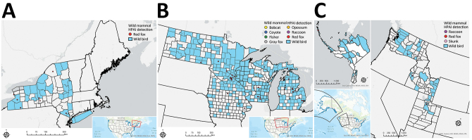
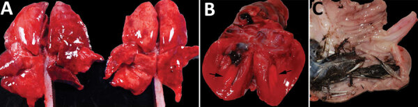
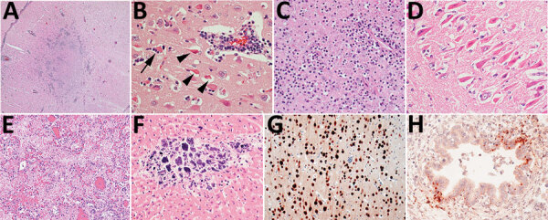
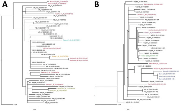
We describe the pathology of natural infection with highly pathogenic avian influenza A(H5N1) virus of Eurasian lineage Goose/Guangdong clade 2.3.4.4b in 67 wild terrestrial mammals throughout the United States during April 1‒July 21, 2022. Affected mammals include 50 red foxes (Vulpes vulpes), 6 striped skunks (Mephitis mephitis), 4 raccoons (Procyon lotor), 2 bobcats (Lynx rufus), 2 Virginia opossums (Didelphis virginiana), 1 coyote (Canis latrans), 1 fisher (Pekania pennanti), and 1 gray fox (Urocyon cinereoargenteus). Infected mammals showed primarily neurologic signs. Necrotizing meningoencephalitis, interstitial pneumonia, and myocardial necrosis were the most common lesions; however, species variations in lesion distribution were observed. Genotype analysis of sequences from 48 animals indicates that these cases represent spillover infections from wild birds.

Keywords: United States; avian influenza; bobcat; clade 2.3.4.4b; coyote; fisher; fox; highly pathogenic avian influenza virus; influenza; influenza A(H5N1); meningitis/encephalitis; opossum; raccoon; respiratory infections; skunk; viruses; wild terrestrial mammals; zoonoses.


Figures

Figure 1
Figure 1
Locations of wild mammals naturally infected with HPAI A(H5N1) virus, United States, April‒June 2022. A) Eastern region; B) midwestern region; C) western region. If exact location could not be identified, the county centroid was used. Blue indicates counties with positive detections of HPAI H5N1 in wild birds during March‒August 2022. HPAI, highly pathogenic avian influenza.
Figure 2,
Figure 2,
Gross photographs of postmortem lesions from red foxes naturally infected with highly pathogenic avian influenza virus, United States. A) Lungs have failed to collapse and are diffusely edematous and mottled pink to dark red. B) Cross section of the left ventricle of the heart showing a focal region of myocardial pallor in the papillary muscle (arrows). C) Stomach contents with feathers.
Figure 3
Figure 3
Histopathology of lesions in red foxes naturally infected with highly pathogenic avian influenza virus, United States. A) Throughout the brain, there are multifocal regions of necrosis and hypercellularity. Original magnification ×4. B) Within the gray matter, there is prominent neuronal necrosis (arrowheads), satellitosis (arrow), and reactive astrocytes. A vessel is surrounded by lymphocytes and plasma cells. Original magnification ×4. C) In areas of necrosis within the brain, there is often abundant, stippled, basophilic karyorrhectic debris. Original magnification ×40. D) Within the hippocampus, there are numerous shrunken, angular, and acidophilic (necrotic) neurons in a laminar pattern. Original magnification ×20. E) Within the lung, there is diffuse vascular congestion. Alveoli contain fibrin, hemorrhage, and edema fluid. Original magnification ×20. F) Regions of cardiomyocyte necrosis in the heart are often mineralized. Original magnification ×40. Panels A‒F, hematoxylin and eosin stain.G) Within the brain, there is positive nuclear and cytoplasmic staining of neuron cell bodies and processes. Avian influenza virus monoclonal immunohistochemical analysis. Original magnification ×40. H) Scattered positive nuclear and cytoplasmic staining of bronchiolar epithelial cells and interstitial macrophages in the lung. Monoclonal immunohistochemical analysis of influenza A virus nucleoprotein. Original magnification ×40.
Figure 4
Figure 4
Single-nucleotide polymorphism phylogenetic trees for highly pathogenic avian influenza A(H5N1) clade 2.3.4.4b virus in mammals and wild birds. A) Genotype B1.2. Data from red fox are shown in red, raccoon in teal, coyote in orange, and Virginia opossum in gold. B) Genotype B3.2. Data from red fox are shown in red, fisher in purple, bobcat in green, and skunk in blue. Trees are rooted to the reference sequence A/Fancy_Ck/NL/FAV33/2021. WB, wild bird. Scale bars indicate nucleotide substitutions per site.

References

    1. Adlhoch C, Fusaro A, Gonzales JL, Kuiken T, Marangon S, Niqueux É, et al.; European Food Safety Authority; European Centre for Disease Prevention and Control; European Union Reference Laboratory for Avian Influenza. Avian influenza overview December 2021 - March 2022. EFSA J. 2022;20:e07289. - PMC - PubMed
    1. Caliendo V, Lewis NS, Pohlmann A, Baillie SR, Banyard AC, Beer M, et al. Transatlantic spread of highly pathogenic avian influenza H5N1 by wild birds from Europe to North America in 2021. Sci Rep. 2022;12:11729. 10.1038/s41598-022-13447-z - DOI - PMC - PubMed
    1. Youk S, Torchetti MK, Lantz K, Lenoch JB, Killian ML, Leyson C, et al. H5N1 highly pathogenic avian influenza clade 2.3.4.4b in wild and domestic birds: Introductions into the United States and reassortments, December 2021-April 2022. Virology. 2023;587:109860. 10.1016/j.virol.2023.109860 - DOI - PubMed
    1. Bevins SN, Shriner SA, Cumbee JC Jr, Dilione KE, Douglass KE, Ellis JW, et al. Intercontinental movement of highly pathogenic avian influenza A(H5N1) clade 2.3.4.4 virus to the United States, 2021. Emerg Infect Dis. 2022;28:1006–11. 10.3201/eid2805.220318 - DOI - PMC - PubMed
    1. US Department of Agriculture. Animal and Plant Health Inspection Service. HPAI 2022 confirmed detections. 2022. [cited 2022 Jun 6]. https://www.aphis.usda.gov/aphis/ourfocus/animalhealth/animal-disease-in...