. 2024 Jan 1;326(1):G78-G93.
doi: 10.1152/ajpgi.00194.2023. Epub 2023 Nov 21.

Characterization of neuromuscular transmission and projections of muscle motor neurons in the rat stomach

Affiliations

Characterization of neuromuscular transmission and projections of muscle motor neurons in the rat stomach

Madeleine R Di Natale et al. Am J Physiol Gastrointest Liver Physiol. .

Abstract

The stomach is the primary reservoir of the gastrointestinal tract, where ingested content is broken down into small particles. Coordinated relaxation and contraction is essential for rhythmic motility and digestion, but how the muscle motor innervation is organized to provide appropriate graded regional control is not established. In this study, we recorded neuromuscular transmission to the circular muscle using intracellular microelectrodes to investigate the spread of the influence of intrinsic motor neurons. In addition, microanatomical investigations of neuronal projections and pharmacological analysis were conducted to investigate neuromuscular relationships. We found that inhibitory neurotransmission to the circular muscle is graded with stimulus strength and circumferential distance from the stimulation site. The influence of inhibitory neurons declined between 1 and 11 mm from the stimulation site. In the antrum, corpus, and fundus, the declines at 11 mm were about 20%, 30%, and 50%, respectively. Stimulation of inhibitory neurons elicited biphasic hyperpolarizing potentials often followed by prolonged depolarizing events in the distal stomach, but only hyperpolarizing events in the proximal stomach. Excitatory neurotransmission influence varied greatly between proximal stomach, where depolarizing events occurred, and distal stomach, where no direct electrical effects in the muscle were observed. Structural studies using microlesion surgeries confirmed a dominant circumferential projection. We conclude that motor neuron influences extend around the gastric circumference, that the effectiveness can be graded by the recruitment of different numbers of motor neuron nerve terminals to finely control gastric motility, and that the ways in which the neurons influence the muscle differ between anatomical regions.NEW & NOTEWORTHY This study provides a detailed mapping of nerve transmission to the circular muscle of the different anatomical regions of rat stomach. It shows that excitatory and inhibitory influences extend around the gastric circumference and that there is a summation of neural influence that allows for finely graded control of muscle tension and length. Nerve-mediated electrical events are qualitatively and quantitatively different between regions, for example, excitatory neurons have direct effects on fundus but not antral muscle.

Keywords: enteric motor neuron; intracellular electrophysiology; neuromuscular junction; smooth muscle; stomach.


Conflict of interest statement

No conflicts of interest, financial or otherwise, are declared by the authors.

Figures

None
Graphical abstract
Figure 1.
Figure 1.
Regions of the rat stomach, based on images of full stomachs dissected from rats after the feeding phase, and fixed whole. A: regions defined by conventional anatomy defining the fundus, corpus, and antrum. B: regions defined by specialization of the mucosa. A stratified squamous (aglandular) epithelial lining occurs in the fundus, proximal corpus, and the esophageal groove that surrounds the opening of the esophagus and extends along a small region of the lesser curvature. It is continuous with the aglandular lining of the esophagus. The remainder of the corpus and the antrum have a glandular lining. The position of the limiting ridge, a boundary between the glandular and aglandular parts, differed between individual rats as indicated by the proximal shaded region in B. Distal shaded region indicates the corpus/antral boundary where there is transition zone of changing glandular cell types. C: regions defined by functional movements. The proximal stomach is the gastric reservoir, and the distal stomach is the region of mixing and proximo-distal propulsion. The pyloric sphincter regulates the timing and particle sizes of aspirates that are propelled into the duodenum. See text for further details.
Figure 2.
Figure 2.
Compound inhibitory junction potentials (IJPs) and associated afterdepolarizations (After-Dep). A: compound IJP and afterdepolarization in response to a single stimulus pulse (at the arrow at cursor 1), applied to intramural axons in an antral muscle strip. Dotted lines indicate the placement of cursors for measurements for the fast and slow components of the IJP and the afterdepolarization. B: a series of spontaneous slow waves (SWs) recorded via an intracellular microelectrode in the antrum. One wave is shown on an expanded timebase matching that of A. C: example traces of the IJP responses and afterdepolarization events seen in the three anatomical regions. D: quantification of the durations of the fast and slow IJP components across the fundus, corpus, and antrum. E: durations of the afterdepolarizations and the duration to peak of the response across all regions. F: amplitudes of the afterdepolarization events post IJP. In these experiments, the measured responses shown were evoked by a single 90-V pulse in cells that were located 3 mm circumferential to stimulus site. Data shown in D as means ± SE; data shown in E and F shown as min to max with line as median. ****P < 0.0001.
Figure 3.
Figure 3.
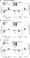
Investigation of chloride conductance dependence of the compound IJP (inhibitory junction potential) and afterdepolarization (After-dep) in the antrum. A: effect of reducing external chloride from 128.6 mM to 9.8 mM by substitution of sodium chloride for sodium gluconate. Although there was no significant reduction of membrane potential, the afterdepolarization and the slow IJPs were almost abolished and the fast IJPs were substantially reduced (n = 14 control and n = 12 chloride substitute cells, 2 rats, 1 female and 1 male). B: niflumic acid (NFA); there was no significant difference in RMP, the afterdepolarization and the slow IJPs were substantially reduced (n = 12 control and n = 8 NFA cells, 5 rats, 2 females and 3 males). C: CaCCinh-AO1 hyperpolarized the smooth muscle (P < 0.0001) and reduced both components of the IJP and the afterdepolarization (n = 7 control and n = 13 CaCCinhA01 cells, 3 rats, 1 female and 2 males). Inset in each panel shows the membrane potential in cells in control (standard physiological solution) and with the chloride substitution or antagonist. Measured responses were evoked by a single 90-V pulse in cells that were located 3 mm circumferential to stimulus sites. Data shown as means ± SE. ****P < 0.0001, ***P = 0.0005, **P = 0.002, *P = 0.01. RMP, resting membrane potential.
Figure 4.
Figure 4.
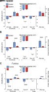
Compound inhibitory junction potential (IJP) and afterdepolarization in the antrum and fundus following small conductance potassium channel (SK) block, or block of nitrergic transmission. A: effect of the SK blocker apamin in the antrum (n = 13 control and n = 6 apamin, 2 male rats). B: effect of NOS inhibitor l-NNA in the antrum (n = 10 control and n = 19 l-NNA, 2 rats, 1 female and 1 male). C: effects of NOS inhibitor l-NNA in the fundus (n = 13 control and n = 16 l-NNA, 2 rats, 1 female and 1 male). The measured responses were evoked by a single 90-V pulse in cells that were located 3 mm circumferential to stimulus sites. Data shown as means ± SE. ****P < 0.0001, ***P = 0.0006, **P = 0.005, *P = 0.01. l-NNA, N-nitro-l-arginine; NOS, nitric oxide synthase.
Figure 5.
Figure 5.
Diminution of inhibitory junction potential (IJP) amplitudes in the circular muscle with increased distances from stimulating electrodes. A: diagram showing in yellow the sites from which strips were removed for investigation of transmission in vitro. B, C, and D: relations between IJP amplitudes and stimulus strength at 1, 3, 5, 7, 9, and 11 mm from the stimulating electrodes in circular muscle strips from the fundus (B), corpus (C), and antrum (D). Data are normalized to the 1-mm 30-V pulse shown as means ± SE with a linear regression line. See Supplemental Figs. S2–S4 for subject numbers.
Figure 6.
Figure 6.
Increase of inhibitory junction potential (IJP) amplitude in the circular muscle with increased stimulus strength. A: superimposed records of individual IJPs in an individual smooth muscle cell of the fundus in response to single transmural electrical pulses (at the arrow, ranging from 5 to 90 V, different stimulus strengths shown in different colors with the 90-V pulse shown in black). B–D: relations between IJP amplitudes and stimulus strength at 1, 5, and 9 mm from the stimulating electrodes in circular muscle strips from the fundus (B), corpus (C), and antrum (D). Data are normalized to the 1-mm 30-V pulse shown as means ± SE. Linear regression lines (5–30 V regression adjusted to cross XY zero, 40–90 V standard linear regression). See Supplemental Figs. S2–S4 for subject numbers.
Figure 7.
Figure 7.
Junction potentials after blocking inhibitory transmission with the NOS inhibitor l-NNA and the P2Y1 purine receptor antagonist MRS2500 elicited by single pulses (dots) or trains of pulses (multiple dots). Junction potentials are shown at either 1 mm (gray shading) or 3 mm away from the stimulating electrodes. The amplitudes and shapes of junction potentials differed between regions. Excitatory junction potentials occurred in the fundus (A) and corpus (B), but not in the antrum (C). l-NNA, N-nitro-l-arginine; NOS, nitric oxide synthase.
Figure 8.
Figure 8.
Influence of excitatory neurons on the membrane potential of the circular muscle in the fundus. A: relations between EJP amplitudes and stimulus strengths at 1, 5, and 9 mm from the stimulating electrodes in circular muscle strips. Linear regression line (5–30 V regression corrected to cross XY zero, 40–90 V standard linear regression). B: relations between EJP amplitudes at 1, 2, 5, 7, 9, and 11 mm distances from the stimulating electrodes in circular muscle strips from the fundus. Data are normalized to the 1-mm 30-V pulse shown as means ± SE. See Supplemental data for subject numbers. EJP, excitatory junction potential.
Figure 9.
Figure 9.
Double myotomy surgery and its consequences for innervation of the circular muscle in the corpus. A: image of the myotomies, in vivo, immediately after completion of the cuts. The myotomy cuts are defined by a small amount of blood that has entered the cuts. B: appearance of the stomach soon after removal from the rat, 10 days later. Colloidal carbon remnants can be seen in the cuts (arrows). C: diagram showing positions of myotomy cuts (purple). Green dotted line indicates the position that sections were taken from. Blue lines indicate the direction of the circular muscle bundles. D–G: representative immunohistochemistry of sections cut perpendicular to the circular muscle, showing cross sections of neural NOS (nNOS; green) and TK (orange) fiber bundles. D: low power image of nNOS and TK fibers in control tissue. E: low-power image of nNOS and TK fibers, at the green dotted line between myotomy lesions (1–2 mm lesion example). F: high-power image of fiber cross sections in control. G: high-power image of fiber cross sections in myotomy. H: quantitative data showing the reduction of fiber bundle area, in the circular muscle from between lesions of 4 mm separation, compared with control, and a reduction between lesions of 1–2 mm separation. ****P < 0.0001, ***P = 0.0005, **P = 0.002, *P = 0.01. CM, circular muscle; LM, longitudinal muscle; MY, myenteric plexus; nNOS, neuronal nitric oxide synthase; TK, tachykinins.
Figure 10.
Figure 10.
Myectomy surgery and its consequences for innervation of the circular muscle in the corpus to investigate proximo-distal fiber distributions. A: representative low-power immunohistochemistry of thick-mount preparation immediately distal to corpus myectomy site (yellow box in B). Immunoreactivity of inhibitory neurons, VIP (green), and Hu C/D (orange) showing neuronal cell bodies. Myectomy example demonstrates a reduction of innervation distal to the lesion site (purple in B) followed by a gradient of increased innervation further from the lesion. B: representation of myectomy lesion (purple) and the location image in A was taken from (yellow box). Further details see Supplemental data. C: high-power image on the edge of the lesion site showing reduced density of nerve fibers. D: high-power image at distance distal from lesion, cell bodies are present, and more fibers are present. VIP, vasoactive intestinal peptide.
Figure 11.
Figure 11.
Depiction of the innervation of the circular muscle deduced from the current study and data in the literature. A: diagram of the anatomy of the rat stomach, showing the directions of muscle bundles of the circular muscle coat (blue lines). B: relationship of a single inhibitory motor neuron to the circular muscle, below showing the approximate distance the axon projects physically (solid bar) and its longer region of influence due to electrotonic spread (indicated by dots). The motor neurons give rise to branching terminals that follow the direction of the muscle. C: multiple inhibitory motor neurons innervate bundles of electrically coupled smooth muscle cells. D: representative cell body of the motor neuron. Its axon runs a short distance orthogonal to the circular muscle to a branch point. E: individual SMCs form bundles of cells that are interconnected by bands of smooth muscle. Different responses to nerve stimulation that are recorded by intracellular electrodes within an individual muscle cell could be affected by the cell’s relation to the muscle bundles, bands, and nerve terminals. For example, if the cell is in the center of a bundle (1), at the edge of a bundle (2), or within connecting bands (3). SMCs, smooth muscle cells.

Similar articles

Cited by

References

    1. Kelly KA. Gastric emptying of liquids and solids: roles of proximal and distal stomach. Am J Physiol Gastrointest Liver Physiol 239: G71–G76, 1980. doi:10.1152/ajpgi.1980.239.2.G71. - DOI - PubMed
    1. Hunt RH, Camilleri M, Crowe SE, El-Omar EM, Fox JG, Kuipers EJ, Malfertheiner P, McColl KEL, Pritchard DM, Rugge M, Sonnenberg A, Sugano K, Tack J. The stomach in health and disease. Gut 64: 1650–1668, 2015. doi:10.1136/gutjnl-2014-307595. - DOI - PMC - PubMed
    1. Di Natale MR, Athavale ON, Wang X, Du P, Cheng LK, Liu Z, Furness JB. Functional and anatomical gastric regions and their relations to motility control. Neurogastroenterol Motil 35: e14560, 2023. doi:10.1111/nmo.14560. - DOI - PubMed
    1. Cannon WB. The movements of the stomach studied by means of the Roentgen rays. Am J Physiol 1: 359–382, 1898. doi:10.1152/ajplegacy.1898.1.3.359. - DOI
    1. O'Grady G, Du P, Cheng LK, Egbuji JU, Lammers WJEP, Windsor JA, Pullan AJ. Origin and propagation of human gastric slow-wave activity defined by high-resolution mapping. Am J Physiol Gastrointest Liver Physiol 299: G585–G592, 2010. doi:10.1152/ajpgi.00125.2010. - DOI - PMC - PubMed

Publication types

LinkOut - more resources