. 2023 Dec;475(12):1409-1419.
doi: 10.1007/s00424-023-02880-2. Epub 2023 Nov 21.

Enzymatic vitamin A2 production enables red-shifted optogenetics

Affiliations

Enzymatic vitamin A2 production enables red-shifted optogenetics

Johanna Gerhards et al. Pflugers Arch. 2023 Dec.

Abstract

Optogenetics is a technology using light-sensitive proteins to control signaling pathways and physiological processes in cells and organs and has been applied in neuroscience, cardiovascular sciences, and many other research fields. Most commonly used optogenetic actuators are sensitive to blue and green light, but red-light activation would allow better tissue penetration and less phototoxicity. Cyp27c1 is a recently deorphanized cytochrome P450 enzyme that converts vitamin A1 to vitamin A2, thereby red-shifting the spectral sensitivity of visual pigments and enabling near-infrared vision in some aquatic species.Here, we investigated the ability of Cyp27c1-generated vitamin A2 to induce a shift in spectral sensitivity of the light-gated ion channel Channelrhodopsin-2 (ChR2) and its red-shifted homolog ReaChR. We used patch clamp to measure photocurrents at specific wavelengths in HEK 293 cells expressing ChR2 or ReaChR. Vitamin A2 incubation red-shifted the wavelength for half-maximal currents (λ50%) by 6.8 nm for ChR2 and 12.4 nm for ReaChR. Overexpression of Cyp27c1 in HEK 293 cells showed mitochondrial localization, and HPLC analysis showed conversion of vitamin A1 to vitamin A2. Notably, the λ50% of ChR2 photocurrents was red-shifted by 10.5 nm, and normalized photocurrents at 550 nm were about twofold larger with Cyp27c1 expression. Similarly, Cyp27c1 shifted the λ50% of ReaChR photocurrents by 14.3 nm and increased normalized photocurrents at 650 nm almost threefold.Since vitamin A2 incubation is not a realistic option for in vivo applications and expression of Cyp27c1 leads to a greater red-shift in spectral sensitivity, we propose co-expression of this enzyme as a novel strategy for red-shifted optogenetics.

Keywords: ChR2; Cyp27c1; Optogenetics; ReaChR; Vitamin A2.


Conflict of interest statement

JCC holds a US patent for the use of Cyp27c1 co-expression to red-shift optogenetic actuators (United States Patent 10047130). The authors have no other financial or non-financial competing interests to disclose.

Figures

Fig. 1
Fig. 1
Spectral sensitivity of ChR2. a Expression of ChR2 in fusion to EYFP (yellow) in HEK 293. b Representative ChR2 currents evoked by illumination (480 nm, 0.5 s, grey line) in ChR2 expressing HEK 293 cells in Panserin 293A medium supplemented with 25 μM vitamin A1 (A1) or vitamin A2 (A2) 24-48 h before experiments. c ChR2 steady-state photocurrent at different wavelengths in relation to the maximal current for each cell in the presence of vitamin A1 (25 μM, n = 15) or A2 (25 μM, n = 17). Spectral difference at 50% photocurrent highlighted by black arrow. d Ratio of normalized ChR2 currents in the presence of vitamin A2 and A1. e,f Quantification of normalized (e) and absolute (f) ChR2 photocurrents at 550 nm in the presence of vitamin A1 and A2. **** p ≤ 0.0001
Fig. 2
Fig. 2
Spectral sensitivity of ReaChR. a Expression of ReaChR in fusion to Cerulean (blue) in HEK 293 cells. b Representative ReaChR currents evoked by illumination (600 nm, 0.5 s, grey line) in ReaChR expressing HEK 293 cells in Panserin 293A medium supplemented with 25 μM vitamin A1 (A1) or vitamin A2 (A2) 24-48 h before experiments. c ReaChR steady-state photocurrent at different wavelengths in relation to the maximal current for each cell in the presence of vitamin A1 (25 μM, n = 10) or A2 (25 μM, n = 10). Spectral difference at 50% photocurrent highlighted by black arrow. d Ratio of normalized ReaChR currents in the presence of vitamin A2 and A1. e,f Quantification of normalized (e) and absolute (f) ReaChR photocurrents at 650 nm in the presence of vitamin A1 and A2. **** p ≤ 0.0001
Fig. 3
Fig. 3
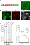
Cyp27c1 expression converts vitamin A1 into vitamin A2. a Plasmid for expression of zebrafish Cyp27c1 and EGFP separated by a 2A self-cleaving peptide. b,c HEK 293 cells stably expressing Cyp17c1-EGFP (green) in overview (b) and stained with antibodies (c) against Cyp27c1 (red) and mitochondrial ATP synthase F1-β (white). d HPLC analysis of HEK 293 cells without (CTR) and with Cyp27c1 (CYP) expression after incubation with 3.5 μM vitamin A1 (all-trans retinol). Specific peaks for all-trans retinol (AT-ROL) and 13-cis retinol (13c-ROL) species of vitamin A1 and A2 highlighted in grey. Note the appearance of vitamin A2 species only in Cyp27c1 expressing cells. e,f Quantification of total amount of vitamin A1 (e, n = 4) or vitamin A2 (f, n = 4) normalized to total protein level in HEK 293 cells without (CTR) and with Cyp27c1 (CYP) expression after incubation with vitamin A1 (pooled data for 3.5 and 35 μM vitamin A1). ** p ≤ 0.01
Fig. 4
Fig. 4
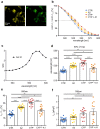
Cyp27c1 co-expression red-shifts the spectral sensitivity of ChR2 a Transient expression of ChR2-EYFP (yellow) in a HEK 293 cell line stably expressing Cyp27c1-EGFP (green). Special fluorescence filters were chosen to discriminate between EYFP and EGFP. b ChR2 photocurrents in relation to the maximal current for each cell in Panserin 293A medium for cells expressing Cyp27c1 without (CYP, n = 14) and with vitamin A1 incubation (CYP + A1, 5 μM, n = 11) and in control cells without (CTR, n = 19) and with vitamin A2 incubation (A2, 25 μM, n = 17). Note that Cyp27c1 expression shifts the spectral sensitivity of ChR2 at 50% photocurrent (black arrow). c Ratio of normalized ChR2 photocurrents in cells expressing Cyp27c1 and control cells shows up to three times higher currents at wavelengths between 550 and 600 nm. d Wavelengths resulting in 50% of ChR2 photocurrents from data shown in b calculated for each cell by linear fitting. e,f Quantification of normalized (e) and absolute (f) ChR2 photocurrents at 550 nm.. * p ≤ 0.05, ** p ≤ 0.01, **** p ≤ 0.0001
Fig. 5
Fig. 5
Cyp27c1 co-expression red-shifts spectral sensitivity of ReaChR a Transient expression of ReaChR-Cerulean (blue) in a HEK 293 cell line stably expressing Cyp27c1-EGFP (green). b ReaChR photocurrents in relation to the maximal current for each cell in Panserin 293A medium for cells expressing Cyp27c1 without (CYP, n = 15) and with vitamin A1 incubation (CYP + A1, 5 μM, n = 14) and in control cells without (CTR, n = 15) and with vitamin A2 incubation (A2, 25 μM, n = 14). Note that Cyp27c1 expression shifts the spectral sensitivity of ReaChR at 50% photocurrent (black arrow). c Ratio of normalized ReaChR photocurrents in cells expressing Cyp27c1 and control cells shows more than three times higher currents at wavelengths above 650 nm. d Wavelengths resulting in 50% of normalized ReaChR photocurrents from data shown in b calculated for each cell by linear fitting e,f Quantification of normalized (e) and absolute (f) ReaChR photocurrents at 650 nm from data shown in b. * p ≤ 0.05, ** p ≤ 0.01, **** p ≤ 0.0001

References

    1. Babino D, Perkins BD, Kindermann A, Oberhauser V, von Lintig J. The role of 11-cis-retinyl esters in vertebrate cone vision. FASEB J. 2015;29:216–226. doi: 10.1096/fj.14-261693. - DOI - PMC - PubMed
    1. Barua AB, Furr HC. Properties of retinoids: Structure, handling, and preparation. Mol Biotechnol. 1998;10:167–182. doi: 10.1007/BF02760863. - DOI - PubMed
    1. Boyden ES, Zhang F, Bamberg E, Nagel G, Deisseroth K. Millisecond-timescale, genetically targeted optical control of neural activity. Nat. Neurosci. 2005;8:1263–1268. doi: 10.1038/nn1525. - DOI - PubMed
    1. Boyle PM, Karathanos TV, Trayanova NA. Cardiac Optogenetics: 2018. JACC: Clin Electrophysiol. 2018;4:155–167. doi: 10.1016/j.jacep.2017.12.006. - DOI - PMC - PubMed
    1. Bruegmann T, Boyle PM, Vogt CC, Karathanos TV, Arevalo HJ, Fleischmann BK, Trayanova NA, Sasse P. Optogenetic defibrillation terminates ventricular arrhythmia in mouse hearts and human simulations. J Clin Investig. 2016;126:3894–3904. doi: 10.1172/JCI88950. - DOI - PMC - PubMed

Publication types