. 2024 Apr 5;73(5):729-740.
doi: 10.1136/gutjnl-2023-330721.

Understanding the malignant potential of gastric metaplasia of the oesophagus and its relevance to Barrett's oesophagus surveillance: individual-level data analysis

Collaborators, Affiliations

Understanding the malignant potential of gastric metaplasia of the oesophagus and its relevance to Barrett's oesophagus surveillance: individual-level data analysis

Emily L Black et al. Gut. .

Abstract

Objective: Whether gastric metaplasia (GM) of the oesophagus should be considered as Barrett's oesophagus (BO) is controversial. Given concern intestinal metaplasia (IM) may be missed due to sampling, the UK guidelines include GM as a type of BO. Here, we investigated whether the risk of misdiagnosis and the malignant potential of GM warrant its place in the UK surveillance.

Design: We performed a thorough pathology and endoscopy review to follow clinical outcomes in a novel UK cohort of 244 patients, covering 1854 person years of follow-up. We complemented this with a comparative genomic analysis of 160 GM and IM specimens, focused on early molecular hallmarks of BO and oesophageal adenocarcinoma (OAC).

Results: We found that 58 of 77 short-segment (<3 cm) GM (SS-GM) cases (75%) continued to be observed as GM-only across a median of 4.4 years of follow-up. We observed that disease progression in GM-only cases and GM+IM cases (cases with reported GM on some occasions, IM on others) was significantly lower than in the IM-only cases (Kaplan-Meier, p=0.03). Genomic analysis revealed that the mutation burden in GM is significantly lower than in IM (p<0.01). Moreover, GM does not bear the mutational hallmarks of OAC, with an absence of associated signatures and driver gene mutations. Finally, we established that GM found adjacent to OAC is evolutionarily distant from cancer.

Conclusion: SS-GM is a distinct entity from SS-IM and the malignant potential of GM is lower than IM. It is questionable whether SS-GM warrants inclusion in BO surveillance.

Keywords: BARRETT'S METAPLASIA; BARRETT'S OESOPHAGUS; GASTRIC METAPLASIA; OESOPHAGEAL CANCER; SURVEILLANCE.


Conflict of interest statement

Competing interests: RCF and MOD are named on patents for Cytosponge and associated technology, licensed to Covidien GI solutions (now Medtronic). RCF and MOD are shareholders of Cyted Ltd., a company working on early detection technology. The other authors declare no competing interests.

Figures

Figure 1
Figure 1
Illustration of the challenge of misdiagnosis due to undersampling of GM+IM segments. BO, Barrett’s oesophagus; GM, gastric metaplasia; IM, intestinal metaplasia.
Figure 2
Figure 2
Study design. Flow chart showing creation of clinical outcomes cohort and sequencing cohorts. BO, Barrett’s oesophagus; GM, gastric metaplasia; IM, intestinal metaplasia; OAC, oesophageal adenocarcinoma.
Figure 3
Figure 3
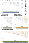
GM is a distinct state, not mis-sampling of IM. Progression to dysplasia and cancer is lower in GM than IM. (A) Surveillance history of 67 index-GM, GM-only outcome cases. (B) Surveillance history of 33 index-GM, GM+IM outcome cases. (C) Surveillance history of 44 index-IM, GM+IM outcome cases. (D) Surveillance history of 100 index-IM, IM-only outcome cases. For (A−D), each vertical trajectory is an individual case history, with the marker shapes denoting the histopathological assessment for each OGD. Each case has its surveillance time indexed to baseline, with the length of the vertical line representing total surveillance time. BO, Barrett’s oesophagus; FIM, focal intestinal metaplasia; GM, gastric metaplasia; HGD, high-grade dysplasia; IM, intestinal metaplasia; IMC, intramucosal carcinoma; IND, indefinite for dysplasia; LGD, low-grade dysplasia; WIM, widespread intestinal metaplasia.
Figure 4
Figure 4
Progression to dysplasia and cancer is lower in GM than IM. Kaplan-Meier curves for progression to LGD or higher and to HGD or higher, separated by classification. GM, gastric metaplasia; HGD, high-grade dysplasia; IM, intestinal metaplasia; LGD, low-grade dysplasia.
Figure 5
Figure 5
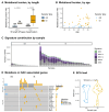
GM does not bear the genomic hallmarks of OAC. (A) Mutational burden by sample type and BO segment length. (B) Mutational burden by age. (C) SBS signature contribution by sample. (D) Mutations in OAC associated genes, split by genes typically mutated early and late in the progression of OAC. Only genes with mutations in this cohort, along with TP53, are shown. (E) SCA load by sample, taken as the length of genome altered by a copy gain, loss or copy neutral loss of heterozygosity. BO, Barrett’s oesophagus; GM, gastric metaplasia; IM, intestinal metaplasia; OAC, oesophageal adenocarcinoma; SCA, somatic chromosomal alterations; SNV, single nucleotide variant.
Figure 6
Figure 6
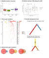
Co-occurring GM and OAC are evolutionarily distant from one another. (A) Mutation burden in OAC tumours, by type of adjacent BO. (B) Mutation burden in BO adjacent to OAC. (C) Mutations in driver genes by sample, across BO samples and adjacent OAC. Only genes with a mutation in a BO sample are shown. (D) Venn diagrams showing the degree of overlap in mutations between different types of BO and the adjacent OAC. (E) Example phylogenetic trees for one case with GM and adjacent OAC, and one case with HGD in an intestinal background, and adjacent OAC. Branch length represents number of mutations. BO, Barrett’s oesophagus; GM, gastric metaplasia; IM, intestinal metaplasia; OAC, oesophageal adenocarcinoma.
Figure 7
Figure 7
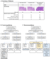
Short-segment gastric metaplasia does not warrant surveillance. (A) Summary of features of GM and IM. (B) Current British Society of Gastroenterology guidelines for management of non-dysplastic BO. Grey boxes denote areas of recommended change. (C) Recommended flow chart for non-dysplastic BO. Yellow boxes denote changes to BO definition, blue boxes denote changes to GM management. BO, Barrett’s oesophagus; GM, gastric metaplasia; IM, intestinal metaplasia; OAC, oesophageal adenocarcinoma; OGD, oesophagogastroduodenoscopy.

Comment in

References

    1. Rastogi A, Puli S, El-Serag HB, et al. . Incidence of esophageal adenocarcinoma in patients with Barrett's esophagus and high-grade dysplasia: a meta-analysis. Gastrointest Endosc 2008;67:394–8. 10.1016/j.gie.2007.07.019 - DOI - PubMed
    1. Kastelein F, van Olphen S, Steyerberg EW, et al. . Surveillance in patients with long-segment Barrett's oesophagus: a cost-effectiveness analysis. Gut 2015;64:864–71. 10.1136/gutjnl-2014-307197 - DOI - PubMed
    1. Hvid-Jensen F, Pedersen L, Drewes AM, et al. . Incidence of adenocarcinoma among patients with Barrett's esophagus. N Engl J Med 2011;365:1375–83. 10.1056/NEJMoa1103042 - DOI - PubMed
    1. Fitzgerald RC, di Pietro M, Ragunath K, et al. . British society of gastroenterology guidelines on the diagnosis and management of Barrett's oesophagus. Gut 2014;63:7–42. 10.1136/gutjnl-2013-305372 - DOI - PubMed
    1. Shaheen NJ, Falk GW, Iyer PG, et al. . ACG clinical guideline: diagnosis and management of Barrett's esophagus. Am J Gastroenterol 2016;111:30–50. 10.1038/ajg.2015.322 - DOI - PMC - PubMed

Publication types

Supplementary concepts