

Immune proteins C1q and CD47 may contribute to aberrant microglia-mediated synapse loss in the aging monkey brain that is associated with cognitive impairment
- PMID: 37989825
- PMCID: PMC10828237
- DOI: 10.1007/s11357-023-01014-x
Immune proteins C1q and CD47 may contribute to aberrant microglia-mediated synapse loss in the aging monkey brain that is associated with cognitive impairment
Abstract
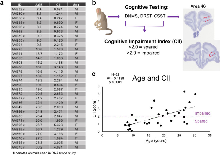
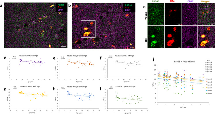
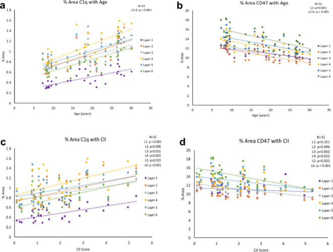
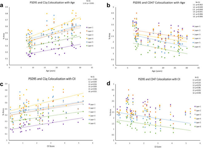
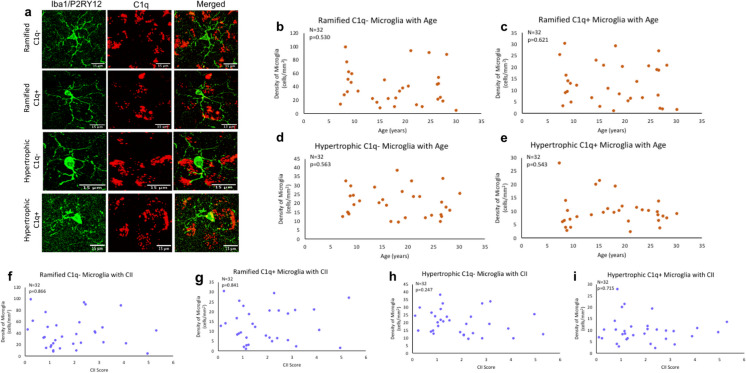
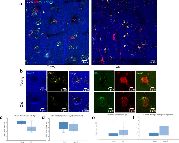
Cognitive impairment in learning, memory, and executive function occurs in normal aging even in the absence of Alzheimer's disease (AD). While neurons do not degenerate in humans or monkeys free of AD, there are structural changes including synapse loss and dendritic atrophy, especially in the dorsolateral prefrontal cortex (dlPFC), and these correlate with cognitive age-related impairment. Developmental studies revealed activity-dependent neuronal properties that lead to synapse remodeling by microglia. Microglia-mediated phagocytosis that may eliminate synapses is regulated by immune "eat me" and "don't eat me" signaling proteins in an activity-dependent manner, so that less active synapses are eliminated. Whether this process contributes to age-related synapse loss remains unknown. The present study used a rhesus monkey model of normal aging to investigate the balance between the "eat me" signal, complement component C1q, and the "don't eat me" signal, transmembrane glycoprotein CD47, relative to age-related synapse loss in dlPFC Area 46. Results showed an age-related elevation of C1q and reduction of CD47 at PSD95+ synapses that is associated with cognitive impairment. Additionally, reduced neuronal CD47 RNA expression was found, indicating that aged neurons were less able to produce the protective signal CD47. Interestingly, microglia do not show the hypertrophic morphology indicative of phagocytic activity. These findings suggest that in the aging brain, changes in the balance of immunologic proteins give microglia instructions favoring synapse elimination of less active synapses, but this may occur by a process other than classic phagocytosis such as trogocytosis.
Keywords: Aging; C1q; CD47; Cognitive decline; Microglia; Synapse.
© 2023. The Author(s), under exclusive licence to American Aging Association.
Conflict of interest statement
The authors declare no competing interests.
Figures
Similar articles
-
Dysregulated C1q and CD47 in the aging monkey brain: association with myelin damage, microglia reactivity, and cognitive decline.Front Immunol. 2024 Sep 27;15:1426975. doi: 10.3389/fimmu.2024.1426975. eCollection 2024. Front Immunol. 2024. PMID: 39399501 Free PMC article.
-
Classical complement cascade initiating C1q protein within neurons in the aged rhesus macaque dorsolateral prefrontal cortex.J Neuroinflammation. 2020 Jan 6;17(1):8. doi: 10.1186/s12974-019-1683-1. J Neuroinflammation. 2020. PMID: 31906973 Free PMC article.
-
CD47 Protects Synapses from Excess Microglia-Mediated Pruning during Development.Neuron. 2018 Oct 10;100(1):120-134.e6. doi: 10.1016/j.neuron.2018.09.017. Neuron. 2018. PMID: 30308165 Free PMC article.
-
Contribution of Neurons and Glial Cells to Complement-Mediated Synapse Removal during Development, Aging and in Alzheimer's Disease.Mediators Inflamm. 2018 Nov 11;2018:2530414. doi: 10.1155/2018/2530414. eCollection 2018. Mediators Inflamm. 2018. PMID: 30533998 Free PMC article. Review.
-
Microglia regulation of synaptic plasticity and learning and memory.Neural Regen Res. 2022 Apr;17(4):705-716. doi: 10.4103/1673-5374.322423. Neural Regen Res. 2022. PMID: 34472455 Free PMC article. Review.
Cited by
-
Knowing the enemy: strategic targeting of complement to treat Alzheimer disease.Nat Rev Neurol. 2025 May;21(5):250-264. doi: 10.1038/s41582-025-01073-y. Epub 2025 Mar 24. Nat Rev Neurol. 2025. PMID: 40128350 Review.
-
KChIP3 fosters neuroinflammation and synaptic dysfunction in the 5XFAD mouse model of Alzheimer's disease.J Neuroinflammation. 2025 Jun 19;22(1):160. doi: 10.1186/s12974-025-03426-2. J Neuroinflammation. 2025. PMID: 40537787 Free PMC article.
-
Pathogenesis and therapeutic applications of microglia receptors in Alzheimer's disease.Front Immunol. 2025 Feb 14;16:1508023. doi: 10.3389/fimmu.2025.1508023. eCollection 2025. Front Immunol. 2025. PMID: 40028337 Free PMC article. Review.
-
Immunological synapse: structures, molecular mechanisms and therapeutic implications in disease.Signal Transduct Target Ther. 2025 Aug 11;10(1):254. doi: 10.1038/s41392-025-02332-6. Signal Transduct Target Ther. 2025. PMID: 40784895 Free PMC article. Review.
-
The Hidden Dangers of Sedentary Living: Insights into Molecular, Cellular, and Systemic Mechanisms.Int J Mol Sci. 2024 Oct 6;25(19):10757. doi: 10.3390/ijms251910757. Int J Mol Sci. 2024. PMID: 39409085 Free PMC article. Review.
References
Publication types
MeSH terms
Substances
Grants and funding
LinkOut - more resources
Full Text Sources
Medical
Research Materials
