. 2023 Nov 21;13(1):20332.
doi: 10.1038/s41598-023-47695-4.

Insight into the on/off switch that regulates expression of the MSMEG-3762/63 efflux pump in Mycobacterium smegmatis

Affiliations

Insight into the on/off switch that regulates expression of the MSMEG-3762/63 efflux pump in Mycobacterium smegmatis

Nicoletta Campolattano et al. Sci Rep. .

Abstract

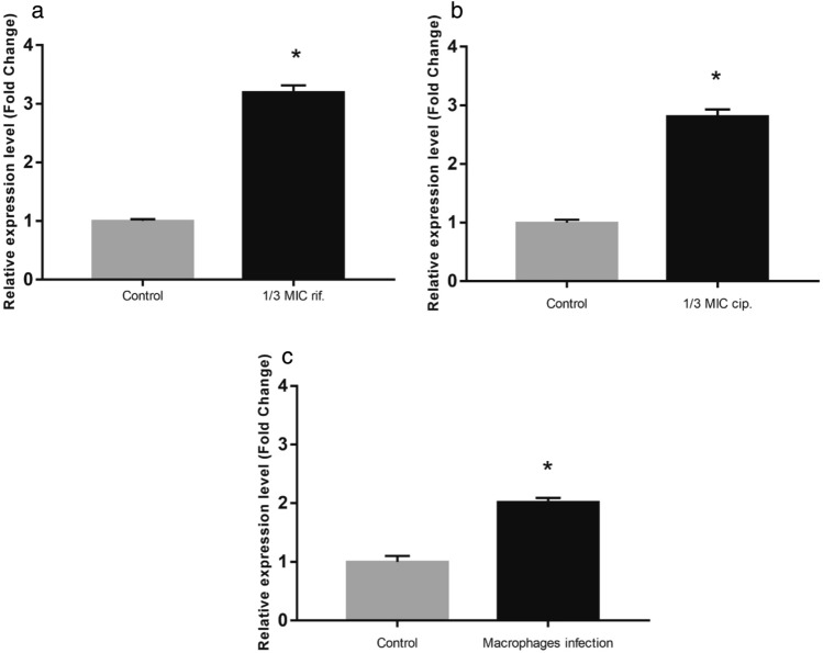
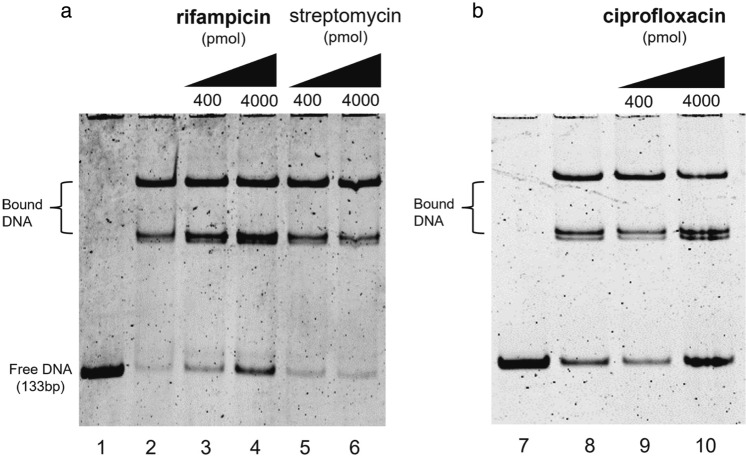
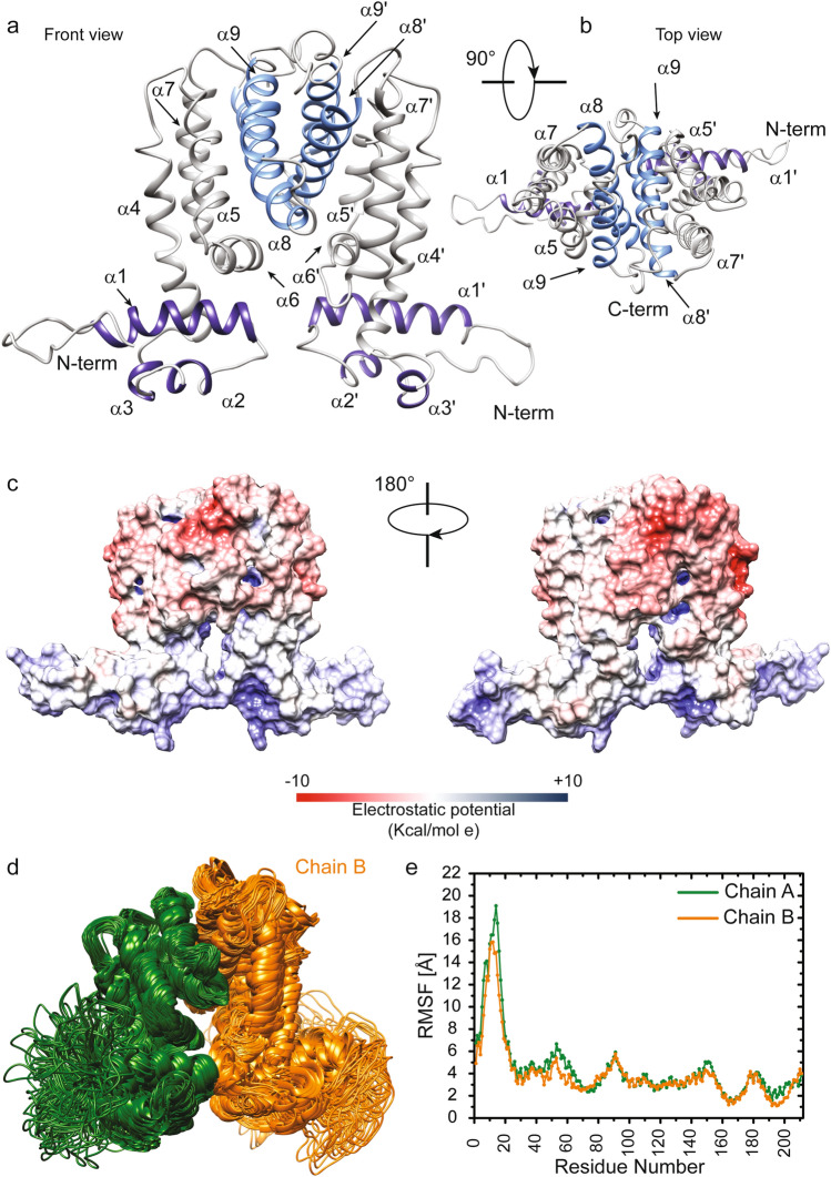
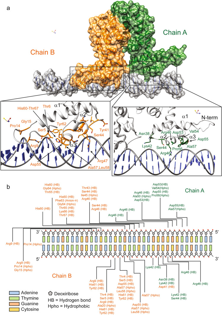
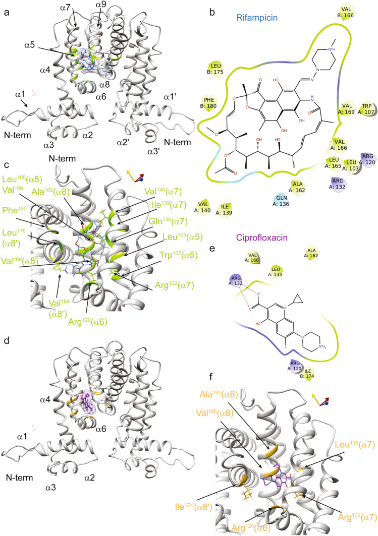
Drug resistance is one of the most difficult challenges facing tuberculosis (TB) control. Drug efflux is among the mechanisms leading to drug resistance. In our previous studies, we partially characterized the ABC-type MSMEG-3762/63 efflux pump in Mycobacterium smegmatis, which shares high percentage of identity with the Mycobacterium tuberculosis Rv1687/86c pump. MSMEG-3762/63 was shown to have extrusion activity for rifampicin and ciprofloxacin, used in first and second-line anti-TB treatments. Moreover, we described the functional role of the TetR-like MSMEG-3765 protein as a repressor of the MSMEG_3762/63/65 operon and orthologous Rv1687/86/85c in M. tuberculosis. Here we show that the operon is upregulated in the macrophage environment, supporting a previous observation of induction triggered by acid-nitrosative stress. Expression of the efflux pump was also induced by sub-inhibitory concentrations of rifampicin or ciprofloxacin. Both these drugs also prevented the binding of the MSMEG-3765 TetR repressor protein to its operator in the MSMEG_3762/63/65 operon. The hypothesis that these two drugs might be responsible for the induction of the efflux pump operon was assessed by bioinformatics analyses. Docking studies using a structural model of the regulator MSMEG-3765 showed that both antibiotics abolished the ability of this transcriptional repressor to recognize the efflux pump operon by interacting with the homodimer at different binding sites within the same binding pocket. Reduced binding of the repressor leads to induction of the efflux pump in M. smegmatis, and reduced efficacy of these two anti-mycobacterial drugs.


Conflict of interest statement

The authors declare no competing interests.

Figures

Figure 1
Figure 1
Comparative analysis of membrane potential. Comparison of the Δψ in M. smegmatis (wt), M. smegmatis (ΔMSMEG_3763) and complemented M. smegmatis (ΔMSMEG_3763 pBD04) (Compl.) was performed using the DiOC2 fluorescent dye and the ratiometric method (Red:Green). Data are the average of biological and technical triplicates in the absence and presence of the depolarizing compound CCCP. Error bars represent the standard deviation of the mean values. Significance of data obtained was tested by the Student’s t test (p < 0.05, compared to wt and Compl.).
Figure 2
Figure 2
Transcriptional analysis in stress conditions. Comparative RT-qPCR analysis showing induction of the MSMEG_3762/63/65 operon in log phase culture after exposure to 1/3rd MIC rifampicin (a) and 1/3rd MIC ciprofloxacin (b) or after macrophage phagocytosis (c). Control in (a) and (b): cells grown in the absence of antibiotics; control in (c): cells grown in RPMI in the absence of macrophages. sigA was used as a reference gene and relative expression levels were calculated with the Pfaffl method (a,b) or the 2−ΔΔCt method (c). Data shown are the mean values of three independent experiments, and each RT-qPCR was carried out in technical triplicate. Error bars represent the standard deviation of the mean values. Significance of data obtained was tested by the Student’s t test (p < 0.05).
Figure 3
Figure 3
Drug interference in repressor/operator interactions. Binding of the TetR-like MSMEG-3765 to the MSMEG_3762/63/65 operator region is perturbed in the presence of rifampicin (a) or ciprofloxacin (b), but not streptomycin (a). Lanes 1 and 7: 133 bp DNA fragment containing the 36 bp palindromic motif upstream of MSMEG_3762. Lanes 2 and 8: DNA and purified MSMEG-3765, showing low abundance of free operator DNA in the presence of the regulator. In the other lanes, increasing concentrations (400 or 4000 pmol) of rifampicin (lanes 3 and 4) or ciprofloxacin (lanes 9 and 10) show an antimicrobial drug dose-dependent increase in free operator DNA, which does not occur after addition of streptomycin (lanes 5 and 6). The experiment was repeated 3 times.
Figure 4
Figure 4
Structure and dynamics of the MSMEG-3765 homodimer. (a,b) The representative 3D structural model of the MSMEG-3765 transcriptional regulator obtained using the MSMEG-3765 primary sequence (https://zhanggroup.org/TACOS/) see “Materials and methods”). The helices 1 to 3 of NTD are depicted in dark violet, whereas helices 8 and 9 from each monomer forming a four-helical bundle that makes up the dimer interface are in light blue. (c) Electrostatic surface potential map of MSMEG-3765. The MSMEG dimer surface is depicted from electropositive (blue; 10 kcal/mol) to electronegative (red; − 10 kcal/mol). (d) Conformational ensemble of the MSMEG-3765 homodimer obtained using NMSim methodology. The two monomers are colored dark green (chain A) and orange (chain B). (e) Cα RMSFs (Root Mean Square Fluctuations) plotted versus the primary sequence for MSMEG-3765 chain A and B.
Figure 5
Figure 5
MSMEG-3765/DNA complex interactions. (a) Surface representation of the MSMEG-3765 homodimer in complex with DNA as reported by docking studies. Monomer A and B are colored dark green and orange, respectively; the DNA is depicted in light grey. Close-up views of side chain-DNA interactions for the NTD residues involved in the complex formation are detailed. (b) A schematic drawing of MSMEG-3765/DNA interactions. Only direct contacts between the protein and the nucleic acid are shown. Hydrogen bond (HB) and hydrophobic interactions (Hpho) are reported. Hydrophobic interactions were determined using a threshold distance of 4.5 Å.
Figure 6
Figure 6
Interactions of the MSMEG-3765 homodimer with rifampicin and ciprofloxacin as revealed by molecular docking. (a) Ribbon drawing representation of the MSMEG-3765 dimer/rifampicin complex, where the ligand surface is highlighted as light blue and the regions of the transcription regulator are depicted in light green. (b) A 2D interaction map of rifampicin in complex with the MSMEG-3765 dimer identifying key interaction sites. (c) The rifampicin binding pocket in chain A of the MSMEG-3765 dimer. The side chains of the MSMEG-3765 residues involved in hydrogen bonds are illustrated as light green, whereas the ligand is colored light blue. The side chains of the residues making hydrophobic interactions are not shown. (d) The ciprofloxacin-binding site in chain A of MSMEG-3765. The ligand is reported in magenta, whereas the residues involved in the interactions are colored gold. The surface of the ligand is also shown. (e) A 2D interaction map of ciprofloxacin in complex with the MSMEG-3765 dimer. (f) Detailed view of the MSMEG-3765/ciprofloxacin complex. The ligand and side chains of the residues involved in the formation of the complex are depicted in magenta and gold, respectively.

Similar articles

References

    1. Bagcchi S. WHO's global tuberculosis report 2022. Lancet Microbe. 2023;4:e20. doi: 10.1016/S2666-5247(22)00359-7. - DOI - PubMed
    1. Chakaya J, et al. The WHO global tuberculosis 2021 report—Not so good news and turning the tide back to End TB. Int. J. Infect. Dis. 2022;124(Suppl 1):S26–S29. doi: 10.1016/j.ijid.2022.03.011. - DOI - PMC - PubMed
    1. Hameed HMA, et al. Molecular targets related drug resistance mechanisms in MDR-, XDR-, and TDR- Front. Cell Infect. Microbiol. 2018;8:114. doi: 10.3389/fcimb.2018.00114. - DOI - PMC - PubMed
    1. Khawbung JL, Nath D, Chakraborty S. Drug resistant tuberculosis: A review. Comp. Immunol. Microbiol. Infect. Dis. 2021;74:101574. doi: 10.1016/j.cimid.2020.101574. - DOI - PubMed
    1. Tang M, Kong X, Hao J, Liu J. Epidemiological characteristics and formation mechanisms of multidrug-resistant hypervirulent. Front. Microbiol. 2020;11:581543. doi: 10.3389/fmicb.2020.581543. - DOI - PMC - PubMed

MeSH terms