Review
. 2023 Nov 21;11(1):61.
doi: 10.1038/s41413-023-00294-5.

Structure and function of the membrane microdomains in osteoclasts

Affiliations
Review

Structure and function of the membrane microdomains in osteoclasts

Jialong Hou et al. Bone Res. .

Abstract

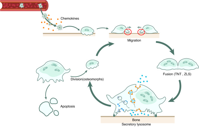
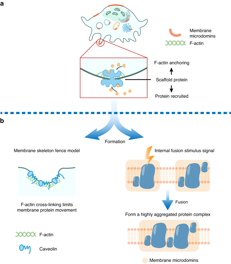
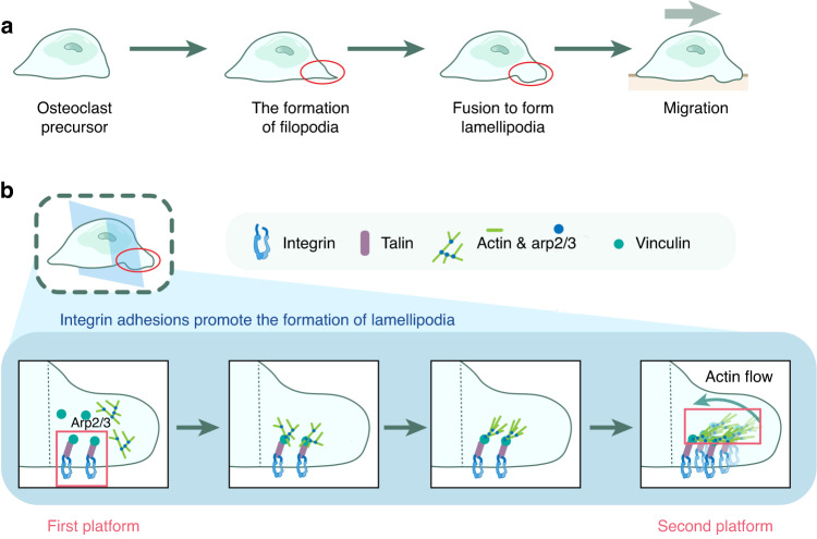
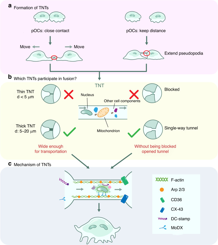
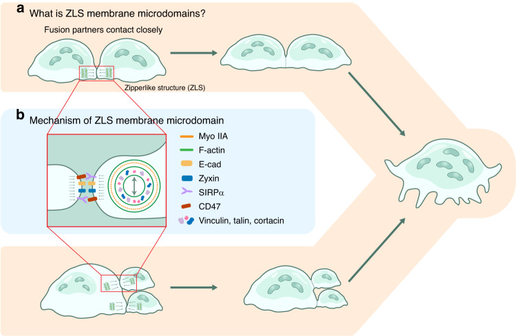
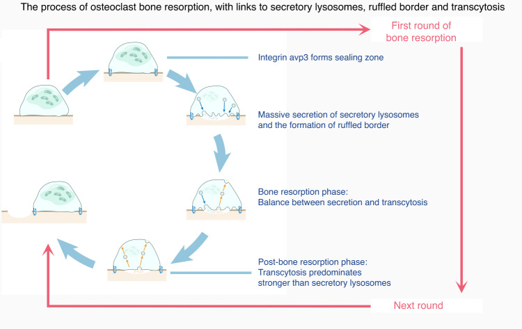
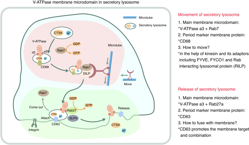
The cell membrane structure is closely related to the occurrence and progression of many metabolic bone diseases observed in the clinic and is an important target to the development of therapeutic strategies for these diseases. Strong experimental evidence supports the existence of membrane microdomains in osteoclasts (OCs). However, the potential membrane microdomains and the crucial mechanisms underlying their roles in OCs have not been fully characterized. Membrane microdomain components, such as scaffolding proteins and the actin cytoskeleton, as well as the roles of individual membrane proteins, need to be elucidated. In this review, we discuss the compositions and critical functions of membrane microdomains that determine the biological behavior of OCs through the three main stages of the OC life cycle.


Conflict of interest statement

The authors declare no competing interests.

Figures

Fig. 1
Fig. 1
The life cycle of OCs. The life cycle of OCs is divided into three phases: (1) hematopoietic stem cells and erythroid-myeloid precursors extend filopodia from their membrane and migrate to the bone matrix;, (2) monocytes form mature OCs (mOCs) through membrane fusion; and (3) mOCs usually continue to be multinucleated and release secretory lysosomes that degrade the bone matrix., In these three phases, special membrane structures are required to mediate OC differentiation and function, from migration to fusion and the release of secretory lysosomes
Fig. 2
Fig. 2
The formation and model of membrane microdomain formation in OCs. a Schematic diagram showing membrane structural domains in OCs. OC scaffolding proteins anchor to the cell membrane and the actin cytoskeleton and recruit proteins to form membrane microdomains. b Two models of membrane microdomain formation, namely, the membrane cytoskeleton fence model and the internal membrane microdomain fusion model, were proposed to provide a reference for the roles of scaffolding proteins and the cytoskeleton in membrane microdomain formation
Fig. 3
Fig. 3
Lamellipodia and their formation. a Schematic diagram at the macroscopic level: the process of lamellipodium formation. pOCs form filamentous pseudopodia, and their fusion drives lamellipodium formation, which determines the direction of cell migration. b Schematic diagram at the microscopic level: the process of integrin adhesion promoting lamellipodium formation. Longitudinal sections of lamellipodia show that integrins recruit the regulatory proteins talin and vinculin, which regulate actin skeleton remodeling mediated via Arp2/3 to initiate reverse actin flow and mediate pseudopod contraction on the basis of the counteracting force provided by the integrin adhesion bodies. In this process, integrins and regulatory proteins form the scaffolds of the pseudopod membrane microdomains and then integrate actin, leading to the formation of membrane macrostructures
Fig. 4
Fig. 4
Early fusion: OCs fuse through TNTs. a Two mechanisms explain TNT formation: filamentous pseudopods extend between fusion partners or nearby fusion partners that have separated from each other by the action of chemokines, and a TNT is formed at the interconnection of the plasma membrane between fusion partners. b Nuclear translocation is possible when a TNT has (1) a diameter in the range of 5–20 µm and (2) an open interconnection inside the duct. c The processes and mechanisms by which membrane microdomains mediate nucleus transport
Fig. 5
Fig. 5
Late fusion: OCs undergo multinucleated cell–multinucleated cell and multinucleated cell–mononuclear cell fusion through ZLS structures. a Fusion partners are closely linked through actin flow, and the actin cytoskeleton forms a ZLS structure at a contact point. b The structure of the ZLS membrane microdomain, including the surface membrane proteins and the internal actin complex
Fig. 6
Fig. 6
mOCs exert their osteolytic function by adopting a secretory lysosomal structure. The process of OC bone resorption is related to secretory lysosome production, the RB and transcytosis. Activation of integrin signaling during initial bone resorption leads to development of a sealing zone for OC bone resorption. Accordingly, many secreted lysosomes are fused to the plasma membrane within the sealing zone, leading to the formation of ruffles. Secretory lysosomes are secreted mainly into peripheral subdomains of RBs and in the central subdomain, which is thought to be the site of transcytosis. Endocytic vesicles are formed in the central subdomain and transported to the apical side of the cell. During bone resorption, secretory lysosomes initially play a key role in facilitating the rapid formation of RBs, whereas transcytosis depletes the ruffles and facilitates the endocytosis and secretion of osteolytic products from the bone resorption lumen to the extracellular surface. At the onset of a new cycle of bone resorption, RBs are formed and depleted again
Fig. 7
Fig. 7
Membrane microstructural domains of V-ATPase secretory lysosomes. Rab7GDP+ binds to the a3 isoform of V-ATPase in the lysosomal membrane, and then, GDP is replaced by GTP. A lysosome binds to motor proteins via Rab7GTP+, which in turn colocalizes with the lysosomal plasma membrane via the action of CD68. Subsequently, Rab7 moves inward along microtubules in collaboration with in-adapter-Rab-interacting lysosomal protein (RILP), while FYVE encoded by FYCO1, the adapter of the kinesin driver protein, moves outward and participates in vesicle transport. Near the plasma membrane, Rab27a preferentially binds to CD63-positive secretory lysosomes, maintains their stability under the action of slp4, and then binds to the plasma membrane with CD63 as a marker, thereby mediating lysosomal content release

Similar articles

Cited by

References

    1. Ng PY, Brigitte Patricia Ribet A, Pavlos NJ. Membrane trafficking in osteoclasts and implications for osteoporosis. Biochem. Soc. Trans. 2019;47:639–650. doi: 10.1042/BST20180445. - DOI - PMC - PubMed
    1. Michigami T, Ozono K. Roles of phosphate in skeleton. Front. Endocrinol. (Lausanne) 2019;10:180. doi: 10.3389/fendo.2019.00180. - DOI - PMC - PubMed
    1. Chen K, et al. Steroid-induced osteonecrosis of the femoral head reveals enhanced reactive oxygen species and hyperactive osteoclasts. Int. J. Biol. Sci. 2020;16:1888–1900. doi: 10.7150/ijbs.40917. - DOI - PMC - PubMed
    1. Guo Q, et al. Rheumatoid arthritis: pathological mechanisms and modern pharmacologic therapies. Bone Res. 2018;6:15. doi: 10.1038/s41413-018-0016-9. - DOI - PMC - PubMed
    1. Krajewska-Wlodarczyk M, et al. Role of microparticles in the pathogenesis of inflammatory joint diseases. Int. J. Mol. Sci. 2019;20:5453. doi: 10.3390/ijms20215453. - DOI - PMC - PubMed

Publication types

MeSH terms

Substances