. 2023 Nov 20;13(1):20336.
doi: 10.1038/s41598-023-47236-z.

Solanum lycopersicum heme-binding protein 2 as a potent antimicrobial weapon against plant pathogens

Affiliations

Solanum lycopersicum heme-binding protein 2 as a potent antimicrobial weapon against plant pathogens

Atefeh Farvardin et al. Sci Rep. .

Abstract

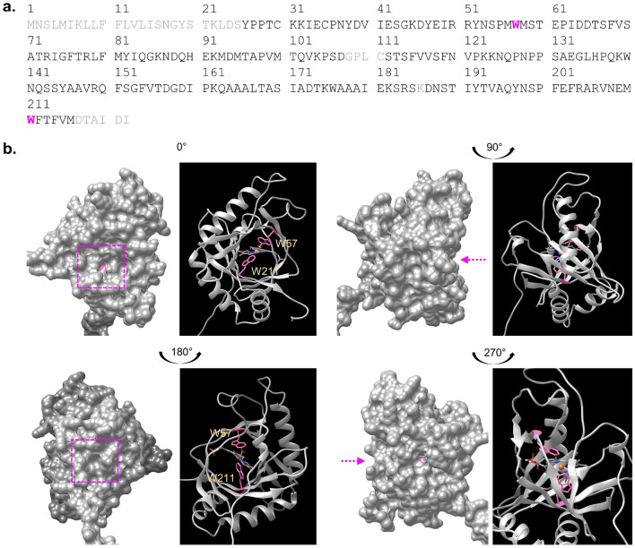
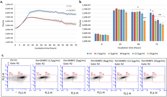
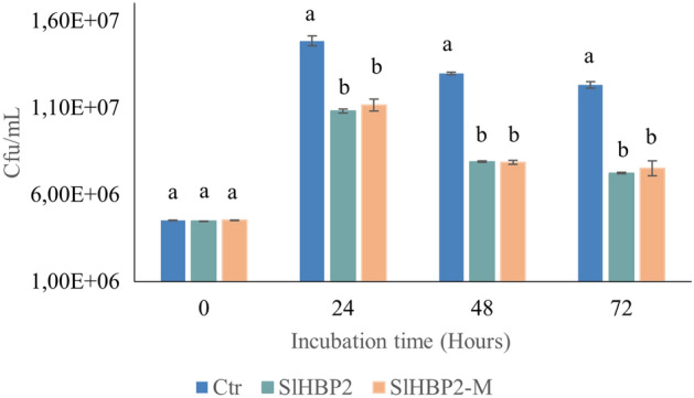
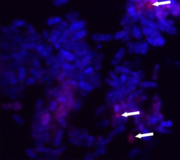
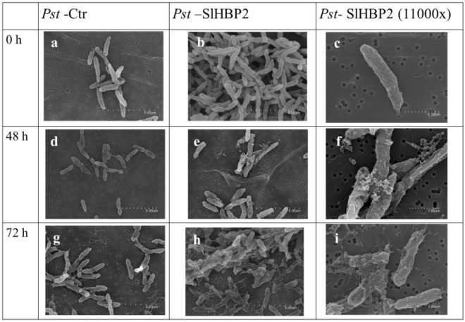
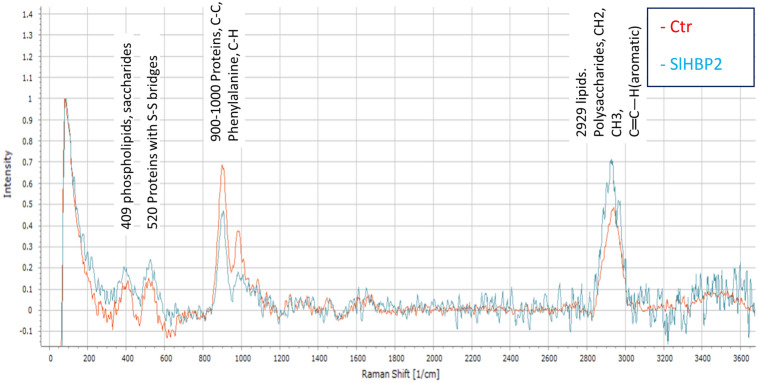
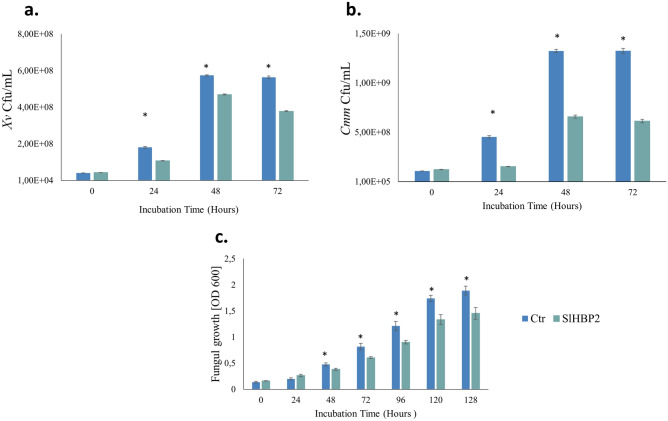
The rise in antibiotic-resistant bacteria caused by the excessive use of antibiotics has led to the urgent exploration of alternative antimicrobial solutions. Among these alternatives, antimicrobial proteins, and peptides (Apps) have garnered attention due to their wide-ranging antimicrobial effects. This study focuses on evaluating the antimicrobial properties of Solanum lycopersicum heme-binding protein 2 (SlHBP2), an apoplastic protein extracted from tomato plants treated with 1-Methyl tryptophan (1-MT), against Pseudomonas syringae pv. tomato DC3000 (Pst). Computational studies indicate that SlHBP2 is annotated as a SOUL heme-binding family protein. Remarkably, recombinant SlHBP2 demonstrated significant efficacy in inhibiting the growth of Pst within a concentration range of 3-25 μg/mL. Moreover, SlHBP2 exhibited potent antimicrobial effects against other microorganisms, including Xanthomonas vesicatoria (Xv), Clavibacter michiganensis subsp. michiganensis (Cmm), and Botrytis cinerea. To understand the mechanism of action employed by SlHBP2 against Pst, various techniques such as microscopy and fluorescence assays were employed. The results revealed that SlHBP2 disrupts the bacterial cell wall and causes leakage of intracellular contents. To summarize, the findings suggest that SlHBP2 has significant antimicrobial properties, making it a potential antimicrobial agent against a wide range of pathogens. Although further studies are warranted to explore the full potential of SlHBP2 and its suitability in various applications.


Conflict of interest statement

The authors Loredana Scalschi; Begonya Vicedo; Eugenio Llorens; Atefeh Farvardin; Gemma Camañes; and Eva Falomir, are listed as inventors in the patent (ES 2 879 643 B2) entitled: Antimicrobial proteins and peptides with stress-resistance-inducing activity. Authors Luisa Liu-Xu, Lorena Sanchez-Gimenez, Aloysius Wong, Elena G. Biosca and Jose M. Pedra, declare no competing interests.

Figures

Figure 1
Figure 1
Sequence and structural analysis of SIHBP2. (a) Full-length amino acid sequence of SIHBP2 (UniProt: A0A3Q7HE03). The key tryptophan residues W57 and W211 identified as crucial for interactions with the heme moiety are represented in pink. Regions with grey colored amino acids have low to very low model confidence (pLDDT < 70) whereas regions with black colored amino acids have high to very high model confidence (pLDDT > 70), based on the protein structure prediction generated by AlphaFold (ref). pLDDT, which is scaled from 0 to 100, is an estimate of the confidence of each amino acid corresponding to the model’s predicted score on the lDDT-Cα metric. Except for the N-terminal 20-amino acid long signal region (marked in grey), the model, which includes the key tryptophan residues at the heme binding pocket, has mostly high to very high confidence score, and is deemed suitable for characterization of ligand or cofactor binding sites. (b) Computational assessment of the heme-binding region of SlHBP2. The SIHBP2 model generated by AlphaFold obtained from https://alphafold.ebi.ac.uk/entry/A0A3Q7HE03 and was used for structural assessment and docking simulation studies. The heme-binding region of SlHBP2 is shown within the pink box as surface model (left) and as ribbon model (right). Docking simulation with AutoDock Vina indicates that the heme-binding region occupies a distinct pocket in the model (pink arrow and pink box), and importantly, this cavity can spatially accommodate heme. The tryptophan residues W57 and W211, are identified as likely amino acids for interaction with the heme moiety. Docking simulations were conducted using AutoDock Vina (version 1.1.2) and structural visualization and image preparation were performed using UCSF Chimera.
Figure 2
Figure 2
Effect of the SlHBP2 protein on the growth of Pst. (a) Antimicrobial effect of SlHBP2 at 75 µg/mL. (b) MIC and (c) MBC results. All the assays were performed in M9 minimal medium in a Multiskan plate reader under constant agitation, at 28 °C, taking measurements at 600 nm every 10 min for 72 h. The asterisks indicate statistically significant differences between groups (P < 0.05; least-significant difference test). After SlHBP2 exposure cell samples were stained with Live/dead BacLight™ and measured by flow cytometry. R3 represents the area with dead bacteria, and R5 represents the area with live bacteria in Ctr, 1,5 µg/mL, 3 µg/mL, 6 µg/mL, 12,5 µg/mL, and 25 µg/mL, respectively. The results of one experiment are shown. Four independent experiments were carried out with similar results.
Figure 3
Figure 3
Comparative antibacterial properties of SlHBP2 and SlHBP2-M (heme group mutant) against Pst. A concentration of 75 µg/mL of SlHBP2 and SlHBP2-M was applied in M9 minimal medium inoculated with Pst, and the culture was kept at 28 °C under continuous agitation for 72 h. Graph shows the means with the standard errors. Different letters represent statistically significant differences (P < 0.05; least-significant difference test). Four independent experiments were carried out with similar results.
Figure 4
Figure 4
Localization of SlHBP2. Fluorescence confocal microscopy images of bacteria treated with SlHBP2 and stained with Alexa Fluor 647-conjugated anti-His secondary antibody and with Hoeschst 33,342. Pink fluorescence indicates the localization of the SlHBP2 in the cells. Blue fluorescence shows cytoplasmic DNA stained with Hoeschst 33,342.
Figure 5
Figure 5
SEM micrographs of untreated Pst (Pst-Ctr) and of SLHBP2 treated Pst (Pst-SlHBP2) after 0, 48 and 72 h of incubation in M9 minimal medium. The untreated Pst cells were long, intact, and evenly shaped at all studied time points (a–d–g). SlHBP2 treated Pst cells show loss of cell wall integrity starting from 48 h while 72 h treatment with the protein caused complete lysis of Pst cells (b–e–h). Higher magnification images of samples (e–f–i).
Figure 6
Figure 6
Raman spectra of control Pst (red line) and SlHBP2 treated Pst (blue line). The assay was repeated three independent times and the spectra are the average of those assays. The y-axis is in arbitrary intensity units and the x-axis is in wavenumbers (cm−1).
Figure 7
Figure 7
Differential gene expression was assessed by RT-qPCR after Pst were cultured in the presence of 50 µg/mL of SlHBP2. (a, b, c) show the synthesis of coronatine (cfa, cmaB, cfl) genes, (d, e) represent T3SS marker genes (hrpA, hrpL), and (f, g) represent fliC and psyI genes respectively. The samples were collected 16 and 24 h Bars represent relative expression changes of target genes in bacteria grown in the presence of the antimicrobial agent , with significance indicated by asterisks (*P < 0.05, Student's t-test).
Figure 8
Figure 8
The effects of SlHBP2 on different plant pathogens. (a) Xv, (b) Cmm, and (c) B. cinerea. Growth was assessed through optical density measurements (OD) at a wavelength of 620 nm and 492 nm for bacteria and fungi respectively. Normality tests were performed, and significant differences (P ≤ 0.05) were determined using one-way ANOVA for parametric data and the Kruskal–Wallis test for non-parametric data. The asterisks indicate significant differences between groups at each time point.

References

    1. Felgueiras HP, Amorim MTP. Functionalization of electrospun polymeric wound dressings with antimicrobial peptides. Colloids Surf. B Biointerfaces. 2017;156:133–148. doi: 10.1016/j.colsurfb.2017.05.001. - DOI - PubMed
    1. Manners, J. M. Hidden weapons of microbial destruction in plant genomes. Genome Biol.8 (2007). - PMC - PubMed
    1. Farvardin A, et al. The apoplast: A key player in plant survival. Antioxidants. 2020;9:1–26. doi: 10.3390/antiox9070604. - DOI - PMC - PubMed
    1. Sels J, Mathys J, De Coninck BMAA, Cammue BPAA, De Bolle MFCC. Plant pathogenesis-related (PR) proteins: A focus on PR peptides. Plant Physiol. Biochem. PPB. 2008;46:941–950. doi: 10.1016/j.plaphy.2008.06.011. - DOI - PubMed
    1. Sitaram N, Nagaraj R. Host-defense antimicrobial peptides: Importance of structure for activity. Curr. Pharm. Des. 2002;8:727–742. doi: 10.2174/1381612023395358. - DOI - PubMed

MeSH terms

Substances

Supplementary concepts