

Brain MRI and EEG overemployment in patients with vasovagal syncope: results from a tertiary syncope unit
- PMID: 37990291
- PMCID: PMC10664686
- DOI: 10.1186/s12872-023-03615-y
Brain MRI and EEG overemployment in patients with vasovagal syncope: results from a tertiary syncope unit
Abstract
Background: The diagnosis of vasovagal syncope (VVS) is mainly based on history-taking and physical examination. However, brain Magnetic Resonance Imaging (MRI) and Electroencephalogram (EEG) are commonly used in the diagnostic course of VVS, despite not being indicated in the guidelines. This study aims to find the possible associated factors with the administration of brain MRI and EEG in patients with VVS.
Methods: Patients with a diagnosis of VVS from 2017 to 2022 were included. Several demographic and syncope features were recorded. The association of these was assessed with undergoing MRI, EEG, and either MRI or EEG. Univariate and multivariable logistic regression models were also used to calculate odds ratios (OR) and 95% confidence intervals (CI).
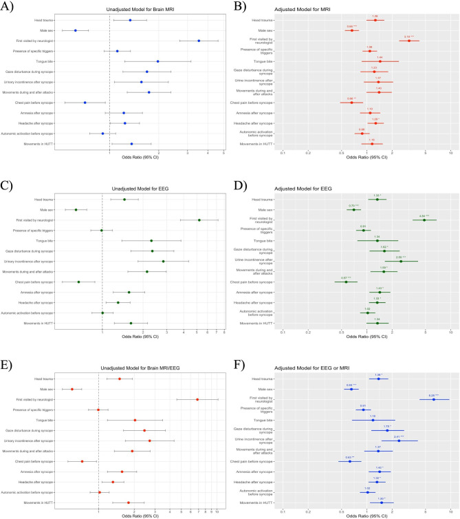
Results: A total of 1882 patients with VVS were analyzed, among which 810 underwent MRI (43.04%), 985 underwent EEG (52.34%), and 1166 underwent MRI or EEG (61.96%). Head trauma (OR 1.38, 95% CI 1.06 to 1.80), previous neurologist visit (OR 6.28, 95% CI 4.24 to 9.64), and gaze disturbance during syncope (OR 1.75, 95% CI 1.13 to 2.78) were all positively associated to the performance of brain MRI/EEG. Similar results were found for urinary incontinence (OR 2.415, 95% CI 1.494 to 4.055), amnesia (OR 1.421, 95% CI 1.053 to 1.930), headache after syncope (OR 1.321, 95% CI 1.046 to 1.672), and tonic-clonic movements in head-up tilt table test (OR 1.501, 95% CI 1.087 to 2.093). However, male sex (OR 0.655, 95% CI 0.535 to 0.800) and chest pain before syncope (OR 0.628, 95% CI 0.459 to 0.860) had significant negative associations with performing brain MRI/EEG.
Conclusion: Based on our findings, performing MRI or EEG was common among VVS patients while it is not indicated in the majority of cases. This should be taken into consideration to prevent inappropriate MRI/EEG when there is a typical history compatible with VVS.
Keywords: Diagnosis; Electroencephalography; Magnetic resonance imaging; Neurologic examination; Vasovagal syncope.
© 2023. The Author(s).
Conflict of interest statement
None declared.
Figures

References
-
- Shen WK, Sheldon RS, Benditt DG, Cohen MI, Forman DE, Goldberger ZD, et al. 2017 ACC/AHA/HRS Guideline for the evaluation and management of patients with Syncope: executive summary: a report of the American College of Cardiology/American Heart Association Task Force on Clinical Practice guidelines and the Heart Rhythm Society. J Am Coll Cardiol. 2017;70(5):620–63. doi: 10.1016/j.jacc.2017.03.002. - DOI - PubMed
MeSH terms
LinkOut - more resources
Full Text Sources
