. 2023 Nov 22;8(22):e171054.
doi: 10.1172/jci.insight.171054.

Platelet factor 4 limits neutrophil extracellular trap- and cell-free DNA-induced thrombogenicity and endothelial injury

Affiliations

Platelet factor 4 limits neutrophil extracellular trap- and cell-free DNA-induced thrombogenicity and endothelial injury

Anh Tp Ngo et al. JCI Insight. .

Abstract

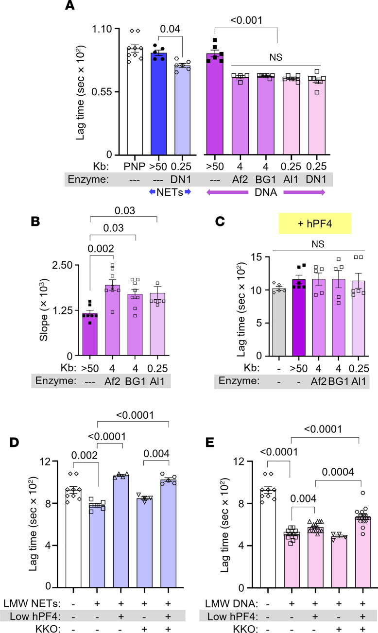
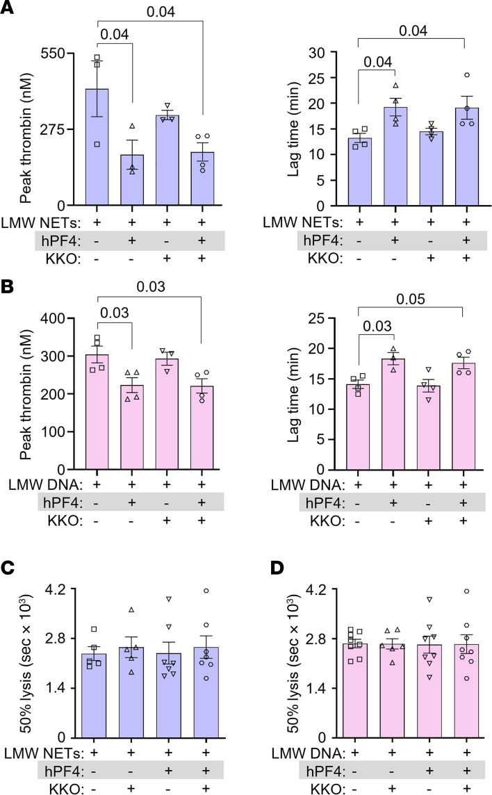
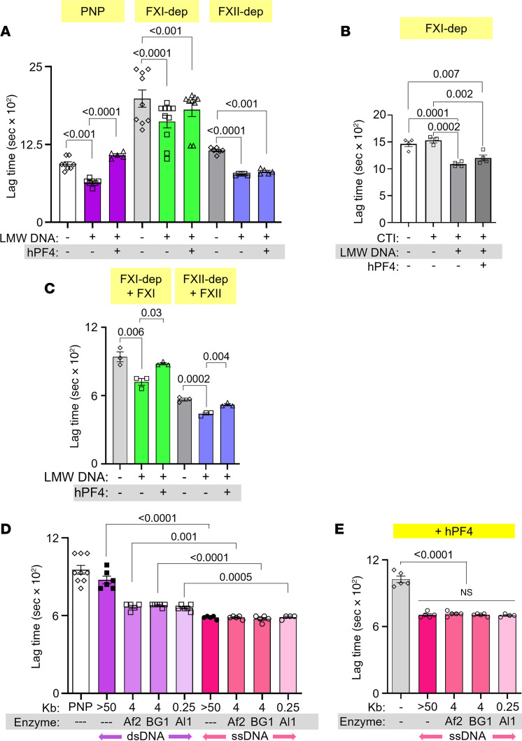
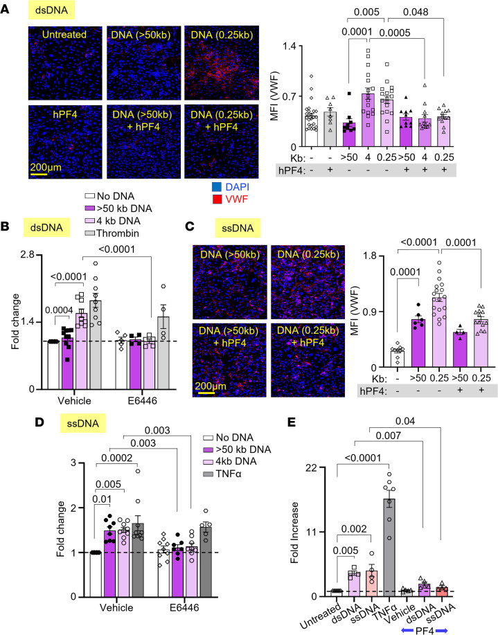
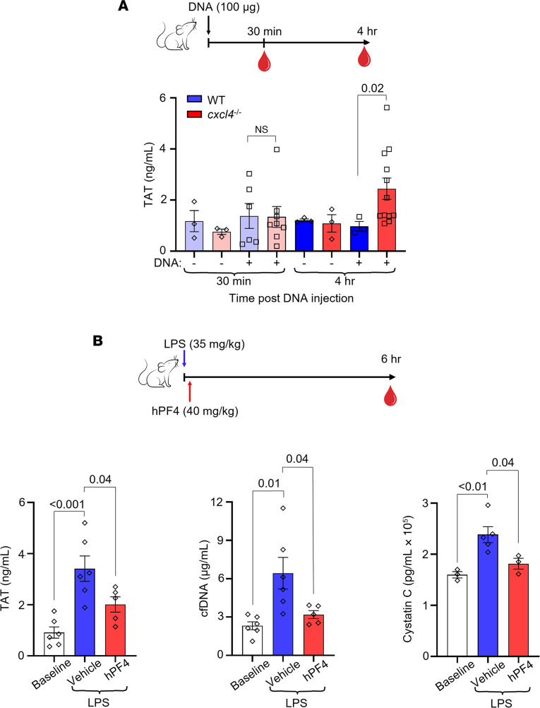
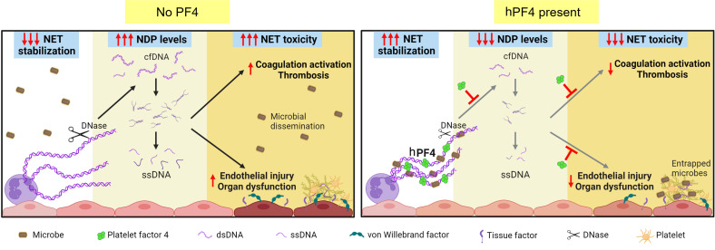
Plasma cell-free DNA (cfDNA), a marker of disease severity in sepsis, is a recognized driver of thromboinflammation and a potential therapeutic target. In sepsis, plasma cfDNA is mostly derived from neutrophil extracellular trap (NET) degradation. Proposed NET-directed therapeutic strategies include preventing NET formation or accelerating NET degradation. However, NET digestion liberates pathogens and releases cfDNA that promote thrombosis and endothelial cell injury. We propose an alternative strategy of cfDNA and NET stabilization with chemokine platelet factor 4 (PF4, CXCL4). We previously showed that human PF4 (hPF4) enhances NET-mediated microbial entrapment. We now show that hPF4 interferes with thrombogenicity of cfDNA and NETs by preventing their cleavage to short-fragment and single-stranded cfDNA that more effectively activates the contact pathway of coagulation. In vitro, hPF4 also inhibits cfDNA-induced endothelial tissue factor surface expression and von Willebrand factor release. In vivo, hPF4 expression reduced plasma thrombin-antithrombin (TAT) levels in animals infused with exogenous cfDNA. Following lipopolysaccharide challenge, Cxcl4-/- mice had significant elevation in plasma TAT, cfDNA, and cystatin C levels, effects prevented by hPF4 infusion. These results show that hPF4 interacts with cfDNA and NETs to limit thrombosis and endothelial injury, an observation of potential clinical benefit in the treatment of sepsis.

Keywords: Hematology; Inflammation; Neutrophils; Platelets; Thrombosis.


Conflict of interest statement

Conflict of interest: The authors have declared that no conflict of interest exists.

Figures

Figure 1
Figure 1. hPF4 and KKO inhibit fibrin generation initiated by LMW NETs and DNA.
Fibrin generation in pooled normal plasma (PNP) with or without added HMW or LMW NETs (blue) or DNA (purple). DN1, DNase I; Af2, AflII; BG1, BsrGI-HF; Al1, AluI. Data are mean ± SEM of at least 3 independent experiments. (A) Lag time determined from kinetic curves of fibrin generation with NETs (blue) and DNA (purple). (B) Slope (rate) of fibrin generation was determined from the same kinetic curves as in A. (C) Lag time of DNA-induced fibrin generation was determined from kinetic curves similarly to A, in the presence of hPF4 (20 μg/mL). (D and E) Lag times are shown for fibrin generation studies of DNase I–digested LMW NETs (D) and DNA (E) in the absence or presence of low hPF4 (1 μg/mL) and/or KKO (10 μg/mL). Data are mean ± SEM of at least 3 independent experiments. Comparative statistical analysis was performed by Kruskal-Wallis 1-way ANOVA.
Figure 2
Figure 2. hPF4 and KKO inhibit thrombin generation but not fibrinolysis.
(A and B) Thrombin generation studies of DNase I–digested LMW NETs (A) and DNA (B) in the absence or presence of hPF4 (20 μg/mL) and/or KKO (10 μg/mL). (C and D) Time to 50% clot lysis of LMW NET–induced (C) and LMW DNA–induced (D) coagulation, as determined based on kinetic curves. Data are mean ± SEM of at least 3 independent experiments. Comparative statistical analysis was performed by Kruskal-Wallis 1-way ANOVA.
Figure 3
Figure 3. Effect of hPF4 on fibrin generation in FXI- and FXII-depleted plasma and on thrombogenicity of ssDNA.
(A) Lag time, determined from kinetic curves, of fibrin generation in PNP or FXI- or FXII-depleted (dep) plasma, induced by DNase I–digested LMW DNA in the absence or presence of hPF4. (B) Fibrin generation lag time in FXI-depleted plasma spiked with CTI, induced by LMW DNA in the absence or presence of hPF4. (C) Lag time of fibrin generation induced by LMW DNA in depleted plasma supplemented with missing coagulation factors. (D) Lag time of ssDNA-induced (pink) fibrin generation compared to dsDNA (purple). (E) Lag time of ssDNA-induced fibrin generation with added hPF4 (20 μg/mL). Data are mean ± SEM of at least 3 independent experiments. Comparative statistical analysis was performed by Kruskal-Wallis 1-way ANOVA.
Figure 4
Figure 4. PF4 protects against LMW dsDNA– and ssDNA–induced procoagulant responses by endothelium.
Mean fluorescence intensity (MFI) of released VWF from HUVECs exposed to fragments of dsDNA (A and B) or ssDNA (C and D), with or without hPF4 or TLR9 inhibitor (E6446). Data were normalized to MFI of untreated cells without exposure to dsDNA or inhibitor (as indicated by dotted lines). Exposure to antithrombin (B) or TNF-α (D) serves as positive controls. (E) TF expression by HUVECs (shown as fold increase from untreated cells without PF4 exposure) induced by dsDNA or ssDNA in the absence or presence of hPF4. TNF-α exposure was used as a positive control. Scale bars: 200 μm. Data are mean ± SEM of at least 3 independent experiments. Comparative statistical analysis was performed by Kruskal-Wallis 1-way ANOVA.
Figure 5
Figure 5. hPF4 attenuates cfDNA-induced coagulability and organ damage in vivo.
(A) Top: Schematic of study in which WT mice or Cxcl4–/– littermates were given normal saline vehicle or normal saline containing digested DNA prior to blood collection at 30 minutes or 4 hours. Bar graph: TAT levels using a commercial ELISA kit in platelet-poor plasma. Data are mean ± SEM of at least 3 independent experiments. (B) Top: Schematic of study in which WT mice received normal saline vehicle or LPS, and a subset of animals was given hPF4 by tail vein injection immediately following LPS injection. TAT, cfDNA, and cystatin C levels were measured in blood drawn at baseline and 6 hours after LPS with and without hPF4 infusion. Bar graphs: TAT levels (left), cfDNA levels (middle), and cystatin C levels (right) 6 hours after LPS challenge. Data are mean ± SEM of at least 5 independent experiments. Comparative statistical analysis was performed by Kruskal-Wallis 1-way ANOVA.
Figure 6
Figure 6. Proposed protective mechanisms of NET stabilization.
Left: NETs are subjected to digestion by DNase I, reducing microbial capture and liberating toxic NDPs, including cfDNA. These cfDNA fragments expose ssDNA at termini that trigger coagulation activation and induce endothelial injury, leading to thrombosis and end-organ dysfunction in sepsis. Right: NET stabilization by hPF4 enhances DNase I resistance, which enhances NET microbial capture, reduces circulating cfDNA levels, and attenuates cfDNA-induced thrombogenicity and toxicity to endothelial cells.

Update of

References

    1. Rudd KE, et al. Global, regional, and national sepsis incidence and mortality, 1990-2017: analysis for the Global Burden of Disease Study. Lancet. 2020;395(10219):200–211. doi: 10.1016/S0140-6736(19)32989-7. - DOI - PMC - PubMed
    1. Mayr FB, et al. Epidemiology of severe sepsis. Virulence. 2014;5(1):4–11. doi: 10.4161/viru.27372. - DOI - PMC - PubMed
    1. Dwivedi DJ, et al. Prognostic utility and characterization of cell-free DNA in patients with severe sepsis. Crit Care. 2012;16(4):R151. doi: 10.1186/cc11466. - DOI - PMC - PubMed
    1. Avriel A, et al. Admission cell free DNA levels predict 28-day mortality in patients with severe sepsis in intensive care. PLoS One. 2014;9(6):e100514. doi: 10.1371/journal.pone.0100514. - DOI - PMC - PubMed
    1. Jing Q, et al. Cell-free DNA as biomarker for sepsis by integration of microbial and host information. Clin Chem. 2022;68(9):1184–1195. doi: 10.1093/clinchem/hvac097. - DOI - PubMed

Publication types