. 2024 Feb;115(2):465-476.
doi: 10.1111/cas.16029. Epub 2023 Nov 22.

NR0B1 augments sorafenib resistance in hepatocellular carcinoma through promoting autophagy and inhibiting apoptosis

Affiliations

NR0B1 augments sorafenib resistance in hepatocellular carcinoma through promoting autophagy and inhibiting apoptosis

Xiao Lan Tan et al. Cancer Sci. 2024 Feb.

Abstract

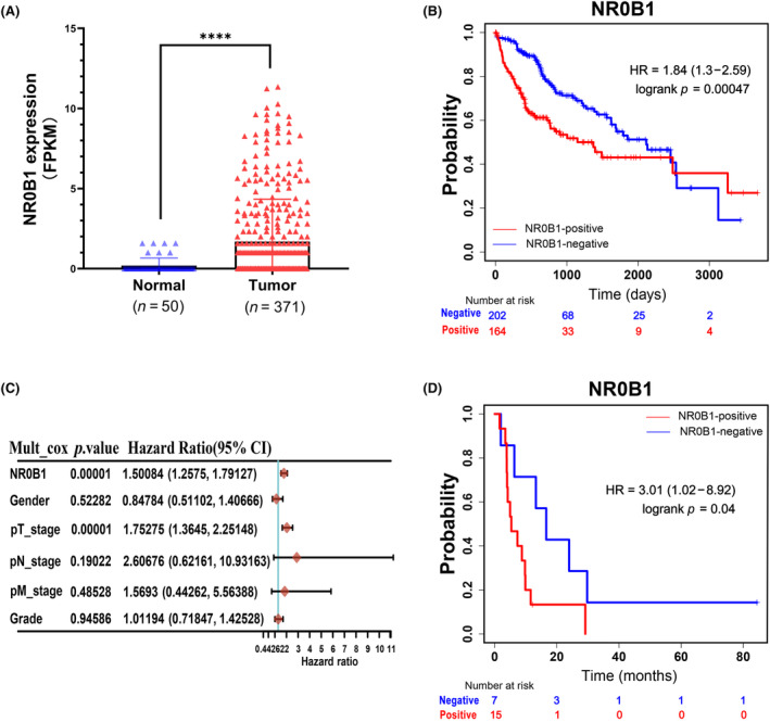
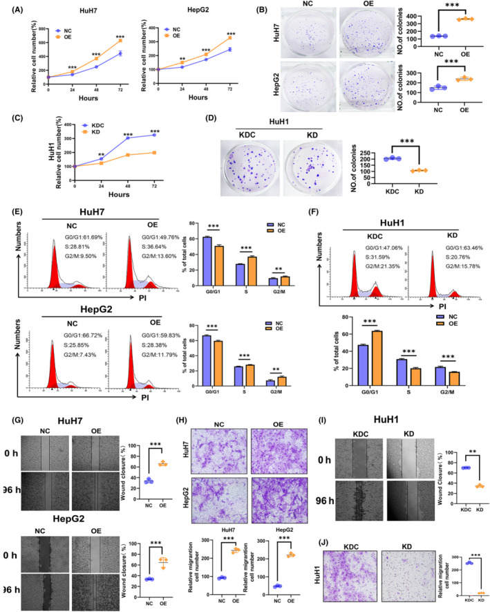
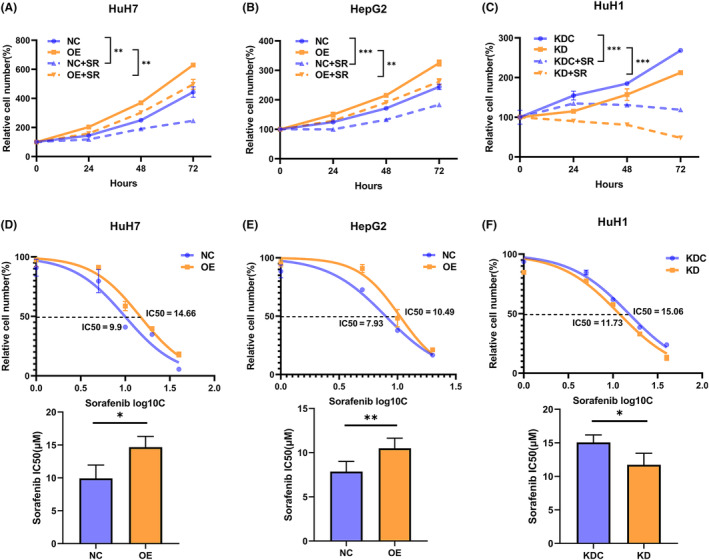
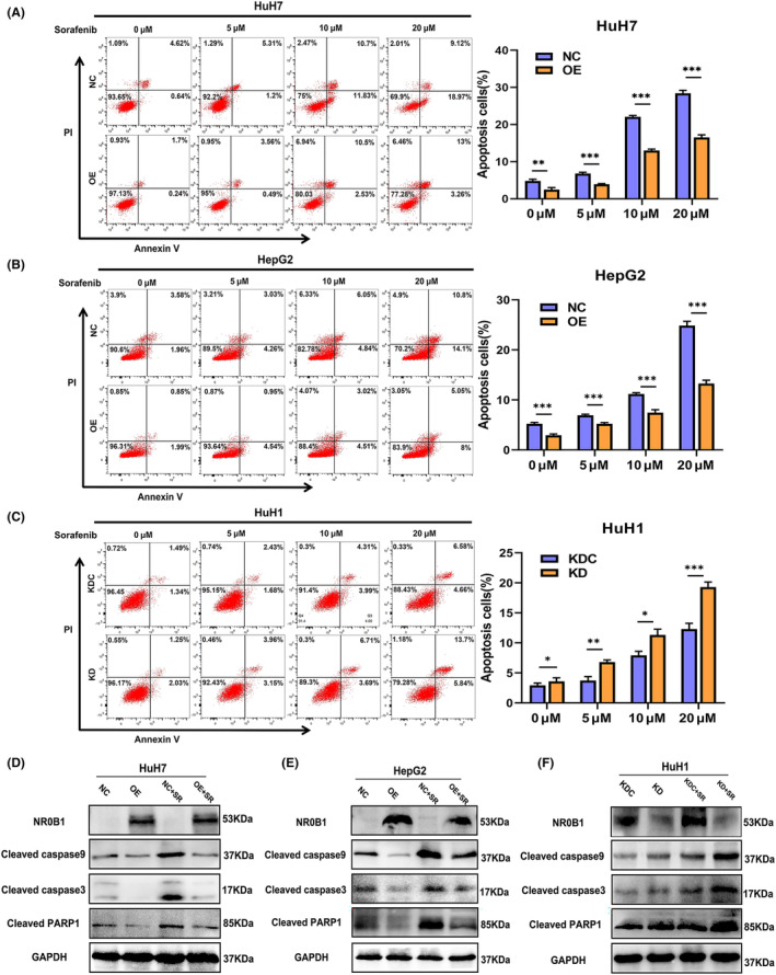
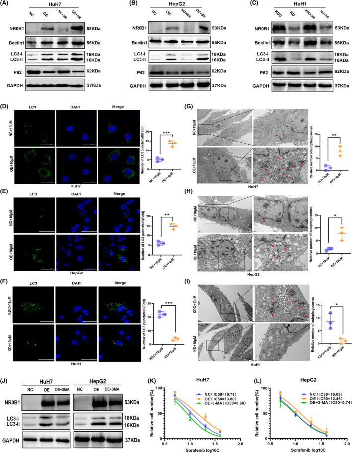
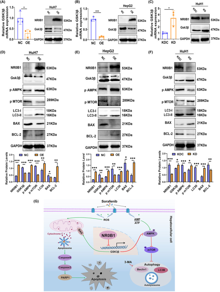
NR0B1 is frequently activated in hepatocellular carcinoma (HCC). However, the role of NR0B1 is controversial in HCC. In this study, we observed that NR0B1 was an independent poor prognostic factor, negatively correlated with the overall survival of HCC and the relapse-free survival of patients treated with sorafenib. Meanwhile, NR0B1 promoted the proliferation, migration, and invasion of HCC cells, inhibited sorafenib-induced apoptosis, and elevated the IC50 of sorafenib in HCC cells. NR0B1 was further displayed to increase sorafenib-induced autophagic vesicles and activate Beclin1/LC3-II-dependent autophagy pathway. Finally, NR0B1 was revealed to transcriptionally suppress GSK3β that restrains AMPK/mTOR-driven autophagy and increases BAX-mediated apoptosis. Collectively, our study uncovered that the ectopic expression of NR0B1 augmented sorafenib-resistance in HCC cells by activating autophagy and inhibiting apoptosis. Our findings supported that NR0B1 was a detrimental factor for HCC prognosis.

Keywords: NR0B1; apoptosis; autophagy; hepatocellular carcinoma (HCC); sorafenib resistance.


Conflict of interest statement

The authors declare no conflict of interest.

Figures

FIGURE 1
FIGURE 1
NR0B1 expression was negatively correlated with the prognosis of hepatocellular carcinoma (HCC). (A) Frequent ectopic expression of NR0B1 in HCC tumors (****p < 0.0001, p < 0.001, data extracted from TCGA database). (B) Kaplan–Meier survival curve showing the negative correlation between NR0B1 activation and overall survival (OS) of HCC (n = 366). (C) Cox multivariate analyses of NR0B1 and various clinicopathological indexes in HCC. (D) Kaplan–Meier survival curve showing the negative correlation between NR0B1 activation and relapse‐free survival (RFS) of HCC patients treated with sorafenib (n = 29).
FIGURE 2
FIGURE 2
Promotion of cell proliferation, cell cycle transition, cell invasiveness, and inhibition of cell apoptosis by NR0B1 in hepatocellular carcinoma (HCC) cells. (A) CCK8 assays showed that NR0B1 overexpression (OE) enhanced the proliferation of HuH7 and HepG2 cells. Relative proliferation was presented as fold change, which was calculated based on the absorbance and was normalized to a control value (n = 3). (B) Colony formation assays showed that NR0B1 OE increased the colony numbers in HuH7 and HepG2 cells. Colony counts were analyzed using ImageJ software (n = 3). (C) NR0B1 knockdown (KD) decreased the proliferation of HuH1 cells. (D) NR0B1 KD reduced the colony numbers in HuH1 cells. (E, F) Cell cycle assays showed that NR0B1 OE promoted G1/S phase transition in HuH7 and HepG2 cells (E), and NR0B1 KD increased the cell ratio of G1 phase in HuH1 cells (F). (G) Scratch‐wound‐healing assays showed that NR0B1 OE promoted the migration of HuH7 and HepG2 cells. (H) Transwell assays showed that NR0B1 OE accelerated the invasion of HuH7 and HepG2 cells. (I) Scratch‐wound‐healing assays showed that NR0B1 KD inhibited the migration of HuH1 cells. (J) Transwell assays showed that NR0B1 KD inhibited the invasion of HuH1 cells. Original magnification, ×400. All data are presented as the mean ± standard deviation (**p < 0.01, ***p < 0.001).
FIGURE 3
FIGURE 3
NR0B1 overexpression (OE) decreases the sensitivity of hepatocellular carcinoma (HCC) cells to sorafenib.(A, B) CCK8 assays showed that sorafenib inhibited the proliferation in HCC cells, and NR0B1 OE reversed the decline of proliferation in HuH7 (A) and HepG2 (B) cells treated with 10 μM sorafenib (SR, n = 5). (C) NR0B1 knockdown (KD) aggravated the decline of proliferation in HuH1 cells treated with 10 μM sorafenib (SR, n = 5). (D, E) NR0B1 OE increased the IC50 of sorafenib in HuH7 (D) and HepG2 (E) cells (n = 3). (F) NR0B1 KD decreased the IC50 of sorafenib in HuH1 cells (n = 3). All data are displayed as the mean ± standard deviation (*p < 0.05, **p < 0.01, ***p < 0.001).
FIGURE 4
FIGURE 4
NR0B1 inhibits sorafenib‐induced apoptosis in hepatocellular carcinoma (HCC) cells. (A, B) NR0B1 overexpression (OE) inhibiting sorafenib‐induced apoptosis in HuH7 (A) and HepG2 (B). (C) NR0B1 knockdown (KD) promoting sorafenib‐induced apoptosis in HuH1 cells. (D–F) Alteration of apoptosis‐related protein expression level in the NR0B1‐OE cells of HuH7 (D) and HepG2 (E) and in the NR0B1‐KD cells of HuH1 (F); sorafenib (SR) concentration = 10 μM. All data are displayed as the mean ± standard deviation (n = 3, *p < 0.05, **p < 0.01, ***p < 0.001).
FIGURE 5
FIGURE 5
NR0B1 promotes autophagy in hepatocellular carcinoma (HCC) cells. (A–C) Alteration of autophagy‐related protein expression level in the NR0B1 overexpression (OE) cells of HuH7 (A) and HepG2 (B) and in the NR0B1 knockdown (KD) cells of HuH1 (C); sorafenib (SR) concentration = 10 μM. (D–F) Immunofluorescence analysis showing the increment of LC3‐II proteins in the NR0B1 OE cells of HuH7 (D) and HepG2 (E) and the decline of LC3‐II puncta in the NR0B1 KD cells of HuH1 (F). Scale size = 50 μm. (G–I) Transmission electron microscopy (TEM) images showing the increment of autophagosomes in the NR0B1 OE cells of HuH7 (G) and HepG2 (H) and the decline of autophagosomes in the NR0B1 KD cells of HuH1 (I). Red arrowheads indicating autophagosomes. Scale size = 2 μm (original) and 500 nm (magnification). (J) Protein levels of LC3‐II in HuH7 and HepG2 cells treated with 3‐MA (5 mM). (K, L) IC50 assays of sorafenib in NR0B1‐OE HuH7 (K) and HepG2 (L) cells treated with 3‐MA (5 mM). All data are displayed as the mean ± standard deviation (n = 3, *p < 0.05, **p < 0.01, ***p < 0.001).
FIGURE 6
FIGURE 6
NR0B1 regulates autophagy and apoptosis through suppressing GSK3β expression in hepatocellular carcinoma (HCC) cells. (A, B) NR0B1 overexpression (OE) suppressed GSK3β expression in the levels of mRNA and protein in HuH7 (A) and HepG2 (B) cells. (C) NR0B1 knockdown (KD) increased GSK3β expression in the levels of mRNA and protein in HuH1 cells. (D, E) The level alteration of proteins involved in GSK3β‐targeted autophagy and apoptosis pathway in NR0B1 OE cells of HuH7 (D) and HepG2 (E) and NR0B1 KD cells of HuH1 (F). All data are displayed as the mean ± standard deviation (n = 3, *p < 0.05, **p < 0.01, ***p < 0.001). (G) Proposed working model for NR0B1‐mediated sorafenib resistance in HCC cells.

Similar articles

Cited by

References

    1. Villanueva A. Hepatocellular Carcinoma. N Engl J Med. 2019;380:1450‐1462. - PubMed
    1. Siegel RL, Miller KD, Jemal A. Cancer statistics, 2019. CA Cancer J Clin. 2019;69:7‐34. - PubMed
    1. Yang JD, Hainaut P, Gores GJ, Amadou A, Plymoth A, Roberts LR. A global view of hepatocellular carcinoma: trends, risk, prevention and management. Nat Rev Gastroenterol Hepatol. 2019;16:589‐604. - PMC - PubMed
    1. Kudo M. Treatment of advanced hepatocellular carcinoma with emphasis on hepatic arterial infusion chemotherapy and molecular targeted therapy. Liver Cancer. 2012;1:62‐70. - PMC - PubMed
    1. Maria R, Alejandro F, Jordi R, et al. BCLC strategy for prognosis prediction and treatment recommendation: the 2022 update. J Hepatol. 2022;76:681‐693. - PMC - PubMed