. 2024 Feb 1;84(3):434-448.
doi: 10.1158/0008-5472.CAN-23-2278.

SUMOylation-Driven mRNA Circularization Enhances Translation and Promotes Lymphatic Metastasis of Bladder Cancer

Affiliations

SUMOylation-Driven mRNA Circularization Enhances Translation and Promotes Lymphatic Metastasis of Bladder Cancer

Yue Zhao et al. Cancer Res. .

Abstract

Aberrant gene expression is a prominent feature of metastatic cancer. Translational initiation is a vital step in fine-tuning gene expression. Thus, exploring translation initiation regulators may identify therapeutic targets for preventing and treating metastasis. Herein, we identified that DHCR24 was overexpressed in lymph node (LN) metastatic bladder cancer and correlated with poor prognosis of patients. DHCR24 promoted lymphangiogenesis and LN metastasis of bladder cancer in vitro and in vivo. Mechanistically, DHCR24 mediated and recognized the SUMO2 modification at lysine 108 of hnRNPA2B1 to foster TBK1 mRNA circularization and eIF4F initiation complex assembly by enhancing hnRNPA2B1-eIF4G1 interaction. Moreover, DHCR24 directly anchored to TBK1 mRNA 3'-untranslated region to increase its stability, thus forming a feed forward loop to elevate TBK1 expression. TBK1 activated PI3K/Akt signaling to promote VEGFC secretion, resulting in lymphangiogenesis and LN metastasis. DHCR24 silencing significantly impeded bladder cancer lymphangiogenesis and lymphatic metastasis in a patient-derived xenograft model. Collectively, these findings elucidate DHCR24-mediated translation machinery that promotes lymphatic metastasis of bladder cancer and supports the potential application of DHCR24-targeted therapy for LN-metastatic bladder cancer.

Significance: DHCR24 is a SUMOylation regulator that controls translation initiation complex assembly and orchestrates TBK1 mRNA circularization to activate Akt/VEGFC signaling, which stimulates lymphangiogenesis and promotes lymph node metastasis in bladder cancer.


Figures

None
Graphical abstract
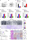
Figure 1. DHCR24 positively correlates with LN metastasis of bladder cancer. A, Heat map of protein-coding genes differentially expressed in bladder cancer tissues and NATs and in bladder cancer tissues with or without LN metastasis. B, Schematic illustration of the screening process of co-upregulated protein-coding genes in both bladder cancer tissues and LN-positive bladder cancer tissues and the TCGA database. C, qRT‒PCR analysis of DHCR24 expression in bladder cancer tissues and NATs (n = 296). D, Representative Western blot images of DHCR24 expression in bladder cancer tissues and NATs (n = 40). E, Quantification of DHCR24 expression by Western blot analysis in 40 paired bladder cancer and NATs. F and G, Kaplan‒Meier survival analysis of the DFS (F) and OS (G) of patients with bladder cancer with low versus high DHCR24 expression. The cutoff value is the median. H, Analysis of DHCR24 expression in bladder cancer tissues and NATs from the TCGA database. I, Kaplan‒Meier survival analysis of the OS of patients with bladder cancer with low versus high DHCR24 expression from the TCGA database. The cutoff value is the median. J, qRT‒PCR analysis of DHCR24 expression in bladder cancer tissues with or without LN metastasis (n = 296). K, Analysis of DHCR24 expression in bladder cancer tissues with or without LN metastasis from the TCGA database. L, Representative IHC images and quantification of DHCR24 expression in NATs and LN-negative and LN-positive bladder cancer tissues. Scale bars, 50 μm. H&E, hematoxylin and eosin. M and N, Representative IHC images and quantification of DHCR24 expression and LYVE1–indicated lymphatic vessels in peritumoral (M) and intratumoral (N) regions of bladder cancer tissues. Scale bars, 50 μm. Significant differences were assessed through the nonparametric Mann–Whitney U test in C, E, H, J, and K and the χ2 test in L and N. *, P < 0.05; **, P < 0.01. BCa, bladder cancer.
Figure 1.
DHCR24 positively correlates with LN metastasis of bladder cancer. A, Heat map of protein-coding genes differentially expressed in bladder cancer tissues and NATs and in bladder cancer tissues with or without LN metastasis. B, Schematic illustration of the screening process of co-upregulated protein-coding genes in both bladder cancer tissues and LN-positive bladder cancer tissues and the TCGA database. C, qRT‒PCR analysis of DHCR24 expression in bladder cancer tissues and NATs (n = 296). D, Representative Western blot images of DHCR24 expression in bladder cancer tissues and NATs (n = 40). E, Quantification of DHCR24 expression by Western blot analysis in 40 paired bladder cancer and NATs. F and G, Kaplan‒Meier survival analysis of the DFS (F) and OS (G) of patients with bladder cancer with low versus high DHCR24 expression. The cutoff value is the median. H, Analysis of DHCR24 expression in bladder cancer tissues and NATs from the TCGA database. I, Kaplan‒Meier survival analysis of the OS of patients with bladder cancer with low versus high DHCR24 expression from the TCGA database. The cutoff value is the median. J, qRT‒PCR analysis of DHCR24 expression in bladder cancer tissues with or without LN metastasis (n = 296). K, Analysis of DHCR24 expression in bladder cancer tissues with or without LN metastasis from the TCGA database. L, Representative IHC images and quantification of DHCR24 expression in NATs and LN-negative and LN-positive bladder cancer tissues. Scale bars, 50 μm. H&E, hematoxylin and eosin. M and N, Representative IHC images and quantification of DHCR24 expression and LYVE1–indicated lymphatic vessels in peritumoral (M) and intratumoral (N) regions of bladder cancer tissues. Scale bars, 50 μm. Significant differences were assessed through the nonparametric Mann–Whitney U test in C, E, H, J, and K and the χ2 test in L and N. *, P < 0.05; **, P < 0.01. BCa, bladder cancer.
Figure 2. DHCR24 promotes lymphangiogenesis and LN metastasis of bladder cancer in vitro and in vivo. A and B, Representative images and quantification of tube formation and migration of HLECs treated with culture media from DHCR24 knockdown or -overexpressing UM-UC-3 cells. Scale bars, 100 μm. C and D, Schematic diagram of the construction of a nude mouse popliteal LN metastasis model. E and F, Representative images and quantification of bioluminescence of the popliteal metastatic LNs (n = 12). Red arrows, footpad tumor and metastatic popliteal LN. G, Representative images and bioluminescence of the popliteal LNs from mice (n = 12). H, Representative IHC images of anti-GFP analysis in the popliteal LNs from mice (n = 12). Red scale bars, 500 μm; black scale bars, 50 μm. I, The table shows the popliteal LN-metastatic rate in different groups (n = 12). J and K, Representative IHC images and quantification of DHCR24 expression and LYVE1–indicated lymphatic vessel density in the intratumoral (J) and peritumoral (K) regions of primary footpad tumor tissues. Scale bar, 50 μm. Significant differences were assessed through one-way ANOVA, followed by the Dunnett test in A; the χ2 test in I; and the two-tailed Student t test in B, F, J, and K. Error bars, SD. *, P < 0.05; **, P < 0.01. H&E, hematoxylin and eosin.
Figure 2.
DHCR24 promotes lymphangiogenesis and LN metastasis of bladder cancer in vitro and in vivo. A and B, Representative images and quantification of tube formation and migration of HLECs treated with culture media from DHCR24 knockdown or -overexpressing UM-UC-3 cells. Scale bars, 100 μm. C and D, Schematic diagram of the construction of a nude mouse popliteal LN metastasis model. E and F, Representative images and quantification of bioluminescence of the popliteal metastatic LNs (n = 12). Red arrows, footpad tumor and metastatic popliteal LN. G, Representative images and bioluminescence of the popliteal LNs from mice (n = 12). H, Representative IHC images of anti-GFP analysis in the popliteal LNs from mice (n = 12). Red scale bars, 500 μm; black scale bars, 50 μm. I, The table shows the popliteal LN-metastatic rate in different groups (n = 12). J and K, Representative IHC images and quantification of DHCR24 expression and LYVE1–indicated lymphatic vessel density in the intratumoral (J) and peritumoral (K) regions of primary footpad tumor tissues. Scale bar, 50 μm. Significant differences were assessed through one-way ANOVA, followed by the Dunnett test in A; the χ2 test in I; and the two-tailed Student t test in B, F, J, and K. Error bars, SD. *, P < 0.05; **, P < 0.01. H&E, hematoxylin and eosin.
Figure 3. DHCR24 induces SUMO2 modification at K108 of hnRNPA2B1. A and B, Detection of the intracellular localization of DHCR24 in bladder cancer cells. Scale bars, 5 μm. C and D, Silver staining and mass spectrometry analysis for the detection of DHCR24-interacting proteins. E, Western blot analysis after co-IP assays with anti-DHCR24 or IgG in UM-UC-3 cells. F, Immunofluorescence assays for the colocalization of DHCR24 and hnRNPA2B1 in bladder cancer cells. Scale bars, 5 μm. G, PLA showing the interaction between hnRNPA2B1 and DHCR24 in UM-UC-3. Scale bars, 5 μm. H, Co-IP assays with DHCR24 to detect the PTM type of hnRNPA2B1 involved in the DHCR24 and hnRNPA2B1 interaction. I, Western blot analysis for the investigation of the SUMOylation type of hnRNPA2B1 in UM-UC-3 cells. J, Western blot analysis for the validation of SUMO2 modification on hnRNPA2B1. K, Western blot analysis to evaluate the effect of DHCR24 on SUMO2 modification on hnRNPA2B1 L. Western blot analysis to confirm the SUMO2 modification site on hnRNPA2B1. M, Western blotting analysis of SUMO2 modification on hnRNPA2B1 in indicated UM-UC-3 cells. N and O, qRT‒PCR of SUMOylation-related enzyme expression in the indicated UM-UC-3 cells. P, Detection of the interaction between DHCR24 and hnRNPA2B1 after hnRNPA2B1K108R mutations or using SENP3 in the indicated cells. Q, Western blot analysis to investigate the interaction between DHCR24 and hnRNPA2B1 after SIM mutation of DHCR24 in the indicated cells. Significant differences were assessed through the two-tailed Student t test in N and one-way ANOVA, followed by the Dunnett test in O. Error bars, SD. *, P < 0.05; **, P < 0.01.
Figure 3.
DHCR24 induces SUMO2 modification at K108 of hnRNPA2B1. A and B, Detection of the intracellular localization of DHCR24 in bladder cancer cells. Scale bars, 5 μm. C and D, Silver staining and mass spectrometry analysis for the detection of DHCR24-interacting proteins. E, Western blot analysis after co-IP assays with anti-DHCR24 or IgG in UM-UC-3 cells. F, Immunofluorescence assays for the colocalization of DHCR24 and hnRNPA2B1 in bladder cancer cells. Scale bars, 5 μm. G, PLA showing the interaction between hnRNPA2B1 and DHCR24 in UM-UC-3. Scale bars, 5 μm. H, Co-IP assays with DHCR24 to detect the PTM type of hnRNPA2B1 involved in the DHCR24 and hnRNPA2B1 interaction. I, Western blot analysis for the investigation of the SUMOylation type of hnRNPA2B1 in UM-UC-3 cells. J, Western blot analysis for the validation of SUMO2 modification on hnRNPA2B1. K, Western blot analysis to evaluate the effect of DHCR24 on SUMO2 modification on hnRNPA2B1 L. Western blot analysis to confirm the SUMO2 modification site on hnRNPA2B1. M, Western blotting analysis of SUMO2 modification on hnRNPA2B1 in indicated UM-UC-3 cells. N and O, qRT‒PCR of SUMOylation-related enzyme expression in the indicated UM-UC-3 cells. P, Detection of the interaction between DHCR24 and hnRNPA2B1 after hnRNPA2B1K108R mutations or using SENP3 in the indicated cells. Q, Western blot analysis to investigate the interaction between DHCR24 and hnRNPA2B1 after SIM mutation of DHCR24 in the indicated cells. Significant differences were assessed through the two-tailed Student t test in N and one-way ANOVA, followed by the Dunnett test in O. Error bars, SD. *, P < 0.05; **, P < 0.01.
Figure 4. DHCR24 promotes TBK1 mRNA stability and translation initiation by forming a SUMO2/hnRNPA2B1/EIF4G1 feed forward loop. A and B, qRT‒PCR analysis of the expression of the indicated genes enriched in the PI3K/Akt signaling pathway in DHCR24-knockdown or DHCR24-overexpressing UM-UC-3 cells. C, Luciferase assays showed the effect of DHCR24 overexpression on the 3′-UTR of TBK1 mRNA. D–G, Quantification and representative agarose electrophoresis images of actinomycin D assays for TBK1 mRNA in DHCR24 knockdown or DHCR24-overexpressing UM-UC-3 cells. H, RIP assay to investigate the interaction between DHCR24 and the 3′-UTR of TBK1 mRNA in UM-UC-3 cells. I, Schematic illustration of the predicted binding region for DHCR24 on the 3’-UTR of TBK1 mRNA from catRAPID analysis. J, RIP assays to investigate the interaction between DHCR24 and the 3′-UTR of TBK1 mRNA after S1 and S2 mutations in the TBK1 3′-UTR. K and L, Quantification and representative agarose electrophoresis images of actinomycin D assays for TBK1 mRNA in DHCR24-overexpressing UM-UC-3 cells with the S1 mutation in the TBK1 3′-UTR. M and N, Quantification and representative agarose electrophoresis images of actinomycin D assays for TBK1 mRNA in DHCR24-overexpressing UM-UC-3 cells with hnRNPA2B1K108R mutation or SENP3 treatment. O, Western blot analysis to investigate TBK1 expression after hnRNPA2B1K108R mutation or SENP3 treatment in DHCR24-overexpressing UM-UC-3 cells. P, Polysome profiles in DHCR24-overexpressing UM-UC-3 cells after hnRNPA2B1K108R mutation or SENP3 treatment. Q, Relative distribution of TBK1 mRNA across the polysome fractions in DHCR24-overexpressing UM-UC-3 cells after hnRNPA2B1K108R mutation or SENP3 treatment. R, Dual-luciferase reporter assays for overexpression of MS2 and MS2–hnRNPA2B1 fusion proteins in UM-UC-3 cells with or without downregulation of DHCR24 in regulating TBK1 mRNA translation. S, Western blot analysis after co-IP assays with anti-hnRNPA2B1 or IgG in UM-UC-3 cells. T, PLA showing the interaction between hnRNPA2B1 and eIF4G1. Scale bars, 5 μm. U, Western blot analysis to investigate the interaction between hnRNPA2B1 and eIF4G1 after hnRNPA2B1 or eIF4G1 sequence mutation in bladder cancer cells. V and W, m7G cap pulldown in hnRNPA2B1WT or hnRNPA2B1KO UM-UC-3 cells confirming DHCR24-mediated hnRNPA2B1 SUMOylation to enhance TBK1 mRNA circularization and eIF4F complex assembly. Significant differences were assessed through one-way ANOVA, followed by the Dunnett test in A, D, K, M, Q, and R; and two-tailed Student t test in B, C, F, H, and J. Error bars, SD. *, P < 0.05; **, P < 0.01.
Figure 4.
DHCR24 promotes TBK1 mRNA stability and translation initiation by forming a SUMO2/hnRNPA2B1/EIF4G1 feed forward loop. A and B, qRT‒PCR analysis of the expression of the indicated genes enriched in the PI3K/Akt signaling pathway in DHCR24-knockdown or DHCR24-overexpressing UM-UC-3 cells. C, Luciferase assays showed the effect of DHCR24 overexpression on the 3′-UTR of TBK1 mRNA. D–G, Quantification and representative agarose electrophoresis images of actinomycin D assays for TBK1 mRNA in DHCR24 knockdown or DHCR24-overexpressing UM-UC-3 cells. H, RIP assay to investigate the interaction between DHCR24 and the 3′-UTR of TBK1 mRNA in UM-UC-3 cells. I, Schematic illustration of the predicted binding region for DHCR24 on the 3’-UTR of TBK1 mRNA from catRAPID analysis. J, RIP assays to investigate the interaction between DHCR24 and the 3′-UTR of TBK1 mRNA after S1 and S2 mutations in the TBK1 3′-UTR. K and L, Quantification and representative agarose electrophoresis images of actinomycin D assays for TBK1 mRNA in DHCR24-overexpressing UM-UC-3 cells with the S1 mutation in the TBK1 3′-UTR. M and N, Quantification and representative agarose electrophoresis images of actinomycin D assays for TBK1 mRNA in DHCR24-overexpressing UM-UC-3 cells with hnRNPA2B1K108R mutation or SENP3 treatment. O, Western blot analysis to investigate TBK1 expression after hnRNPA2B1K108R mutation or SENP3 treatment in DHCR24-overexpressing UM-UC-3 cells. P, Polysome profiles in DHCR24-overexpressing UM-UC-3 cells after hnRNPA2B1K108R mutation or SENP3 treatment. Q, Relative distribution of TBK1 mRNA across the polysome fractions in DHCR24-overexpressing UM-UC-3 cells after hnRNPA2B1K108R mutation or SENP3 treatment. R, Dual-luciferase reporter assays for overexpression of MS2 and MS2–hnRNPA2B1 fusion proteins in UM-UC-3 cells with or without downregulation of DHCR24 in regulating TBK1 mRNA translation. S, Western blot analysis after co-IP assays with anti-hnRNPA2B1 or IgG in UM-UC-3 cells. T, PLA showing the interaction between hnRNPA2B1 and eIF4G1. Scale bars, 5 μm. U, Western blot analysis to investigate the interaction between hnRNPA2B1 and eIF4G1 after hnRNPA2B1 or eIF4G1 sequence mutation in bladder cancer cells. V and W, m7G cap pulldown in hnRNPA2B1WT or hnRNPA2B1KO UM-UC-3 cells confirming DHCR24-mediated hnRNPA2B1 SUMOylation to enhance TBK1 mRNA circularization and eIF4F complex assembly. Significant differences were assessed through one-way ANOVA, followed by the Dunnett test in A, D, K, M, Q, and R; and two-tailed Student t test in B, C, F, H, and J. Error bars, SD. *, P < 0.05; **, P < 0.01.
Figure 5. DHCR24 activates the TBK1/PI3K/Akt signaling pathway to promote VEGFC secretion by bladder cancer. A and B, Western blot analysis was performed to evaluate the expression of associated genes in the PI3K/Akt signaling pathway in DHCR24 knockdown or DHCR24-overexpressing UM-UC-3 cells. C and D. ELISA of VEGFC secretion in DHCR24 knockdown or DHCR24-overexpressing UM-UC-3 cells. E–H, qRT‒PCR analysis of VEGFC expression and ELISA analysis of VEGFC secretion in DHCR24-overexpressing UM-UC-3 or T24 cells with or without treatment with LY294002. I, Representative images and quantification of tube formation and migration of HLECs treated with CM from DHCR24-overexpressing UM-UC-3 cells with or without αVEGFC treatment. Scale bars, 100 μm. J, Quantification of bioluminescence of the popliteal metastatic LNs (n = 12) in mice of the indicated groups. K, The table shows the popliteal LN-metastatic rate in different groups (n = 12). L–N, Representative IHC images and quantification of DHCR24 and VEGFC expression and LYVE1-indicated lymphatic vessel density in footpad tumor tissues from the indicated mice. Scale bar, 50 μm. Significant differences were assessed through one-way ANOVA, followed by the Dunnett test in C, E–J, M, and N; two-tailed Student t test in D; and the χ2 test in K. Error bars , SD. *, P < 0.05; **, P < 0.01.
Figure 5.
DHCR24 activates the TBK1/PI3K/Akt signaling pathway to promote VEGFC secretion by bladder cancer. A and B, Western blot analysis was performed to evaluate the expression of associated genes in the PI3K/Akt signaling pathway in DHCR24 knockdown or DHCR24-overexpressing UM-UC-3 cells. C and D. ELISA of VEGFC secretion in DHCR24 knockdown or DHCR24-overexpressing UM-UC-3 cells. E–H, qRT‒PCR analysis of VEGFC expression and ELISA analysis of VEGFC secretion in DHCR24-overexpressing UM-UC-3 or T24 cells with or without treatment with LY294002. I, Representative images and quantification of tube formation and migration of HLECs treated with CM from DHCR24-overexpressing UM-UC-3 cells with or without αVEGFC treatment. Scale bars, 100 μm. J, Quantification of bioluminescence of the popliteal metastatic LNs (n = 12) in mice of the indicated groups. K, The table shows the popliteal LN-metastatic rate in different groups (n = 12). L–N, Representative IHC images and quantification of DHCR24 and VEGFC expression and LYVE1-indicated lymphatic vessel density in footpad tumor tissues from the indicated mice. Scale bar, 50 μm. Significant differences were assessed through one-way ANOVA, followed by the Dunnett test in C, E–J, M, and N; two-tailed Student t test in D; and the χ2 test in K. Error bars , SD. *, P < 0.05; **, P < 0.01.
Figure 6. Inhibition of DHCR24 suppresses tumor growth in PDXs from LN-metastatic bladder cancer. A, Schematic illustration of the construction of the PDX model. B–D, Images and quantification of the tumor volume in mice treated with sh-DHCR24 or sh-NC (n = 6 per patient). E, Representative images of immunofluorescence staining of DHCR24, TBK1, and VEGFC expression and LYVE1–indicated lymphatic vessel density in the tumor tissues from PDXs. F and G, qRT‒PCR analysis of TBK1 expression in bladder cancer tissues versus NATs and LN-positive versus LN-negative bladder cancer tissues (n = 296). H and I, Kaplan‒Meier survival analysis of OS (H) and DFS (I) in patients with bladder cancer with high versus low TBK1 expression. The cutoff value is the median. J–L, Representative IHC images and correlation analysis of DHCR24, TBK1, and VEGFC expression and LYVE1–indicated lymphatic vessel density in both intratumoral and peritumoral regions of bladder cancer tissues (n = 296). Scale bars, 50 μm. H&E, hematoxylin and eosin.The significant difference was assessed through two-tailed Student t test in C and D; the nonparametric Mann–Whitney U test in F and G; and the χ2 test in K and L. **, P < 0.01.
Figure 6.
Inhibition of DHCR24 suppresses tumor growth in PDXs from LN-metastatic bladder cancer. A, Schematic illustration of the construction of the PDX model. B–D, Images and quantification of the tumor volume in mice treated with sh-DHCR24 or sh-NC (n = 6 per patient). E, Representative images of immunofluorescence staining of DHCR24, TBK1, and VEGFC expression and LYVE1–indicated lymphatic vessel density in the tumor tissues from PDXs. F and G, qRT‒PCR analysis of TBK1 expression in bladder cancer tissues versus NATs and LN-positive versus LN-negative bladder cancer tissues (n = 296). H and I, Kaplan‒Meier survival analysis of OS (H) and DFS (I) in patients with bladder cancer with high versus low TBK1 expression. The cutoff value is the median. J–L, Representative IHC images and correlation analysis of DHCR24, TBK1, and VEGFC expression and LYVE1–indicated lymphatic vessel density in both intratumoral and peritumoral regions of bladder cancer tissues (n = 296). Scale bars, 50 μm. H&E, hematoxylin and eosin.The significant difference was assessed through two-tailed Student t test in C and D; the nonparametric Mann–Whitney U test in F and G; and the χ2 test in K and L. **, P < 0.01.

References

    1. Hautmann RE, de Petriconi RC, Pfeiffer C, Volkmer BG. Radical cystectomy for urothelial carcinoma of the bladder without neoadjuvant or adjuvant therapy: long-term results in 1,100 patients. Eur Urol 2012;61:1039–47. - PubMed
    1. Zahoor H, Mir MC, Barata PC, Stephenson AJ, Campbell SC, Fergany A, et al. . Phase II trial of continuous treatment with sunitinib in patients with high-risk (BCG-refractory) non-muscle invasive bladder cancer. Invest New Drugs 2019;37:1231–8. - PubMed
    1. Richters A, Aben KKH, Kiemeney L. The global burden of urinary bladder cancer: an update. World J Urol 2020;38:1895–904. - PMC - PubMed
    1. Sun L, Wang W, Han C, Huang W, Sun Y, Fang K, et al. . The oncomicropeptide APPLE promotes hematopoietic malignancy by enhancing translation initiation. Mol Cell 2021;81:4493–508e9. - PubMed
    1. Choe J, Lin S, Zhang W, Liu Q, Wang L, Ramirez-Moya J, et al. . mRNA circularization by METTL3–eIF3h enhances translation and promotes oncogenesis. Nature 2018;561:556–60. - PMC - PubMed

Publication types

MeSH terms

Substances

Grants and funding