. 2023 Dec 15;4(4):102735.
doi: 10.1016/j.xpro.2023.102735. Epub 2023 Nov 21.

Protocol for establishing primary human lung organoid-derived air-liquid interface cultures from cryopreserved human lung tissue

Affiliations

Protocol for establishing primary human lung organoid-derived air-liquid interface cultures from cryopreserved human lung tissue

Diana Cadena Castaneda et al. STAR Protoc. .

Abstract

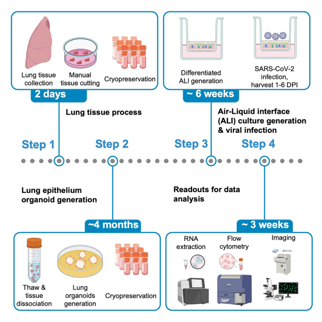
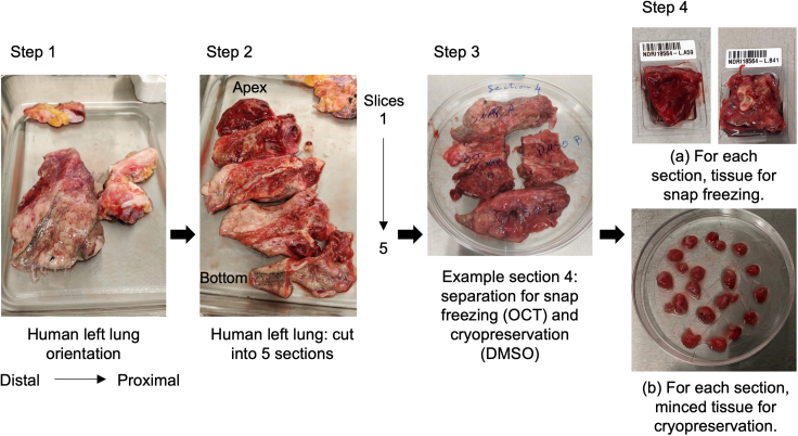
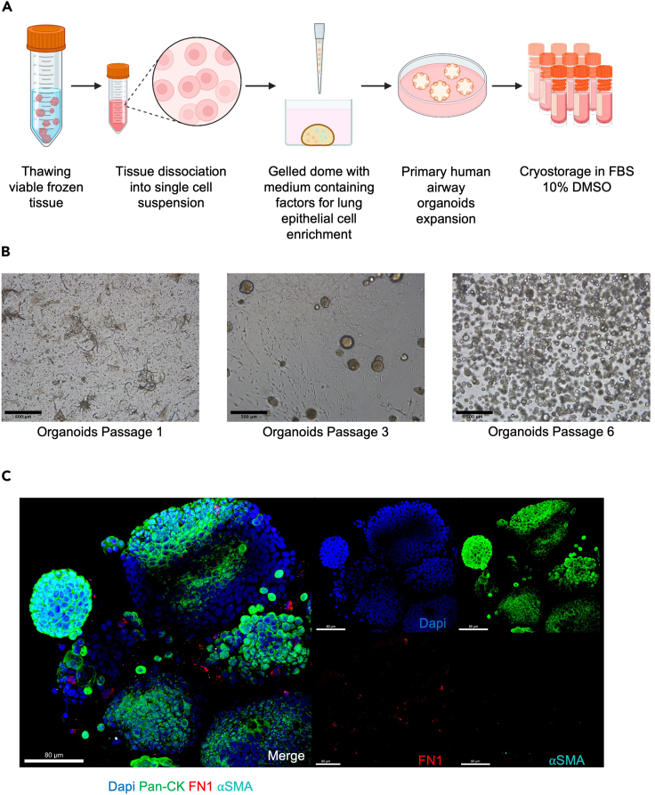
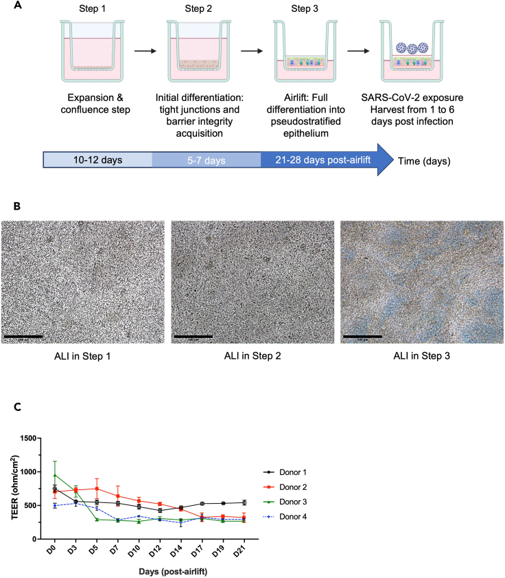
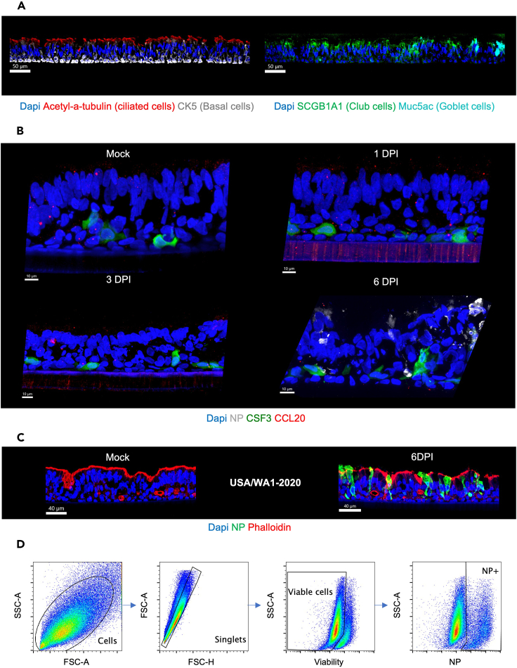
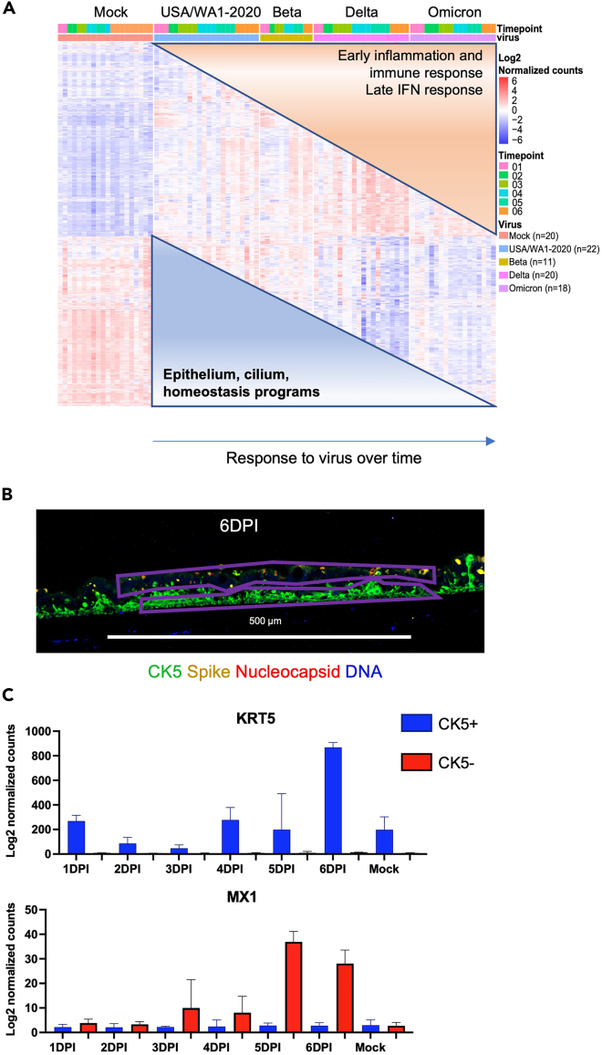
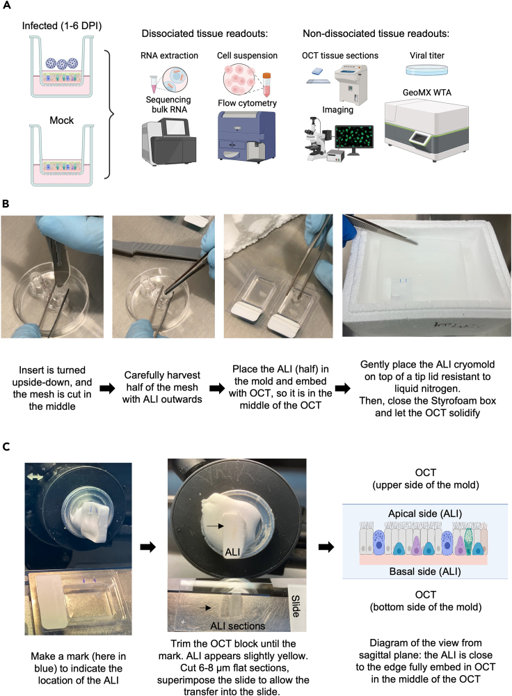
Primary human lung organoid-derived air-liquid interface (ALI) cultures serve as a physiologically relevant model to study human airway epithelium in vitro. Here, we present a protocol for establishing these cultures from cryopreserved human lung tissue. We describe steps for lung tissue cryostorage, tissue dissociation, lung epithelial organoid generation, and ALI culture differentiation. We also include quality control steps and technical readouts for monitoring virus response. This protocol demonstrates severe acute respiratory syndrome coronavirus 2 infection in these cultures as an example of their utility. For complete details on the use and execution of this protocol, please refer to Diana Cadena Castaneda et al. (2023).1.

Keywords: Immunology; Microscopy; Organoids.


Conflict of interest statement

Declaration of interests The A.G.-S. laboratory has received research support from GSK, Pfizer, Senhwa Biosciences, Kenall Manufacturing, Blade Therapeutics, Avimex, Johnson & Johnson, Dynavax, 7 Hills Pharma, PharmaMar, ImmunityBio, Accurius, nanoComposix, Hexamer Therapeutics, N-fold LLC, Model Medicines, Atea Pharmaceuticals, Applied Biological Laboratories, and Merck, outside of the reported work. A.G.-S. has consulting agreements for the following companies involving cash and/or stock: CastleVax, Amovir, Vivaldi Biosciences, ContraFect, 7 Hills Pharma, Avimex, Pagoda, Accurius, Esperovax, Farmak, Applied Biological Laboratories, PharmaMar, CureLab Oncology, CureLab Veterinary, Synairgen, Paratus, and Pfizer, outside of the reported work. A.G.-S. has been an invited speaker in meeting events organized by Seqirus, Janssen, Abbott, and AstraZeneca. A.G.-S. is an inventor on patents and patent applications on the use of antivirals and vaccines for the treatment and prevention of virus infections and cancer, owned by the Icahn School of Medicine at Mount Sinai, New York, outside of the reported work. The M.S. laboratory has received unrelated funding support in sponsored research agreements from Phio Pharmaceuticals, 7 Hills Pharma, argenx, and Moderna. K.P. is a stockholder in Cue Biopharma and Guardian Bio, scientific advisor to Cue Biopharma and Guardian Bio, and co-founder of Guardian Bio. K.P. declares unrelated funding support from Guardian Bio (current) and Merck (past).

Figures

None
Graphical abstract
Figure 1
Figure 1
Setup for human lung tissue processing and cryopreservation: Representative pictures for lung tissue processing (Step 1) The left lung, including a partial portion of the trachea, is used for the processing. The tissue is oriented with the distal (alveolar) portion on the left and the proximal (bronchial) portion on the right. (Step 2) Cut the tissue into five sections from the apex (upper part) to the lower part. (Step 3–4) Cut each section into smaller pieces. Track the pieces from different areas of the lung as follows: for example, those from the alveolar region are named LA, and those from the bronchial region are named LB. From each region, either snap-freeze small pieces in OCT for histology or subject them to cryopreservation (10% FBS and DMSO).
Figure 2
Figure 2
Viable frozen tissue processing and generation of primary airway lung organoids (A) Schematic experimental design for primary human lung organoid generation: viable frozen tissue is thawed and digested to obtain a single cell lung suspension containing airway cell progenitors. The cell lung suspension is resuspended in Cultrex, dispensed as a dome, and cultured in a medium containing factors allowing airway organoid expansion and lung epithelium enrichment. After each passage dissociated organoids are cryopreserved. (B) Representative photomicrographs of primary lung organoids at different passages were captured using a bright-field microscope. Images were generated using ImageJ. Scale bars 500 μm, in black on the left corner. (C) Representative immunofluorescent (IF) images of whole mounted lung organoids showing markers for epithelial cells (PAN-CK, green), extracellular matrix (Fibronectin, red), and nuclei (DAPI, blue). Pan-CK is present exclusively in epithelial cells and fibronectin allows visualization of the presence of the remaining extracellular matrix from tissue dissociation. The fibronectin disappears after four passages and characterizes the enrichment of lung epithelial cells. Scale bar 80 μm, in white in the left corner. Note: Figure 2C reprinted with permission from Diana Cadena Castaneda et al. 2023 (Cell Press, Open Access).
Figure 3
Figure 3
Generation of primary human lung organoid-derived ALI cultures to study response to virus (A) Schematic experimental design for primary human lung organoid-derived ALI culture generation: (1) cell expansion in submerged culture to obtain confluence at 100%; (2) initial differentiation in submerged cultures to foster tight junctions and barrier integrity, monitored by TEER values (>500 Ω cm2). (3) TEER goals are achieved, and cultures are transitioned to airlift by removal of apical media, which initiates final differentiation into pseudo-stratified epithelia. Cultures are monitored for a minimum of 4 weeks for the presence of beating cilia and mucus production. (B) Representative photomicrographs of primary lung organoids-derived ALI cultures at Step 1 (3–4 days after seeding), Step 2 (at confluence approximately 12–14 days after seeding) and Step 3 (at 34 days post-airlift) captured using a bright-field microscope. Images were generated using ImageJ. Scale bars 500 μm, in black on the left corner. (C) Measurement of trans-epithelial electrical resistance (TEER, Ω cm2) with error bars (mean ± SD), 3 measurements per time-point performed 3 times per week starting when the epithelium is confluent, one representative experiment per donor (four donors). Fully pseudo-stratified differentiated ALI cultures are obtained from primary lung organoid progenitors within 3–4 weeks (post-airlift). Note: Figure 3C reprinted with permission from Diana Cadena Castaneda et al. 2023 (Cell Press, Open Access).
Figure 5
Figure 5
Examples of expected outcomes of immunofluorescent images and flow cytometry gating strategy (A) Representative immunofluorescent (IF) section (8 μm) of differentiated lung organoid-derived ALI cultures. The left panel, merged figures, shows markers for ciliated cells (acetylated a-tubulin, red) and goblet cells (MUC5AC, cyan). Right panel, merged figures, showing club cells (SCGB1A1, green), and basal cells (CK5, white). Nuclei (DAPI, blue). Scale bar 50 μm, in white on the left corner. (B) Representative images of ALI cultures mock-infected at 6 days and ALI infected with SARS-CoV-2 USA/WA1-2020 (105 PFU) at 1, 3, and 6 days post-infection (DPI) stained for nuclei (DAPI, blue), viral NP (white) to reveal the effective viral replication, CSF3 (green) and CCL20 (red). Scale bars 10 μm, in white on the left corner. (C) Representative images of donor 3 of mock-infected (control media without virus at 6 days) and infected ALI cultures with SARS-CoV-2 (105 PFU) at 6 days post-infection (DPI) stained for nuclei (DAPI), viral nucleoprotein (NP, green) to reveal the effective viral replication and phalloidin (Actin filament, red) to reveal tissue structure. Scale bar 40 μm, in white on the left corner. (D) Gating strategy example for flow cytometry analysis. A single cell suspension was prepared from SARS-CoV-2 infected ALI cultures. Cell suspensions were stained for viability and viral infection using an anti-NP antibody specific to SARS-CoV-2. Plots from left to right show serial gating to identify percentages of infected (NP-positive) viable cells. Note: Figure 5 reprinted with permission from Diana Cadena Castaneda et al. 2023 (Cell Press, Open Access).
Figure 6
Figure 6
Examples of expected outcomes of transcriptional response to SARS-CoV-2 variants and ROI selection for GeoMx data (A) Heatmap representing differentially expressed genes over time in response to SARS-CoV-2 variants. ALI cultures from four donors were infected with SARS-CoV-2 and harvested for sequencing at 1, 2, 3, 4, 5, and 6 dpi, and mock-infected (control media without virus) samples were collected from days 1 through 6 days as well. The sequencing was performed in multiple batches with at least 2 independent experiments at each time-point, the cut-off used for defining differentially expressed genes: |logFC| > 1; adjustedpvalue < 0.01; normalized counts > 10. Rows represent individual transcripts and columns represent individual biological replicates ordered by time-points and SARS-CoV-2 variants. Batch effect was removed using SVAseq R package. All variants induced expression of genes associated with the viral response at later time-points. This response to the virus from 1 to 6 DPI is depicted by the schematic covering the heatmap, with two “clusters”: one on the lower part “down-regulation” from 1 to 6 DPI, enriched for cilia and epithelium maintenance signatures whereas the upper part showed “up-regulation” of signatures enriched for inflammatory, immune and IFN response. (B) ROI selection for GeoMx data: Representative image of infected ALI culture section with SARS-CoV-2 (105 PFU), at 6DPI stained for nuclei (DAPI), viral nucleoprotein (red), spike viral protein (yellow) and cytokeratin 5 (CK5). The thick purple polygons represent selected ROIs for the apical cytokeratin 5- cells (CK5-) vs. basal side CK5+. Scale bar 500 μm (white). (C) GeoMx data (one representative experiment): Bar graphs with error bars (mean ± SD) were generated using Graphpad (Prism 5) and illustrate the gene expression (log 2 normalized counts) over time within selected ROIs (at least 3 ROIs per condition) based on cytokeratin 5 protein expression (CK5 +) through KRT5 gene expression and based on cytokeratin 5 negative expression (CK5 -) of infected cells positive for SARS-CoV-2 Spike and NP, through MX1 as part of the anti-viral response which is mainly increased at later time-points. Note: Figure 6 reprinted with permission from Diana Cadena Castaneda et al. 2023 (Cell Press, Open Access).
Figure 4
Figure 4
Readouts to assess response to a virus, ALI culture OCT embedding, and OCT cutting (A) Schematic workflow presenting five methods to assess response to virus. Flow cytometry and RNA extraction require tissue dissociation versus Immunofluorescence, plaque assay (viral titer on apical supernatant) and GeoMx WTA do not require tissue dissociation. (B) Procedure of embedding ALIs in OCT and cryopreservation. First, turn the insert upside-down, and with a scalpel cut the mesh in the middle and partially around the edges, enough to hold the mesh and pull out from the insert. Be careful not to damage the cell layer. Place the ALI mesh on top of a layer of OCT and cover it with OCT to fully embed the ALI insert. Gently, with a pipette tip make sure the ALI is not curved or too close to the bottom or surface of the cryomold. Then, snap freeze in liquid nitrogen. To ensure no liquid nitrogen (LN) enters the tissue, the cryomold should be placed on top of a plastic lid resistant to LN. (C) ALI section cutting and transfer to Superfrost plus slides. Make a mark with a Sharpie pen to indicate the location of the ALI culture on the cryomold then on the solidify OCT block. This tip will facilitate the OCT trimming until the mark and visualize the ALI (slightly in yellow). The diagram allows to picture the configuration of the ALI culture in the OCT block. Finally, ALI culture sections could be transferred into Superfrost plus slides. These steps will ensure good-quality sections for further experiments.

References

    1. Castaneda D.C., Jangra S., Yurieva M., Martinek J., Callender M., Coxe M., Choi A., García-Bernalt Diego J., Lin J., Wu T.-C., et al. Spatiotemporally organized immunomodulatory response to SARS-CoV-2 virus in primary human broncho-alveolar epithelia. iScience. 2023;26 - PMC - PubMed
    1. Fulcher M.L., Gabriel S., Burns K.A., Yankaskas J.R., Randell S.H. Well-differentiated human airway epithelial cell cultures. Methods Mol. Med. 2005;107:183–206. - PubMed
    1. Rayner R.E., Makena P., Prasad G.L., Cormet-Boyaka E. Optimization of Normal Human Bronchial Epithelial (NHBE) Cell 3D Cultures for in vitro Lung Model Studies. Sci. Rep. 2019;9:500. - PMC - PubMed
    1. Ghosh B., Park B., Bhowmik D., Nishida K., Lauver M., Putcha N., Gao P., Ramanathan M., Hansel N., Biswal S., Sidhaye V.K. Strong correlation between air-liquid interface cultures and in vivo transcriptomics of nasal brush biopsy. Am. J. Physiol. Lung Cell Mol. Physiol. 2020;318:L1056–L1062. - PMC - PubMed
    1. Nishida K., Brune K.A., Putcha N., Mandke P., O’Neal W.K., Shade D., Srivastava V., Wang M., Lam H., An S.S., et al. Cigarette smoke disrupts monolayer integrity by altering epithelial cell-cell adhesion and cortical tension. Am. J. Physiol. Lung Cell Mol. Physiol. 2017;313:L581–L591. - PMC - PubMed

Publication types

LinkOut - more resources