. 2023 Nov 24;9(47):eadg2263.
doi: 10.1126/sciadv.adg2263. Epub 2023 Nov 22.

ATM deficiency confers specific therapeutic vulnerabilities in bladder cancer

Affiliations

ATM deficiency confers specific therapeutic vulnerabilities in bladder cancer

Yuzhen Zhou et al. Sci Adv. .

Abstract

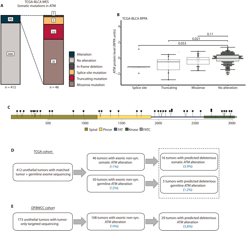
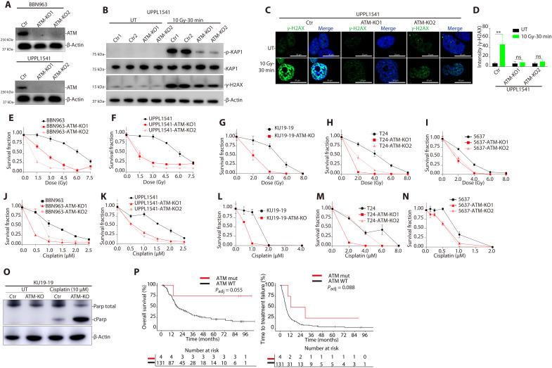
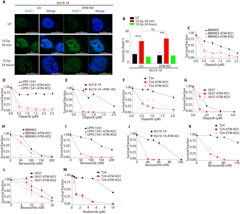
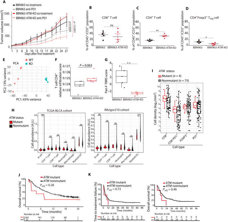
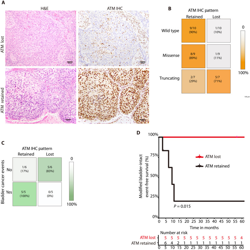
Ataxia-telangiectasia mutated (ATM) plays a central role in the cellular response to DNA damage and ATM alterations are common in several tumor types including bladder cancer. However, the specific impact of ATM alterations on therapy response in bladder cancer is uncertain. Here, we combine preclinical modeling and clinical analyses to comprehensively define the impact of ATM alterations on bladder cancer. We show that ATM loss is sufficient to increase sensitivity to DNA-damaging agents including cisplatin and radiation. Furthermore, ATM loss drives sensitivity to DNA repair-targeted agents including poly(ADP-ribose) polymerase (PARP) and Ataxia telangiectasia and Rad3 related (ATR) inhibitors. ATM loss alters the immune microenvironment and improves anti-PD1 response in preclinical bladder models but is not associated with improved anti-PD1/PD-L1 response in clinical cohorts. Last, we show that ATM expression by immunohistochemistry is strongly correlated with response to chemoradiotherapy. Together, these data define a potential role for ATM as a predictive biomarker in bladder cancer.


Figures

Fig. 1.
Fig. 1.. ATM alterations in bladder cancer.
(A) Frequency and classes of somatic ATM alterations in the TCGA-BLCA WES cohort. (B) Tumors with ATM splice sites or truncating alterations had lower protein levels (as quantified by RPPA) in the TCGA-BLCA cohort (Wilcoxon rank sum test). (C) Distribution of all observed non-synonymous somatic missense mutations in TCGA-BLCA cohort. FAT, FRAP-ATM-TRRAP domain; FATC, FRAP-ATM-TRRAP C-terminal domain. (D) ATM alterations in TCGA-BLCA WES cohort. (E) ATM alterations in the DFBWCC urothelial tumor cohort. RPPA, reverse phase protein array; DFBWCC, Dana-Farber/Brigham and Women’s Cancer Center.
Fig. 2.
Fig. 2.. ATM deficiency results in altered DNA damage signaling and increased sensitivity to DNA-damaging agents in bladder cancer preclinical models.
(A) Immunoblot demonstrating loss of ATM protein in ATM-deleted murine bladder cancer cell lines BBN963 and UPPL1541 (see also fig. S6). (B) Immunoblot demonstrating a decrease in radiation-induced KAP1 and γ-H2AX phosphorylation in ATM-deleted compared to WT ATM bladder cancer cells. (C) Immunofluorescence images demonstrating a decrease in radiation-induced γ-H2AX foci formation in ATM-deleted compared to WT ATM cells. (D) Quantification of γ-H2AX foci. There is a significant increase in γ-H2AX foci following radiation in WT ATM cells but not in ATM-deleted cells (**P < 0.01, Student’s t test). (E to I) Cell survival curves for WT ATM and ATM-deleted human and mouse bladder cancer cell lines following ionizing radiation. There was a significant increase in radiation sensitivity in ATM-deleted cells compared to WT ATM lines across all models (see also fig. S7). (J to N) Cell survival curves for WT ATM and ATM-deleted human and mouse bladder cancer cell lines following cisplatin treatment. There was a significant increase in cisplatin sensitivity in ATM-deleted cells compared to WT ATM lines across all models. (O) Cisplatin treatment resulted in higher levels of cleaved PARP in ATM-deleted compared to WT ATM bladder cancer cells, consistent with increased cisplatin-induced apoptosis. (P) Time to treatment failure (TTF; left) and OS (right) of patients with ATM-mutant and nonmutant DFBWCC metastatic urothelial cancer treated with cisplatin-based chemotherapy. All cell line data are plotted as the means ± SD (n = 5) unless otherwise specified. Ctr, control; KO, knockout; UT, untreated; Gy, gray; ns, not significant.
Fig. 3.
Fig. 3.. ATM loss drives sensitivity to inhibitors of PARP, ATR, and DNA-PK in bladder cancer preclinical models.
(A) Immunofluorescence images showing radiation-induced Rad51 foci formation in ATM-deleted and WT ATM KU19-19 bladder cancer cells. (B) Quantification of radiation-induced Rad51 foci shows no difference in ATM-deleted compared to WT ATM KU19-19 cells. Data are displayed as means ± SD, n = 5, ***P < 0.001 and ****P < 0.0001. (C to G) Cell survival curves for WT ATM and ATM-deleted human and mouse bladder cancer cell lines following olaparib treatment. There was a significant increase in olaparib sensitivity in ATM-deleted cells compared to WT ATM lines across all models (see also fig. S8). (H to L) Cell survival curves for WT ATM and ATM-deleted human and mouse bladder cancer cell lines following berzosertib treatment. There was a significant increase in berzosertib sensitivity in ATM-deleted cells compared to WT ATM lines across nearly all models. (M) Cell survival curves for WT ATM and ATM-deleted bladder cancer cells following nedisertib treatment.
Fig. 4.
Fig. 4.. ATM loss is sufficient to drive immune changes and improve anti-PD1 response in preclinical bladder models but ATM mutations are not associated with immune properties or anti-PD1 response in clinical bladder cohorts.
(A) Growth curves for ATM-deleted and WT ATM tumor xenografts. The ATM-deleted tumors had a larger response to anti-PD1 treatment than WT ATM xenografts (linear regression model). (B) Flow cytometry showed no differences in the number of CD8+ T cells in ATM-deleted versus WT ATM tumors. Data are plotted as the means ± SD, n = 5 tumors. (C) Flow cytometry showed a significantly higher number of CD4+ T cells in ATM-deleted compared to WT ATM tumors, **P < 0.01. (D) Flow cytometry showed significantly fewer FOXP3+ CD4+ T cells in ATM-deleted compared to WT ATM tumors, *P < 0.05. (E) Principal components analysis (PCA) of RNA sequencing (RNA-seq) counts from ATM-deleted and WT ATM tumors. (F) RNA-seq–based immune cell fraction estimation using TIMER revealed a trend toward increased CD4+ T cells in ATM-deleted compared to WT ATM tumors (Student’s t test; BH correction). (G) A gene expression signature of transforming growth factor–β signaling in fibroblasts was higher in WT ATM tumors than in ATM-deleted tumors. (H) RNA-seq analysis of immune cell subsets in ATM-mutant versus ATM nonmutant tumors from the TCGA (left) and IMvigor210 (right) bladder cancer cohorts. There were no significant differences in any immune cell subsets (Wilcoxon rank sum test). (I) Quantification of multiplexed immunofluorescence data from institutional bladder cancer cases showed no difference in CD8, PD1, or FOXP3 staining between ATM-mutant and nonmutant bladder tumors. (J) OS of patients with ATM-mutant and nonmutant bladder tumors in the Imvigor210 cohort (Kaplan-Meier method). (K) TTF (left) and OS (right) for ATM-mutant and ATM nonmutant institutional bladder cancer cases following anti-PD1/PD-L1 treatment (Kaplan-Meier method).
Fig. 5.
Fig. 5.. Correlations among ATM mutational status, ATM IHC expression, and chemoradiotherapy response.
(A) Representative photomicrographs of urothelial carcinoma and corresponding ATM IHC demonstrating loss of ATM expression (top row) or retained expression (bottom row). In tumors with ATM loss, non-neoplastic cells (e.g. endothelial cells and fibroblasts) retain ATM expression and serve as an internal positive control. (B) Correlation between ATM mutational status and ATM IHC pattern in a cohort of bladder tumors from the DFBWCC. Most tumors with WT ATM or an ATM missense mutation have retained ATM staining by IHC, whereas most tumors with an ATM nonsense or frameshift mutation have ATM loss by IHC. (C) Correlation between ATM IHC pattern and bladder cancer events in the MGH cohort. (D) Modified bladder-intact event-free survival (mBI-EFS) in MGH cases with ATM protein expression retained versus lost (Kaplan-Meier method). There was significantly longer mBI-EFS in patients with loss of ATM expression. MGH, Massachusetts General Hospital.

References

    1. Y. Shiloh, Y. Ziv, The ATM protein kinase: Regulating the cellular response to genotoxic stress, and more. Nat. Rev. Mol. Cell Biol. 14, 197–210 (2013). - PubMed
    1. A. Ciccia, S. J. Elledge, The DNA damage response: Making it safe to play with knives. Mol. Cell 40, 179–204 (2010). - PMC - PubMed
    1. C. Rothblum-Oviatt, J. Wright, M. A. Lefton-Greif, S. A. McGrath-Morrow, T. O. Crawford, H. M. Lederman, Ataxia telangiectasia: A review. Orphanet J. Rare Dis. 11, 159 (2016). - PMC - PubMed
    1. A. G. Robertson, J. Kim, H. Al-Ahmadie, J. Bellmunt, G. Guo, A. D. Cherniack, T. Hinoue, P. W. Laird, K. A. Hoadley, R. Akbani, M. A. A. Castro, E. A. Gibb, R. S. Kanchi, D. A. Gordenin, S. A. Shukla, F. Sanchez-Vega, D. E. Hansel, B. A. Czerniak, V. E. Reuter, X. Su, B. de Sa Carvalho, V. S. Chagas, K. L. Mungall, S. Sadeghi, C. S. Pedamallu, Y. Lu, L. J. Klimczak, J. Zhang, C. Choo, A. I. Ojesina, S. Bullman, K. M. Leraas, T. M. Lichtenberg, C. J. Wu, N. Schultz, G. Getz, M. Meyerson, G. B. Mills, D. J. M. Conkey; TCGA Research Network, J. N. Weinstein, D. J. Kwiatkowski, S. P. Lerner, Comprehensive molecular characterization of muscle-invasive bladder cancer. Cell 171, 540–556.e25 (2017). - PMC - PubMed
    1. M. Choi, T. Kipps, R. Kurzrock, ATM mutations in cancer: Therapeutic implications. Mol. Cancer Ther. 15, 1781–1791 (2016). - PubMed

MeSH terms

Substances