. 2023 Nov 24;9(47):eadh8362.
doi: 10.1126/sciadv.adh8362. Epub 2023 Nov 22.

Rapidly determining the 3D structure of proteins by surface-enhanced Raman spectroscopy

Affiliations

Rapidly determining the 3D structure of proteins by surface-enhanced Raman spectroscopy

Hao Ma et al. Sci Adv. .

Abstract

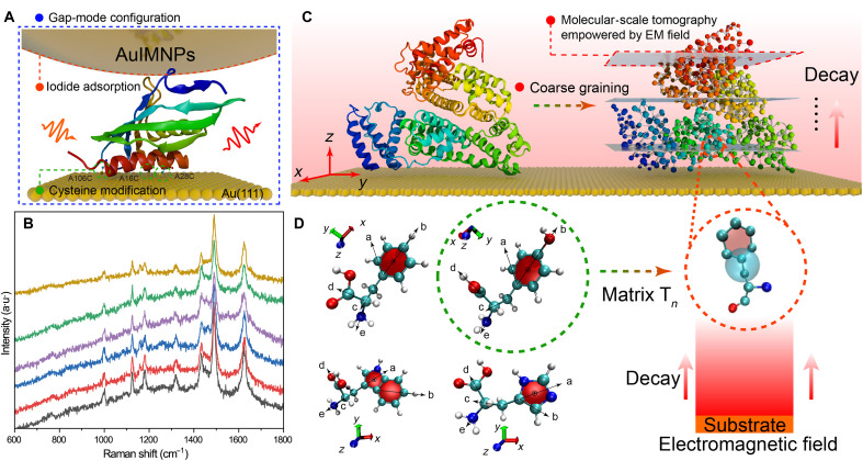
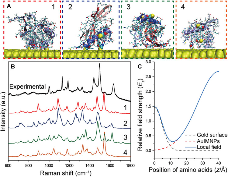
Despite great advances in protein structure analysis, label-free and ultrasensitive methods to obtain the natural and dynamic three-dimensional (3D) structures are still urgently needed. Surface-enhanced Raman spectroscopy (SERS) can be a good candidate, whereas the complexity originated from the interactions between the protein and the gradient surface electric field makes it extremely challenging to determine the protein structure. Here, we propose a deciphering strategy for accurate determination of 3D protein structure from experimental SERS spectra in seconds by simply summing SERS spectra of isolated amino acids in electric fields of different strength with their orientations in protein. The 3D protein structure can be reconstructed by comparing the experimental spectra obtained in a well-defined gap-mode SERS configuration with the simulated spectra. The gradient electric field endows SERS with a unique advantage to section biomolecules with atomic precision, which makes SERS a competent tool for monitoring biomolecular events under physiological conditions.


Figures

Fig. 1.
Fig. 1.. Scheme of the proposed strategy for deciphering 3D protein structure.
(A) Schematic diagram of protein in the nanocavity of the gap-mode configuration constructed by Au(111) surface and AuIMNPs, where every protein in the gap is subjected to the highly localized plasmonic field. Ketosteroid isomerase (KSI) was used as a model system (Protein Data Bank: 1buq), in which the positions of 16, 28, and 106 are replaced by cysteine. These cysteines can bind to the Au surface through thiol-Au interaction, allowing for orientation-controlled immobilization. (B) SERS spectra of KSI underneath six different NPs on a same single-crystal surface, indicating high reproducibility of the gap-mode configuration. a.u., arbitrary units. (C) On the basis of the coarse-grained model, the POI (bovine serum albumin as a model) on the surface can be sectioned into different layers, where each amino acid would experience different strengths in the decay electromagnetic field. The gray planes, indicating different field strengths, are similar to the sectioning plane in computed tomography but at the molecular scale, giving SERS the additional power to extract the amino acids position with atomic precision. (D) Every two vectors could define a bond rotation in amino acids. Thus, amino acids require at least three rotation matrices (Tn) describing the dynamic structure of amino acids, which would then be used to transform PDs under specific orientation to real orientation.
Fig. 2.
Fig. 2.. Testifying the performance of SPARC.
(A) Schematic diagram of the proposed system by Li et al. as well as local electric field distribution for viologen in the gap. Surface-enhanced Raman spectra of viologen with different alkane lengths (n) to the Au(111) surface in the gap of silica coating AuNPs and Au(111) surfaces. (B) Experimental SERS spectra. (C) Simulated SERS spectra by SPARC. (D) Simulated SERS spectra in the literature. (B and D) Reproduced with original data by courtesy of Li et al. (18).
Fig. 3.
Fig. 3.. Demonstration of the deciphering strategy by TERS.
(A) Schematic diagram of TERS detection of the self-assembled peptide on MoS2. (B) AFM image of the sample, showing a full monolayer of order structure of peptides in the bottom layer and some multilayer features on top. (C) Interfacial repeating unit of peptide on MoS2 in the literature (40). (D) Simulated TERS spectrum (red) by SPARC and experimental TERS spectrum of the monolayer of peptide (black). The peaks in the light blue region are from MoS2, and the light red region is from peptides.
Fig. 4.
Fig. 4.. MD simulation and experimental and theoretical SERS spectra of peptides by SPARC.
(A) Schematic diagram of single NP gap-mode SERS. (B) An example of the initial configuration and final configuration of the heptapeptide on a Au surface. (C) Dark-field scattering image of the sample. (D) Theoretical SERS spectra by SPARC (black spectrum) in comparison with experimental SERS spectra (red spectrum) obtained from the NPs as indicated in (C) with white dashed circles. Insets are corresponding configuration scrutinized by our strategy.
Fig. 5.
Fig. 5.. Scrutinization of the KSI structure from the gap-mode SERS spectrum.
(A) Four representative orientations of KSI obtained by MD simulation. (B) Experimental gap-mode SERS spectrum of KSI in comparison with theoretical SERS spectra of four representative conformations calculated by SPARC. (C) Local EM field from contributions of Au surface and AuIMNPs.

Similar articles

Cited by

References

    1. Panda P. K., Arul M. N., Patel P., Verma S. K., Luo W., Rubahn H.-G., Mishra Y. K., Suar M., Ahuja R., Structure-based drug designing and immunoinformatics approach for SARS-CoV-2. Sci. Adv. 6, eabb8097 (2020). - PMC - PubMed
    1. Bravo J. P. K., Liu M.-S., Hibshman G. N., Dangerfield T. L., Jung K., McCool R. S., Johnson K. A., Taylor D. W., Structural basis for mismatch surveillance by CRISPR–Cas9. Nature 603, 343–347 (2022). - PMC - PubMed
    1. Cao L., Coventry B., Goreshnik I., Huang B., Sheffler W., Park J. S., Jude K. M., Marković I., Kadam R. U., Verschueren K. H. G., Verstraete K., Walsh S. T. R., Bennett N., Phal A., Yang A., Kozodoy L., DeWitt M., Picton L., Miller L., Strauch E.-M., DeBouver N. D., Pires A., Bera A. K., Halabiya S., Hammerson B., Yang W., Bernard S., Stewart L., Wilson I. A., Ruohola-Baker H., Schlessinger J., Lee S., Savvides S. N., Garcia K. C., Baker D., Design of protein-binding proteins from the target structure alone. Nature 605, 551–560 (2022). - PMC - PubMed
    1. Chen H., Simoska O., Lim K., Grattieri M., Yuan M., Dong F., Lee Y. S., Beaver K., Weliwatte S., Gaffney E. M., Minteer S. D., Fundamentals, applications, and future directions of Bioelectrocatalysis. Chem. Rev. 120, 12903–12993 (2020). - PubMed
    1. Sesterhenn F., Yang C., Bonet J., Cramer J. T., Wen X., Wang Y., Chiang C.-I., Abriata L. A., Kucharska I., Castoro G., Vollers S. S., Galloux M., Dheilly E., Rosset S., Corthésy P., Georgeon S., Villard M., Richard C.-A., Descamps D., Delgado T., Oricchio E., Rameix-Welti M.-A., Más V., Ervin S., Eléouët J.-F., Riffault S., Bates J. T., Julien J.-P., Li Y., Jardetzky T., Krey T., Correia B. E., De novo protein design enables the precise induction of RSV-neutralizing antibodies. Science 368, (2020). - PMC - PubMed