. 2024 May 3;147(5):1680-1695.
doi: 10.1093/brain/awad398.

Insulin-like growth factor binding protein-2 in at-risk adults and autopsy-confirmed Alzheimer brains

Collaborators, Affiliations

Insulin-like growth factor binding protein-2 in at-risk adults and autopsy-confirmed Alzheimer brains

Marc James Quesnel et al. Brain. .

Abstract

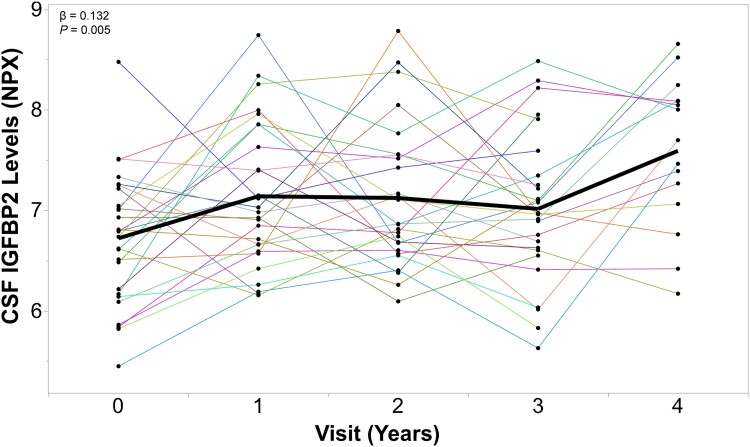
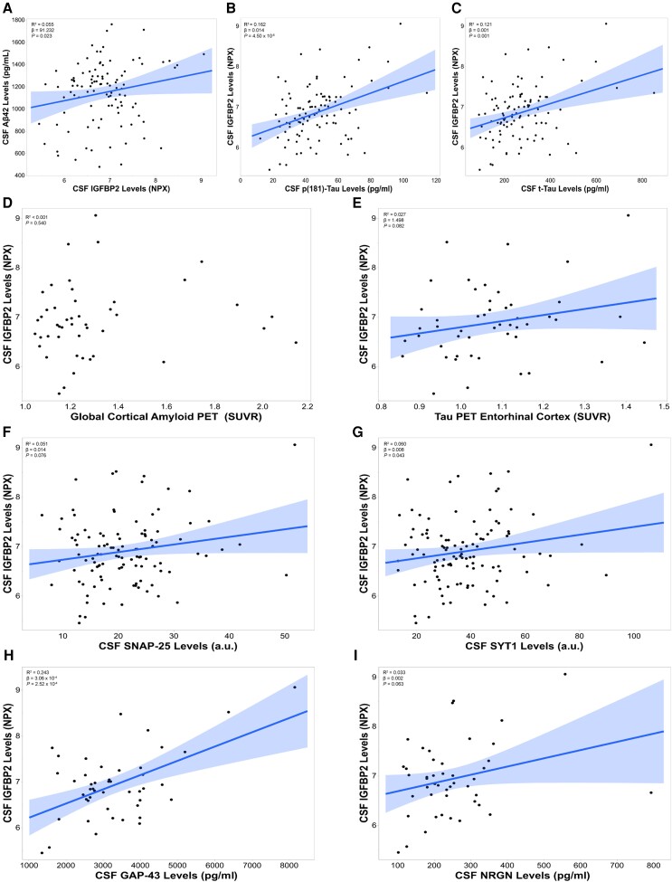
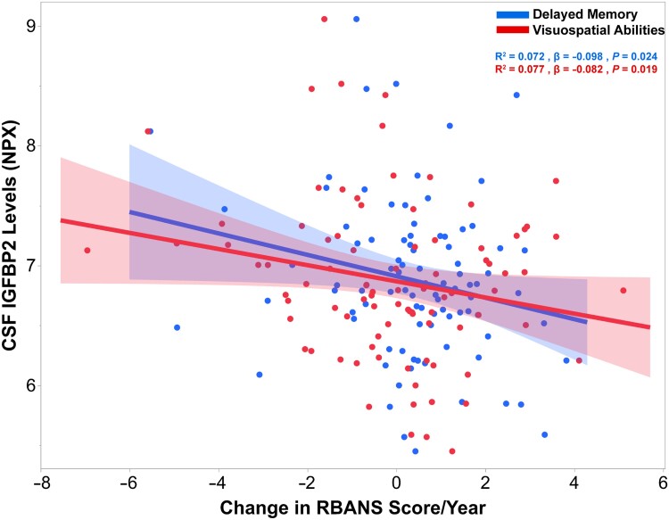
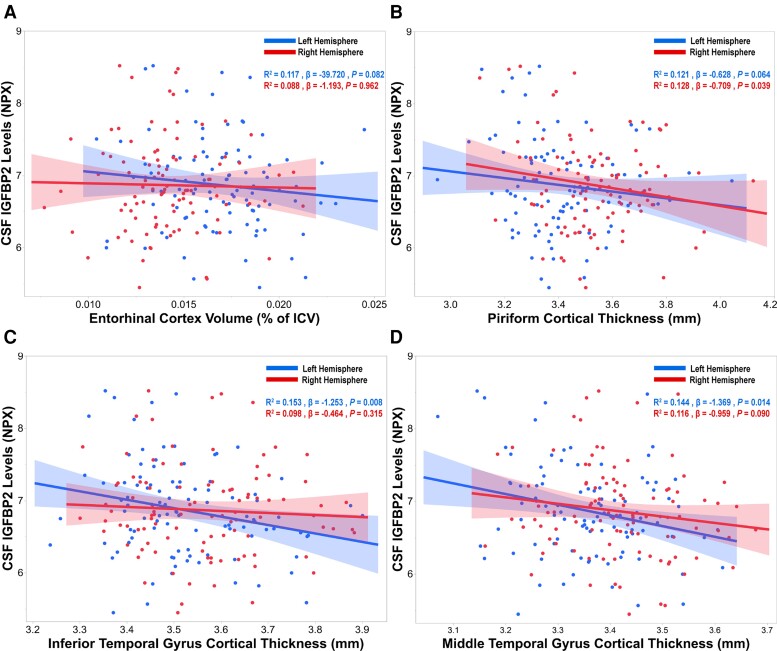
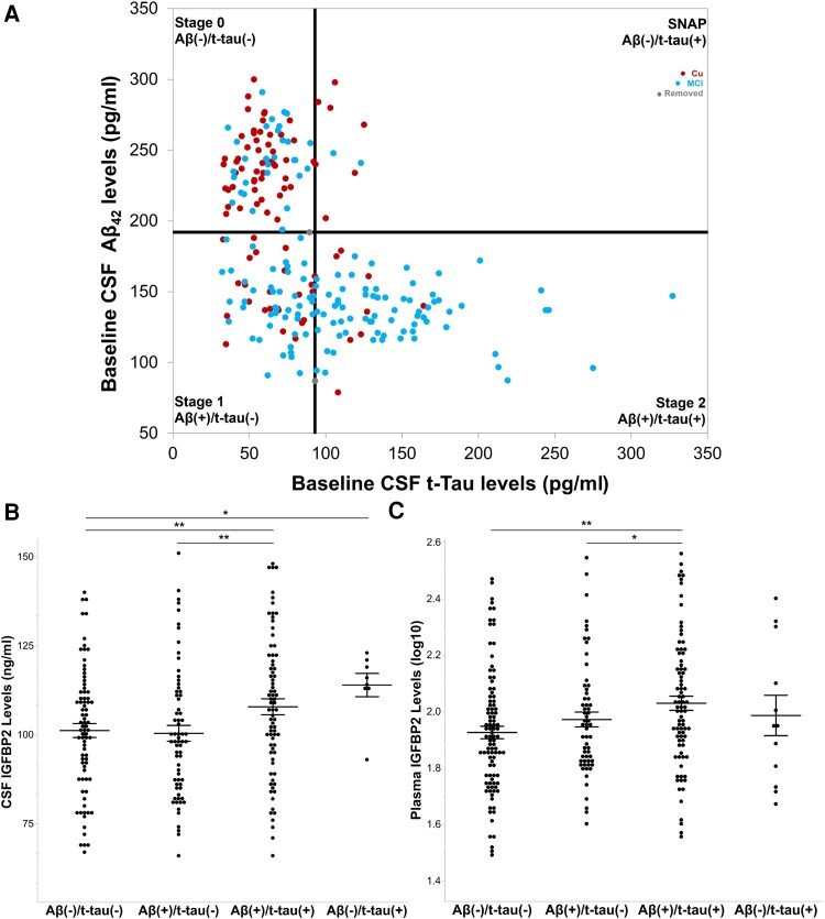
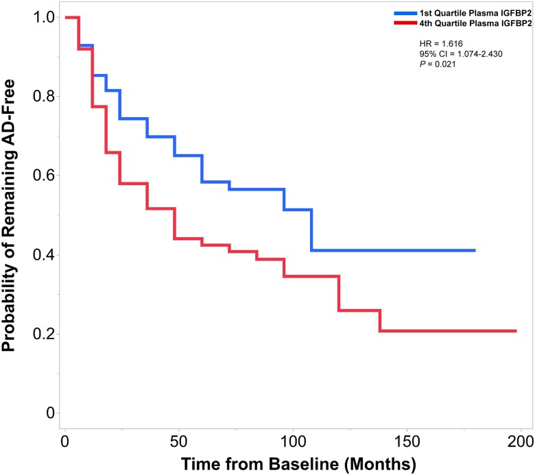
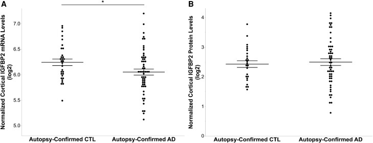
Insulin, insulin-like growth factors (IGF) and their receptors are highly expressed in the adult hippocampus. Thus, disturbances in the insulin-IGF signalling pathway may account for the selective vulnerability of the hippocampus to nascent Alzheimer's disease (AD) pathology. In the present study, we examined the predominant IGF-binding protein in the CSF, IGFBP2. CSF was collected from 109 asymptomatic members of the parental history-positive PREVENT-AD cohort. CSF levels of IGFBP2, core AD and synaptic biomarkers were measured using proximity extension assay, ELISA and mass spectrometry. Cortical amyloid-beta (Aβ) and tau deposition were examined using 18F-NAV4694 and flortaucipir. Cognitive assessments were performed during up to 8 years of follow-up, using the Repeatable Battery for the Assessment of Neuropsychological Status. T1-weighted structural MRI scans were acquired, and neuroimaging analyses were performed on pre-specified temporal and parietal brain regions. Next, in an independent cohort, we allocated 241 dementia-free ADNI-1 participants into four stages of AD progression based on the biomarkers CSF Aβ42 and total-tau (t-tau). In this analysis, differences in CSF and plasma IGFBP2 levels were examined across the pathological stages. Finally, IGFBP2 mRNA and protein levels were examined in the frontal cortex of 55 autopsy-confirmed AD and 31 control brains from the Quebec Founder Population (QFP) cohort, a unique population isolated from Eastern Canada. CSF IGFBP2 progressively increased over 5 years in asymptomatic PREVENT-AD participants. Baseline CSF IGFBP2 was positively correlated with CSF AD biomarkers and synaptic biomarkers, and negatively correlated with longitudinal changes in delayed memory (P = 0.024) and visuospatial abilities (P = 0.019). CSF IGFBP2 was negatively correlated at a trend-level with entorhinal cortex volume (P = 0.082) and cortical thickness in the piriform (P = 0.039), inferior temporal (P = 0.008), middle temporal (P = 0.014) and precuneus (P = 0.033) regions. In ADNI-1, CSF (P = 0.009) and plasma (P = 0.001) IGFBP2 were significantly elevated in Stage 2 [CSF Aβ(+)/t-tau(+)]. In survival analyses in ADNI-1, elevated plasma IGFBP2 was associated with a greater rate of AD conversion (hazard ratio = 1.62, P = 0.021). In the QFP cohort, IGFBP2 mRNA was reduced (P = 0.049); however, IGFBP2 protein levels did not differ in the frontal cortex of autopsy-confirmed AD brains (P = 0.462). Nascent AD pathology may induce an upregulation in IGFBP2 in asymptomatic individuals. CSF and plasma IGFBP2 may be valuable markers for identifying CSF Aβ(+)/t-tau(+) individuals and those with a greater risk of AD conversion.

Keywords: Alzheimer's disease; RBANS; cerebrospinal fluid; insulin-like growth factor; insulin-like growth factor-binding protein-2; post-mortem brain tissue.


Conflict of interest statement

J.P. serves as a scientific advisor to the Alzheimer Society of France. H.Z. has served at scientific advisory boards and/or as a consultant for Abbvie, Acumen, Alector, Alzinova, ALZPath, Annexon, Apellis, Artery Therapeutics, AZTherapies, CogRx, Denali, Eisai, Nervgen, Novo Nordisk, Optoceutics, Passage Bio, Pinteon Therapeutics, Prothena, Red Abbey Labs, reMYND, Roche, Samumed, Siemens Healthineers, Triplet Therapeutics, and Wave, has given lectures in symposia sponsored by Cellectricon, Fujirebio, Alzecure, Biogen and Roche, and is a co-founder of Brain Biomarker Solutions in Gothenburg AB (BBS), which is part of the GU Ventures Incubator Program (outside submitted work). K.B. has served as a consultant and at advisory boards for Acumen, ALZPath, BioArctic, Biogen, Eisai, Lilly, Moleac Pte. Ltd, Novartis, Ono Pharma, Prothena, Roche Diagnostics and Siemens Healthineers; has served at data monitoring committees for Julius Clinical and Novartis; has given lectures, produced educational materials and participated in educational programs for AC Immune, Biogen, Celdara Medical, Eisai and Roche Diagnostics; and is a co-founder of Brain Biomarker Solutions in Gothenburg AB (BBS), which is a part of the GU Ventures Incubator Program, outside the work presented in this paper.

Figures

Figure 1
Figure 1
CSF IGFBP2 levels progressively increase over 5years in asymptomatic PREVENT-AD participants. IGFBP2 was measured in the CSF of a subset of PREVENT-AD participants (n = 27) at baseline and at follow-up visits, using the Olink Proximity Extension Assay. Linear mixed models accounting for participant-specific trajectories demonstrate CSF IGFBP2 levels increase in a subset of at-risk individuals that have been followed for 5 years. β- and P-values are located in the top left corner. IGFBP2 = insulin-like growth factor binding protein-2; PREVENT-AD = PRe-symptomatic EValuation of Experimental or Novel Treatments for Alzheimer's Disease; NPX = Normalized Protein eXpression.
Figure 2
Figure 2
CSF IGFBP2 is associated with CSF and PET Alzheimer’s disease biomarkers in the asymptomatic PREVENT-AD cohort. CSF IGFBP2 levels were measured using the Olink Proximity Extension Assay (n = 109). CSF Alzheimer’s disease (AD) biomarkers (A) amyloid-beta 42 (Aβ42) (B) phosphorylated tau 181 (p181-tau) and (C) total tau (t-tau) were measured using validated Innotest ELISA kits, following the standardized protocols established by the BIOMARKAPD consortium (n = 101). (D) The global cortical amyloid standardized uptake value ratio (SUVR) was measured using 18F-NAV4694 (n = 46). (E) Tau deposition in the entorhinal cortex was measured with flortaucipir (n = 49). The synaptic markers (F) SNAP25 (n = 106), (G) SYT1 (n = 106), (H) GAP43 (n = 46) and (I) NRGN (n = 46) were quantified using immunoprecipitation followed by mass spectrometry. Significant or trend-level linear regressions are represented with a confidence region of the fitted line. R2 , β- and P-values are located in the top left corners of each panel. Analyses were adjusted for age, sex and APOE ɛ4 carrier status. a.u. = arbitrary units; BIOMARKAPD = Biomarkers for Alzheimer's and Parkinson's disease; IGFBP2 = insulin-like growth factor binding protein-2; NPX = Normalized Protein eXpression; PREVENT-AD = PRe-symptomatic EValuation of Experimental or Novel Treatments for Alzheimer's Disease.
Figure 3
Figure 3
CSF IGFBP2 is associated with longitudinal changes in delayed memory and visuospatial abilities over 5–8years in PREVENT-AD. CSF IGFBP2 levels were measured using the Olink Proximity Extension Assay (n = 109). Cognitive performance trajectory slopes were computed for each cognitive domain of the Repeatable Battery for the Assessment of Neuropsychological Status (RBANS; delayed memory, visuospatial abilities, language, immediate memory and attention) in a subset of PREVENT-AD participants that were followed for 5–8 years (n = 89). Significant linear regressions are represented with a confidence region of the fitted line. R2, β- and P-values are located in the top right corner. Analyses were adjusted for age, sex, APOE ɛ4 carrier status and years of education. IGFBP2 = insulin-like growth factor binding protein-2; NPX = Normalized Protein eXpression; PREVENT-AD = PRe-symptomatic EValuation of Experimental or Novel Treatments for Alzheimer's Disease.
Figure 4
Figure 4
CSF IGFBP2 is associated with atrophy in Alzheimer’s disease-related brain regions in PREVENT-AD. CSF IGFBP2 levels were measured using the Olink Proximity Extension Assay (n = 109). T1-weighted structural MRI scans were performed on a subset of PREVENT-AD participants (n = 104). The imaging processing pipeline CIVET 1.1.12 was used to analyse neuroimaging data. (A) Entorhinal cortex volumes were normalized by total intracranial volumes (ICV). Cortical thickness measurements were acquired from Alzheimer’s disease (AD)-related brain regions, such as the (B) piriform cortex, (C) inferior temporal gyrus and (D) middle temporal gyrus. Significant or trend-level linear regressions are represented with a confidence region of the fitted line. R2 , β- and P-values are located in the top right corner of each panel. Analyses were adjusted for age, sex and APOE ɛ4 carrier status. IGFBP2 = insulin-like growth factor binding protein-2; NPX = Normalized Protein eXpression; PREVENT-AD = PRe-symptomatic EValuation of Experimental or Novel Treatments for Alzheimer's Disease.
Figure 5
Figure 5
CSF and plasma IGFBP2 is elevated in CSF Aβ(+)/t-tau(+) individuals from the ADNI-1 cohort. (A) Cognitively unaffected participants (n = 92) and participants with mild cognitive impairment (MCI; n = 149) from the ADNI-1 cohort were staged as CSF amyloid-β and/or CSF total tau-positive according to the recommended thresholds of 192 pg/ml and 93 pg/ml, respectively. Linear models, adjusted for age, sex and APOE ɛ4 carrier status were used to examine mean differences in IGFBP2 protein levels across stages. (B) CSF IGFBP2 was elevated at Stage 2 (n = 77) relative to Stage 0 (n = 80) and Stage 1 (n = 68). Furthermore, CSF IGFBP2 was elevated in suspected non-Alzheimer pathology (SNAP, n = 8) compared with Stage 0. (C) Plasma IGFBP2 was elevated at Stage 2 (n = 84) relative to Stage 0 (n = 98) and Stage 1 (n = 60). However, plasma IGFBP2 did not significantly differ between SNAP (n = 12) and Stage 0. The data are represented as mean ± standard error of the mean. *P < 0.05, **P < 0.01. Aβ42 = amyloid-beta 42; ADNI = Alzheimer's Disease Neuroimaging Initiative; IGFBP2 = insulin-like growth factor binding protein-2; MCI = mild cognitive impairment; SNAP = suspected non-Alzheimer pathology; t-tau = total tau.
Figure 6
Figure 6
Elevated plasma IGFBP2 is associated with a greater rate of conversion to Alzheimer’s disease in individuals from the ADNI-1 cohort. Cox proportional hazards models examined the association between baseline plasma IGFBP2 levels and rate of conversion to Alzheimer’s disease (AD). The first quartile and fourth values of plasma IGFBP2 were contrasted. Participants were followed from the baseline visit to the time of diagnosis (of AD), or to the time the participant was last confirmed to be free of AD (mean follow-up, 3.8 years; range, 0.5–16.5 years). Of the 226 individuals that were followed longitudinally, 107 individuals progressed to AD. Individuals with plasma IGFBP2 values in the fourth quartile exhibited a greater rate of conversion to AD, compared with the first quartile. Hazard ratio (HR) and P-values are located in the top right corner. Cox models were adjusted for age, gender and APOE ɛ4 carrier status. ADNI = Alzheimer's Disease Neuroimaging Initiative; IGFBP2 = insulin-like growth factor binding protein-2.
Figure 7
Figure 7
Frontal cortex IGFBP2 gene expression is reduced in autopsy-confirmed Alzheimer’s disease brains, however protein levels do not differ from elderly controls. (A) Microarray technology was used to measure IGFBP2 mRNA levels in the frontal cortex of autopsy-confirmed Alzheimer’s disease brains (AD; n = 55) and elderly controls (CTL; n = 31) from the Quebec Founder Population (QFP) cohort. (B) IGFBP2 protein levels in the frontal cortex were measured in AD (n = 53) and control (n = 25) brains using a commercially available ELISA kit. Analyses were adjusted for age, sex, APOE ɛ4 carrier status and post-mortem interval. The data are represented as mean ± standard error of the mean. *P < 0.05. IGFBP2 = insulin-like growth factor binding protein-2.

Comment in

References

    1. GBD 2019 Dementia Forecasting Collaborators . Estimation of the global prevalence of dementia in 2019 and forecasted prevalence in 2050: An analysis for the global burden of disease study 2019. Lancet Public Health. 2022;7:e105–e125. - PMC - PubMed
    1. Nandi A, Counts N, Chen S, et al. Global and regional projections of the economic burden of Alzheimer’s disease and related dementias from 2019 to 2050: A value of statistical life approach. EClinicalMedicine. 2022;51:101580. - PMC - PubMed
    1. Alzheimer’s Association . 2022 Alzheimer’s disease facts and figures. Alzheimers Dement. 2022;18:700–789. - PubMed
    1. Serrano-Pozo A, Frosch MP, Masliah E, Hyman BT. Neuropathological alterations in Alzheimer disease. Cold Spring Harbor Perspect Med. 2011;1:a006189. - PMC - PubMed
    1. Jack CR Jr, Knopman DS, Jagust WJ, et al. Hypothetical model of dynamic biomarkers of the Alzheimer’s pathological cascade. Lancet Neurol. 2010;9:119–128. - PMC - PubMed

Publication types

MeSH terms