. 2024 Jan;625(7994):360-365.
doi: 10.1038/s41586-023-06855-2. Epub 2023 Nov 22.

Structural basis of Gabija anti-phage defence and viral immune evasion

Affiliations

Structural basis of Gabija anti-phage defence and viral immune evasion

Sadie P Antine et al. Nature. 2024 Jan.

Abstract

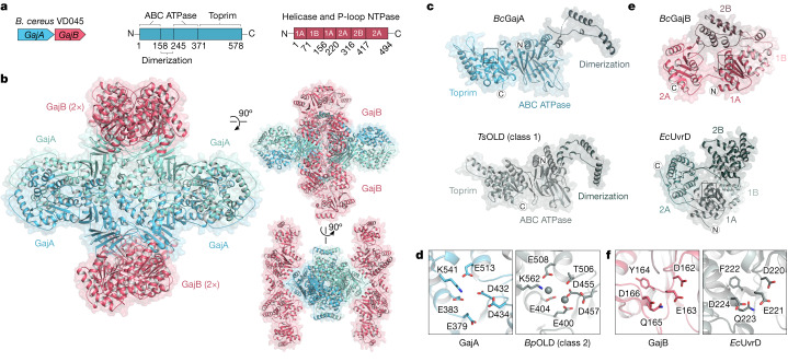
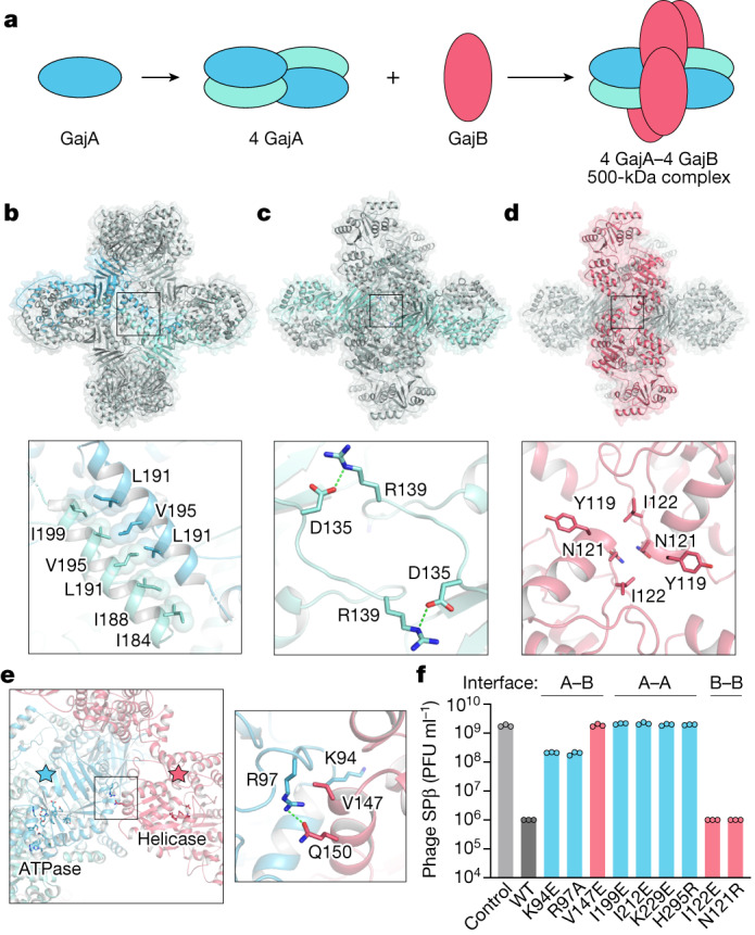
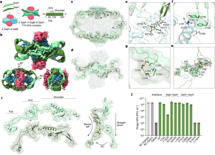
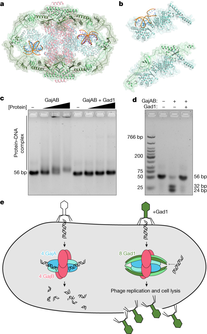
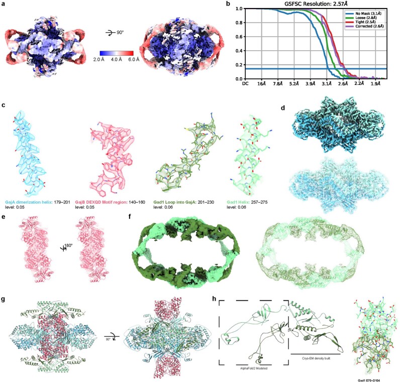
Bacteria encode hundreds of diverse defence systems that protect them from viral infection and inhibit phage propagation1-5. Gabija is one of the most prevalent anti-phage defence systems, occurring in more than 15% of all sequenced bacterial and archaeal genomes1,6,7, but the molecular basis of how Gabija defends cells from viral infection remains poorly understood. Here we use X-ray crystallography and cryo-electron microscopy (cryo-EM) to define how Gabija proteins assemble into a supramolecular complex of around 500 kDa that degrades phage DNA. Gabija protein A (GajA) is a DNA endonuclease that tetramerizes to form the core of the anti-phage defence complex. Two sets of Gabija protein B (GajB) dimers dock at opposite sides of the complex and create a 4:4 GajA-GajB assembly (hereafter, GajAB) that is essential for phage resistance in vivo. We show that a phage-encoded protein, Gabija anti-defence 1 (Gad1), directly binds to the Gabija GajAB complex and inactivates defence. A cryo-EM structure of the virally inhibited state shows that Gad1 forms an octameric web that encases the GajAB complex and inhibits DNA recognition and cleavage. Our results reveal the structural basis of assembly of the Gabija anti-phage defence complex and define a unique mechanism of viral immune evasion.


Conflict of interest statement

R.S. is a scientific cofounder and advisor of BiomX and EcoPhage. The remaining authors declare no competing interests.

Figures

Fig. 1
Fig. 1. Structure of the Gabija anti-phage defence complex.
a, Schematic of B. cereus (Bc) Gabija defence operon and domain organization of GajA and GajB. b, Overview of the GajAB X-ray crystal structure shown in three orientations. GajA protomers are depicted in two shades of blue and GajB protomers are in red. c, Isolated GajA monomer (top) and comparison with a TsOLD nuclease monomer (bottom) (Protein Data Bank (PDB) ID:6P74). d, Magnified views of Toprim catalytic residues in GajA (left) and BpOLD (right) (PDB ID: 6NK8). The location of the GajA cut-away image is indicated with a box in c and magnesium ions are depicted as grey spheres. e, Isolated GajB monomer (top) and comparison with E. coli (Ec) UvrD (bottom) (PDB ID: 2IS2). f, Magnified views of the DEXQD-box motif in GajB (left) and EcUvrD (right). The locations of the GajB and UvrD cut-away images are indicated with boxes in e.
Fig. 2
Fig. 2. Mechanism of Gabija supramolecular complex assembly.
a, Schematic model of GajAB complex formation by GajA tetramerization and GajB docking. b, Overview of the GajA α2D–α2D dimerization interface and detailed view of interacting residues. For clarity, each GajA monomer is depicted in two shades of blue. c, Overview of the GajA–GajA ATPase interaction and detailed view of the inter-subunit D135–R139 interaction. d, Overview of the minimal GajB–GajB dimer interface and detailed view of GajB–GajB hydrophobic interactions centred around Y119, N121 and I122. e, Left, overview of the GajA–GajB interface, highlighting the proximity of GajA ABC ATPase and GajB helicase active-site residues. Right, the box indicates the location of GajA R97 and GajB V147 and Q150 interaction. f, Analysis of mutations in the GajA–GajB (A–B), GajA–GajA (A–A), and GajB–GajB (B–B) multimerization interfaces. GajA and GajB mutations were selected by identifying central residues with well-defined protein–protein contacts within each multimerization interface, and were tested to determine their effects on the ability of the B. cereus Gabija operon to defend cells against phage infection. Data represent the phage SPβ average plaque-forming units (PFU) ml−1 of three biological replicates, with individual data points shown. WT, wild type. Source Data
Fig. 3
Fig. 3. Structural basis of viral evasion of Gabija defence.
a, Schematic model of GajAB–Gad1 co-complex formation and domain organization of phage Phi3T Gad1. b, Cryo-EM density map of BcGajAB in complex with Phi3T Gad1, shown in three different orientations. The map is coloured by the model, with Gad1 monomers depicted in two shades of green. c,d, Side view of the complete Gad1 octameric complex (c) and top-down view of the Gad1 tetrameric interface (d), with boxes highlighting views that are magnified in eh. e,f, Magnified views of major Gad1–GajA interface contacts including a Gad1 positively charged loop (e) and hydrophobic interactions with GajA α2D (f). g,h, Magnified views of major Gad1–Gad1 oligomerization interactions including disulfide bonds in the C-terminal shoulder domain (g) and fist–fist domain contacts modelled by rigid-body placement of an AlphaFold2 fist-domain structure prediction into the cryo-EM map (h). i, Two distinct conformations of Gad1 observed in the GajAB–Gad1 co-complex structure. Differences in the rotation of the Gad1 arm domain are highlighted on the right. j, Analysis of the effect of Gad1 mutations in the GajA–Gad1 and Gad1–Gad1 multimerization interfaces on the ability of Gad1 to enable evasion of Gabija defence. Data represent PFU ml−1 of phage SPβ infecting cells expressing BcGabija and Shewanella sp. phage 1/4 Gad1, or negative control (NC) cells expressing empty vector for either plasmid. Shewanella sp. phage 1/4 Gad1 residues are numbered according to the Phi3T Gad1 structure. Shewanella sp. phage 1/4 Gad1 N-terminal and C-terminal truncations (N-term and C-term, respectively) are M1–L152 and V153–E348, respectively. Data are the average of three biological replicates, with individual data points shown. Source Data
Fig. 4
Fig. 4. Inhibition of Gabija DNA binding and cleavage enables viral evasion.
a, Cartoon representation of the GajAB–Gad1 co-complex structure with modelled DNA (orange), based on structural homology with E. coli MutS (PDB ID 3K0S). b, Top, isolated GajA protomer with modelled DNA (orange) bound to the Toprim domain. Bottom, the same GajA promoter with Gad1, showing substantial steric clashes between Gad1 and the path of the DNA. c,d, Biochemical analysis of GajAB 56-bp target DNA binding (c) and target cleavage (d) shows that Gad1 potently inhibits the activity of GajAB. Data are representative of three independent experiments. e, Model of Gabija anti-phage defence and mechanism of Gad1 immune evasion.
Extended Data Fig. 1
Extended Data Fig. 1. GajA and GajB form a supramolecular complex that cleaves phage lambda DNA in vitro.
a, Size-exclusion chromatography (16/600 S200) analysis of recombinant BcGajA and BcGajB proteins, and the co-expressed BcGajAB complex. Brackets indicate fractions collected for biochemical and structural analysis with A260/280 of the final purified proteins listed above. b, SDS–PAGE analysis of purified GajA, GajB, and GajAB. Asterisk indicates minor contamination with the E. coli protein ArnA. Data are representative of at least 3 independent experiments. c, Agarose gel analysis of the ability of GajA, GajB, and GajAB to cleave a 56-bp target and scrambled dsDNA demonstrates that GajA alone and the GajAB complex can cleave target DNA only. The sequence-specific GajA target dsDNA with cleavage site described in Cheng et al. and the scrambled 15-bp sequence are shown below. d, Catalytic dead GajA[E379A]–GajB complex binding to target dsDNA (left) and scrambled dsDNA (right). e, Structural comparison of GajB and EcRep (PDB ID 1UAA) demonstrates the GajB 2B domain is rotated in a partially active intermediate position in the GajAB complex structure. f, SDS–PAGE analysis of BcGajAB mutant protein complex formation after co-expression and Ni-NTA pull-down demonstrates that mutations to the GajA–GajB interface disrupt complex formation. The GajA and GajB homo-oligomerization interfaces are not required for GajA–GajB interaction, but it is not known if these mutants remain competent at forming the wild-type 4:4 complex. Data in c,d,f are representative of 3 independent experiments.
Extended Data Fig. 2
Extended Data Fig. 2. Structural characterization of GajA.
a, Structure-guided alignment of GajA proteins from indicated bacteria coloured according to amino acid conservation. The determined Bacillus cereus VD045 GajA secondary structure is displayed, and active-site and oligomerization interface residues are annotated according to the key below. Secondary structure abbreviations include ABC ATPase domain (ABC), dimerization domain (D), and Toprim domain (T). b,c, Zoomed-in views of GajA–GajA oligomerization interactions including dimerization domain interactions (b) and ABC ATPase domain interactions (c).
Extended Data Fig. 3
Extended Data Fig. 3. Structural characterization of GajB.
a, Structure-guided alignment of GajB proteins from indicated bacteria coloured according to amino acid conservation. The determined Bacillus cereus VD045 GajB secondary structure is displayed, and active-site and oligomerization interface residues are annotated according to the key below.
Extended Data Fig. 4
Extended Data Fig. 4. Size comparison of Gad1 with known phage immune-evasion proteins and biochemical characterization of Gad1 for binding to the GajAB complex.
a, Analysis of known phage immune-evasion proteins according to function and molecular weight demonstrates that Gad1 is atypically large for an evasion protein that functions through protein–protein interactions with a host anti-phage defence system. Phage immune-evasion proteins are categorized and exhibited as coloured dots coloured according to the key below. Notable evasion proteins are indicated with text labels–,,–. b, Top, size-exclusion chromatography analysis (16/600 S200) of SUMO2-tagged BcGajAB with or without phage Phi3T Gad1 used for cryo-EM structural studies. Bottom, size-exclusion chromatography analysis (16/600 S300) of BcGajAB with or without Shewanella phage 1/4 Gad1 used for biochemical studies. Brackets indicate fractions collected and the A260/280 of the final purified proteins is indicated above. Shewanella phage 1/4 Gad1 was used preferentially for biochemical studies due to less toxicity during E. coli expression. c, SDS–PAGE analysis of purified SUMO2-tagged GajAB, SUMO2-tagged GajAB in complex with phage Phi3T Gad1, untagged GajAB, and untagged GajB in complex with Shewanella phage 1/4 Gad1. d, SDS–PAGE analysis of Ni-NTA co-purified GajA, GajB, and GajAB with Shewanella phage 1/4 Gad1 indicates that Gad1 only binds the fully assembled GajAB complex. Asterisk indicates minor contamination with the E. coli protein ArnA. Data in bd are representative of 3 independent experiments.
Extended Data Fig. 5
Extended Data Fig. 5. Cryo-EM data processing for the GajAB–Gad1 co-complex.
a, Section of a representative electron micrograph (n = 9,243) of SUMO2–GajAB in complex with phage Phi3T Gad1. Scale bar, 50 nm. b, Data-processing scheme used to generate the final 2.57-Å map.
Extended Data Fig. 6
Extended Data Fig. 6. Cryo-EM map quality of the GajAB–Gad1 co-complex and model to map fitting.
a, Reconstruction of the GajAB–Gad1 co-complex coloured by local resolution. b, Fourier shell correlation (FSC) of the EM map. c, GajA, GajB, and Gad1 map to model fit for designated regions. df, Isolated GajA (d), GajB (e) and Gad1 (f) density maps with model fitting. g, GajAB–Gad1 model that was used for refining the cryo-EM map for Extended Data Table 2. h, Left, sections of Gad1 chains that were built de novo from the cryo-EM density and built using rigid-body placement of AlphaFold2 modelled residues. Right, cryo-EM density used to fit placement of Gad1 fist–fist domain contacts that complete protomer interactions.
Extended Data Fig. 7
Extended Data Fig. 7. Biochemical and structural characterization of the GajAB–Gad1 co-complex.
a, Structure-guided alignment of Gad1 proteins from indicated phage or prophage genomes coloured according to amino acid conservation. The Bacillus phage Phi3T Gad1 secondary structure is displayed according to the two different conformations observed in the GajAB–Gad1 co-complex structure. Oligomerization interface residues are annotated according to the key below. b,c, Magnified views of Gad1–GajA interface contacts including hydrophobic interactions in AlphaFold2 arm domain structure of Gad1 and the Toprim domain of GajA (b) and Gad1 shoulder domain residue Q244 interaction with GajA dimerization domain residue E277 (c). d, Magnified view of Gad1–Gad1 oligomerization interactions between shoulder domains of Gad1 protomers. e, SDS–PAGE analysis of the ability of Shewanella phage 1/4 Gad1 mutant proteins to interact with the GajAB complex. Shewanella phage 1/4 Gad1 mutant proteins were co-expressed with SUMO2-tagged GajAB (GajA-tagged) and co-purified by Ni-NTA pull-down. Shewanella sp. phage 1/4 Gad1 residues are numbered according to the Phi3T Gad1 structure. To measure high stringency of GajAB–Gad1 interactions, complexes were washed with a 1 M NaCl buffer prior to elution and co-purification. Notably, the Gad1 mutant C282E is no longer able to interact with GajAB in vitro under these stringent conditions, but retains the ability to disrupt Gabija defence in vivo, suggesting that lower-affinity interactions still occur. f, SDS–PAGE analysis of the ability of Shewanella phage 1/4 Gad1 fist–fist interface mutant proteins to interact with the GajAB complex. Shewanella phage 1/4 Gad1 mutant proteins were co-expressed with SUMO2-tagged GajAB (GajA-tagged), co-purified by Ni-NTA pull-down, and treated with SENP2 to cleave the SUMO2 tag prior to SDS–PAGE gel loading. Shewanella sp. phage 1/4 Gad1 residues are numbered according to the Phi3T Gad1 structure. g, Agarose gel analysis of the ability of GajAB–Gad1 mutant complexes to cleave target 56-bp dsDNA after a 1 min or 20 min incubation. h, Superposition of the GajAB crystal structure and GajAB from the GajAB–Gad1 cryo-EM structure demonstrates no significant conformational change after Gad1 binding. Data in eg are representative of 3 independent experiments.
Extended Data Fig. 8
Extended Data Fig. 8. Modelling DNA-bound GajA.
a,b, Isolated GajA protomer modelled with DNA bound to the Toprim domain shown with surface electrostatic potential (a) and in cartoon format (b). DNA modelling was performed using structural homology with the E. coli MutS–DNA complex (PDB ID 3K0S). c, Magnified view of the GajA Toprim active site with modelled DNA.

References

    1. Doron S, et al. Systematic discovery of antiphage defense systems in the microbial pangenome. Science. 2018;359:eaar4120. doi: 10.1126/science.aar4120. - DOI - PMC - PubMed
    1. Gao L, et al. Diverse enzymatic activities mediate antiviral immunity in prokaryotes. Science. 2020;369:1077–1084. doi: 10.1126/science.aba0372. - DOI - PMC - PubMed
    1. Millman A, et al. An expanded arsenal of immune systems that protect bacteria from phages. Cell Host Microbe. 2022;30:1556–1569. doi: 10.1016/j.chom.2022.09.017. - DOI - PubMed
    1. Rousset F, et al. Phages and their satellites encode hotspots of antiviral systems. Cell Host Microbe. 2022;30:740–753. doi: 10.1016/j.chom.2022.02.018. - DOI - PMC - PubMed
    1. Vassallo CN, Doering CR, Littlehale ML, Teodoro GIC, Laub MT. A functional selection reveals previously undetected anti-phage defence systems in the E. coli pangenome. Nat. Microbiol. 2022;7:1568–1579. doi: 10.1038/s41564-022-01219-4. - DOI - PMC - PubMed

MeSH terms