. 2023 Nov 22;14(1):7638.
doi: 10.1038/s41467-023-43075-8.

Horizontal acquisition of a DNA ligase improves DNA damage tolerance in eukaryotes

Affiliations

Horizontal acquisition of a DNA ligase improves DNA damage tolerance in eukaryotes

Emilien Nicolas et al. Nat Commun. .

Abstract

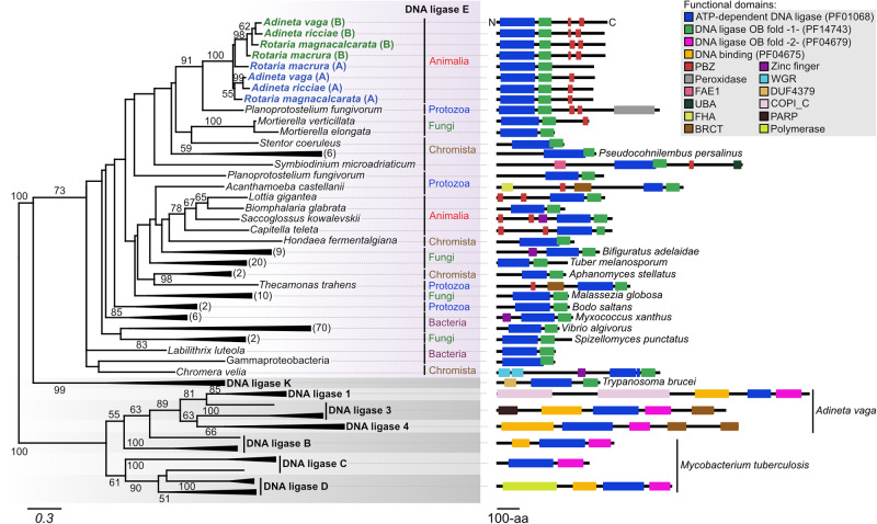
Bdelloid rotifers are part of the restricted circle of multicellular animals that can withstand a wide range of genotoxic stresses at any stage of their life cycle. In this study, bdelloid rotifer Adineta vaga is used as a model to decipher the molecular basis of their extreme tolerance. Proteomic analysis shows that a specific DNA ligase, different from those usually involved in DNA repair in eukaryotes, is strongly over-represented upon ionizing radiation. A phylogenetic analysis reveals its orthology to prokaryotic DNA ligase E, and its horizontal acquisition by bdelloid rotifers and plausibly other eukaryotes. The fungus Mortierella verticillata, having a single copy of this DNA Ligase E homolog, also exhibits an increased radiation tolerance with an over-expression of this DNA ligase E following X-ray exposure. We also provide evidence that A. vaga ligase E is a major contributor of DNA breaks ligation activity, which is a common step of all important DNA repair pathways. Consistently, its heterologous expression in human cell lines significantly improves their radio-tolerance. Overall, this study highlights the potential of horizontal gene transfers in eukaryotes, and their contribution to the adaptation to extreme conditions.


Conflict of interest statement

The authors declare no competing interests.

Figures

Fig. 1
Fig. 1. Upregulation of a DNA ligase upon irradiation of the bdelloid rotifer Adineta vaga at 1 kGy of X-ray.
a Global proteomic analysis. Total proteins were extracted from around 100,000 rotifers at t0h, t4h, t24h, and t72h post-irradiation. Differential expression analysis of each protein was performed with Peaks studio. This panel shows the proteins that are either over-represented (≥2.5x, blue) or down-represented (≤2.5x, green) in comparison to the non-irradiated control (NI) (see also Supplementary Table S1). b Single qualitative Western-blot analysis showing the expression pattern of the DNA ligase copies A and B upon irradiation. c Subcellular localization analysis of the induced DNA ligase (copy B) by immuno-fluorescence at different timepoints post-irradiation. White lines delineate the bdelloid rotifer A. vaga individual and dotted lines delimit the ovaries of A. vaga in the middle part of their body, where less to no staining of DNA ligase-B was observed. Nuclei were stained with DAPI but were displayed in green to ease the interpretation of the result. A second replicate of this analysis is presented in Supplementary Fig. S1d.
Fig. 2
Fig. 2. Phylogeny of the induced DNA ligase in Adineta vaga, deriving from DNA ligase E.
The sequence of the DNA ligase over-represented in A. vaga following exposure to X-ray was compared to the available bdelloid rotifer sequences and to the uniref50 database using blastp (evalue 1e-05). The best 155 hits were extracted to perform an alignment using mafft and a phylogenetic analysis using iq-tree. Representative sequences of DNA ligases K, 1, 3, 4, B, C, and D were added separately from the initial blast search. The functional domains of the proteins were identified using interproscan and the structures were manually drawn for each representative case. Only boostrap values > 50 are indicated on the tree. Numbers between brackets represent the number of species of the same kingdom that are collapsed at each branch.
Fig. 3
Fig. 3. Relative mRNA expression analysis of DNA ligases upon irradiation in bdelloid species Adineta vaga and Adineta ricciae and in the fungus species Mortierella verticillata.
a, b, d Kinetics expression analysis of DNA ligase E from A. vaga, A. ricciae and M. verticillata, respectively. The bdelloid rotifer species were irradiated at 1 kGy and M. verticillata at 0.2 kGy of X-ray. The mRNA level at t0h, t4h, t24h, t48h and t72h post-irradiation was determined by qPCR in triplicate. The relative expression were measured in comparison to the non-irradiated control (NI). In d the IR survival rate of M. verticillata was determined by its growth efficiency after X-ray irradiation to doses ranging from 0.05 to 1 kGy. A significant growth reduction was observed for radiation >0.2 kGy. M. verticillata has only one copy of DNA ligase E (MvLigE). c Relative expression analysis of AvLigE in comparison to the other DNA ligases of A. vaga. The rotifers were irradiated at 1 kGy and the mRNA levels were measured at t4h post-irradiation. Indicated fold changes correspond to the level of expression of each mRNA in comparison to the level of the same mRNA in the non-irradiated condition (NI). AvLig3 and AvLig4 have only one copy within the genome of A. vaga. The histograms represent the average values and standard deviations of three technical replicates.
Fig. 4
Fig. 4. In vitro DNA ligation assay to assess the functionality of AvLigE protein.
a Graphical scheme representing the workflow of immuno-depletion of AvLigE from A. vaga protein extracts. The rotifers were irradiated at 1 kGy to stimulate the production of AvLigE-B and the proteins were extracted at t72h post-irradiation. After extraction, AvLigE-B was immuno-depleted from the extracts. A non-irradiated control was also added to the analysis (NI). b Single qualitative Western-blot analysis demonstrating the efficiency of AvLigE-B immuno-depletion. See text for details. c Principle of the DNA ligase assay used. A complex of two oligonucleotides is assembled to form a hairpin loop containing a DNA nick to mimick a single-strand break (SSB). A fluorescein moeity is incorporated at the opposite end of the oligonucleotide structure. The complex is then incubated in the presence of increasing concentrations of A. vaga protein extracts, followed by a denaturation of the oligonucleotide structure. In the absence of any ligation activity, the oligonucleotide carrying fluorescein is lost from the wells during the denaturation process. Conversely, in the presence of a ligation activity, the DNA nick is sealed and the fluorescein moiety is kept in the wells after denaturation. The enzyme activity is determined by quantifying the amount of fluorescein retained in the wells as a result of the denaturation step. Figure adapted from Healing et al. . Flc, fluorescein. d DNA ligation activities with increasing concentrations of the various A. vaga protein extracts. Data represent the mean ± SD of four technical replicates. Differential activities were measured by comparison of the slopes of the relative absorbance at 450 nm in the linear portions of the curves. Statistical analysis in comparison to NI condition: one-sided Multiple unpaired t test, *p value ≤ 0.05. Exact pvalues for NI-AvLigEB-depl. condition at concentrations from 0.0 to 6.4 µg*10−1/reaction: >0.999999, 0.870033, 0.889157, 0.511038, 0.001045, 0.962556, 0.977686, 0.977686. Exact p values for IR condition at concentrations from 0.0 to 6.4 µg*10−1/reaction: 0.750223, 0.113340, 0.000260, 0.000260, 0.000114, 0.000001, 0.000027, 0.000016. Exact pvalues for IR-AvLigEB-depl. condition at concentrations from 0.0 to 6.4 µg*10−1/reaction: >0.999999, 0.572836, 0.159447, 0.003717, <0.000001, 0.000012, 0.000002, 0.000154. IR, protein extract from irradiated A. vaga rotifers; IR (AvLigE-B depl.), protein extract from irradiated A. vaga rotifers and immuno-depleted for AvLigE-B; NI, protein extract from non-irradiated A. vaga rotifers; NI (AvLigE-B depl.), protein extract from non-irradiated A. vaga rotifers and immuno-depleted for AvLigE-B.
Fig. 5
Fig. 5. AvLigE improves X-ray tolerance of human cell lines.
a Graphical scheme describing the HEK 293T derivative cell lines expressing the AvLigE variants. The control cell line (HEK SCR) includes the empty pDNR plasmid. The other two cell lines express either AvLigE-B gene or AvLigE-A gene under the constitutive CMV promoter. All constructed cell lines are resistant to puromycin. The cell lines expressed (tested by RT-qPCR) both genes at a similar level: 1:1.1 ratio of relative mRNA concentration for AvLigE-A vs AvLigE-B. b In vitro DNA ligation assay (same assay as in Fig. 4c) with the different variants of the HEK 293T cell line. Differential activities were measured by doing a ratio of the slopes in the linear portions of the activity curves. Data represent the mean ± SD of four technical replicates (n = 4). Statistical analysis in comparison to HEK SCR condition: one-sided Multiple unpaired t test, *p value ≤ 0.05. Exact pvalues for HEK-AvLigE-B at concentrations from 0.0 to 12.8 µg*10−1/reaction: 0.373901, 0.218750, 0.078090, 0.076832, 0.091878, 0.007156, 0.027471, 0.136681. Exact p values for HEK-AvLigE-A at concentrations from 0.0 to 12.8 µg*10−1/reaction: 0.373901, 0.000508, 0.000170, 0.000053, 0.000152, 0.000035, 0.000437, 0.001109. c Survival fraction (logarithmic scale) of the different human cell lines upon X-ray irradiation as assessed by colony formation assay. Cells were irradiated at 0.002 or 0.004 kGy. Survival was normalized to the fraction of surviving cells in the non-irradiated condition. Statistical analysis was performed to compare the survival fraction in comparison to the HEK SCR control condition using one-sided multiple t-tests. Significant values: *p value ≤ 0.05; ***p value ≤ 0.001; ****p value ≤ 0.0001. Exact pvalues for HEK-AvLigE-B condition: >0.999999 in NI condition, 0.273588 at 0.02-kGy, and 0.001981 at 0.04-kGy. Exact pvalues for HEK-AvLigE-A condition: >0.999999 in NI condition, 0.022496 at 0.02-kGy, and 0.000071 at 0.04-kGy. Data represent the mean ± SD of two biological replicates, each including three technical replicates (n = 6). NI non-irradiated control. d Representative images of the colonies formed by the variable human cell lines in the different conditions of irradiation. NI non-irradiated control.

References

    1. Ciccia A, Elledge SJ. The DNA damage response: making it safe to play with knives. Mol. Cell. 2010;40:179–204. doi: 10.1016/j.molcel.2010.09.019. - DOI - PMC - PubMed
    1. Friedberg, E. C. et al. DNA Repair and Mutagenesis, 2nd ed., (ASM Press, 2006).
    1. Eccles LJ, O’Neill P, Lomax ME. Delayed repair of radiation induced clustered DNA damage: friend or foe? Mutat. Res Fund. Mol. Mech. Mutagen. 2011;711:134–141. doi: 10.1016/j.mrfmmm.2010.11.003. - DOI - PMC - PubMed
    1. Byrne RT, et al. Evolution of extreme resistance to ionizing radiation via genetic adaptation of DNA repair. eLIFE. 2014;3:e01322. doi: 10.7554/eLife.01322. - DOI - PMC - PubMed
    1. Jin M, et al. The diversity and commonalities of the radiation-resistance mechanisms of Deinococcus and its up-to-date applications. AMB Express. 2019;9:138–12. doi: 10.1186/s13568-019-0862-x. - DOI - PMC - PubMed

Publication types