. 2023 Nov 22;13(1):20451.
doi: 10.1038/s41598-023-47516-8.

Lipidure-based micropattern fabrication for stereotyping cell geometry

Affiliations

Lipidure-based micropattern fabrication for stereotyping cell geometry

Drew B Grespin et al. Sci Rep. .

Abstract

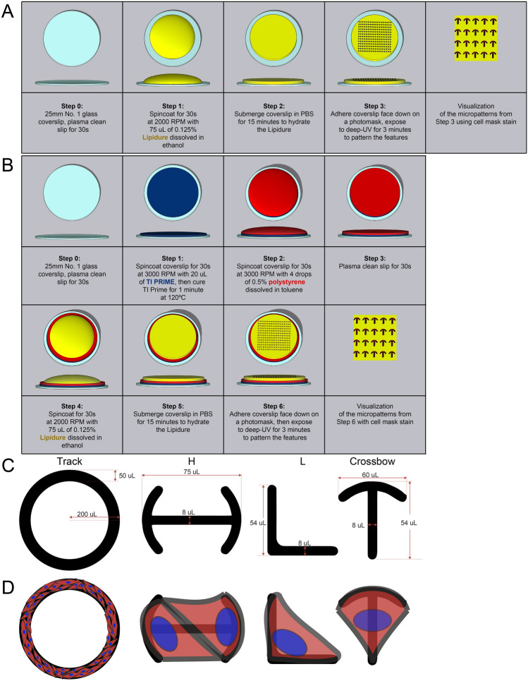
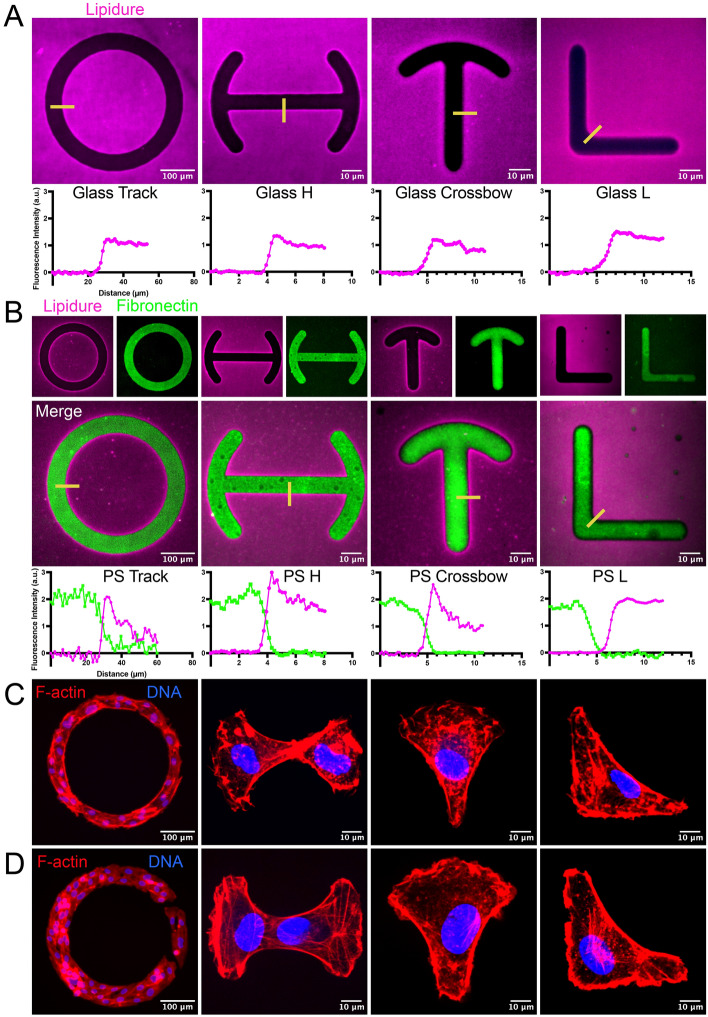
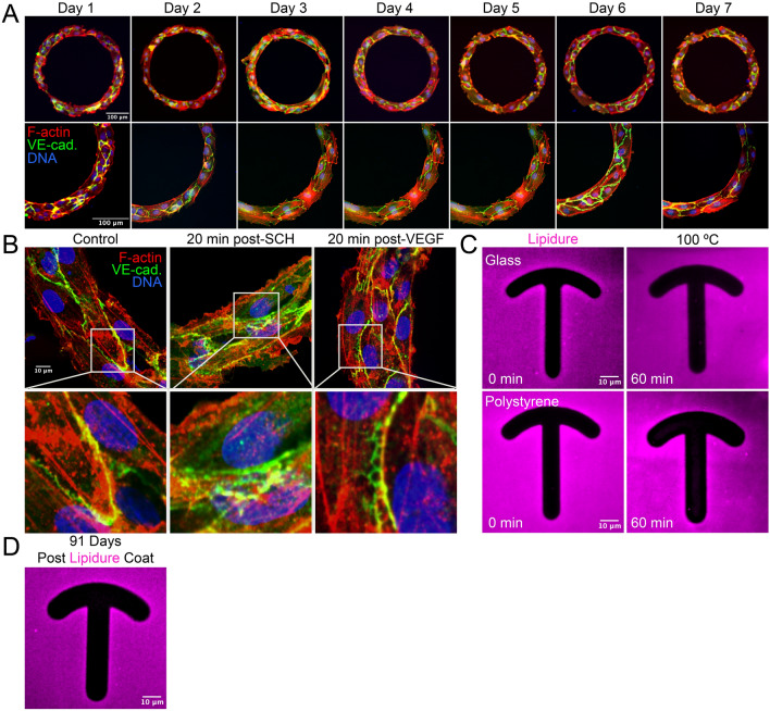
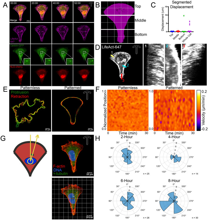
Cell autonomous behaviors such as migration and orchestration of cell polarity programs are required for physiological tissue formation. Micropatterns are cell-adhesive shapes that confine cell(s) to a user defined geometry. This biophysical confinement allows researchers to standardize the cell shape, and in doing so, stereotype organelle and cytoskeletal systems that can have an arbitrary organization. Thus, micropatterning can be a powerful tool in interrogation of polarity programs by enforcing a homogenous cell shape and cytoskeletal organization. A major drawback of this approach is the equipment and reagent costs associated with fabrication. Here, we provide a characterization of a compound called Lipidure (2-Methacryloyloxy ethyl phosphorylcholine) that is up to 40X less expensive than other cell repulsive coating agents. We found that Lipidure is an effective cell-repulsive agent for photolithography-based micropattern fabrication. Our results demonstrate that Lipidure is sensitive to deep UV irradiation for photolithography masking, stable in both benchtop and aqueous environments, non-toxic in prolonged culture, and effective at constraining cell geometry for quantification of cytoskeletal systems.


Conflict of interest statement

The authors declare no competing interests.

Figures

Figure 1
Figure 1
Overview of micropatterning surface preparation and masking protocol. (A) Schematic of adhering Lipidure to a glass coverslip and UV-based micropatterning. (B) Schematic of adhering polystyrene and Lipidure to a glass coverslip. Coated coverslips were subjected to masking and UV exposure to create micropattern features. (C) Diagram of micropattern features with dimensions in µm. (D) Predicted cell orientation on indicated micropatterns.
Figure 2
Figure 2
Lipidure is sensitive to deep-UV exposure and capable of micropatterning endothelial cells. (A) Representative images of coverslip coated with 0.125% Lipidure after masking/ultraviolet (UV) exposure and then stained with Cell Mask™ dye to highlight the UV-voided areas. Line scans correspond to yellow lines in top panels. (B) Representative images of coverslips coated with polystyrene and then coated with 0.125% Lipidure. After masking and UV exposure, fluorescent-dye conjugated fibronectin was grafted and then Lipidure was stained with Cell Mask dye. Line scan graphs correspond to yellow lines in top panels. (C) Human umbilical vein endothelial cells (ECs) adhered to glass-only micropatterns, stained for actin and DNA as indicated. (D) ECs adhered to polystyrene and fibronectin-coated micropatterns. ECs were stained for actin and DNA as indicated.
Figure 3
Figure 3
Lipidure is stable in prolong cell culture. (A) Representative images of Human umbilical vein endothelial cells (ECs) plated on ‘track’ micropatterns cultured between 1 and 7 days. Bottom panels are areas of higher magnification. ECs were stained as indicated in bottom panels. (B) Representative images of ‘track’ micropatterns treated with DMSO (control), ERK inhibitor SCH772984 (10 μM) or vascular-endothelial growth factor (VEGF, 20 ng/μl) 165 ligand for 20 min (min). Thereafter, ECs were stained for indicated proteins. Bottom panels indicate areas of higher magnification.
Figure 4
Figure 4
Using Lipidure-based micropatterns to stereotype endothelial cell geometry. (A) Representative live-cell imaging of a Human umbilical vein endothelial cell (EC) plated on a ‘crossbow’ micropattern expressing LifeAct-TagRFP647, Centrin-GFP, and NLSmCherry. (B) Binary image of micropatterned cell trisected into labeled regions. (C) Quantification of membrane displacement of trisected regions noted in panel B or whole cell analysis. (D) Representative cell and associated kymographs of actin (LifeAct) dynamics over time. (E) Non-micropatterned (patternless) and micropatterned EC with protrusive (green) and retractive (red) cell membrane movements marked over time as analyzed by APAPT software. (F) Heatmap derived from cells in panel E for visualization of cell membrane velocity over time. (G) Diagram and example of nuclear-centrosome axis angle quantification. ECs on crossbow micropatterns were aligned with the bow portion up and a central vertical line in the middle of the cell. Nuclear-centrosome axis was determined by drawing a line originating from the centroid of the nucleus passing through the centrosome cluster. (H) Compass plots of nuclear-centrosome axis over various plating times.

References

    1. Francis CR, Kushner EJ. Capturing membrane trafficking events during 3D angiogenic development in vitro. Microcirculation. 2021;29:e12726. doi: 10.1111/micc.12726. - DOI - PMC - PubMed
    1. Thery M, et al. Anisotropy of cell adhesive microenvironment governs cell internal organization and orientation of polarity. Proc. Natl. Acad. Sci. U. S. A. 2006;103(52):19771–19776. doi: 10.1073/pnas.0609267103. - DOI - PMC - PubMed
    1. Théry M. Micropatterning as a tool to decipher cell morphogenesis and functions. J. Cell Sci. 2010;123(24):4201–4213. doi: 10.1242/jcs.075150. - DOI - PubMed
    1. Azioune A, et al. Simple and rapid process for single cell micro-patterning. Lab Chip. 2009;9(11):1640–1642. doi: 10.1039/b821581m. - DOI - PubMed
    1. Thery M, Piel M. Adhesive micropatterns for cells: a microcontact printing protocol. Cold Spring Harb Protoc. 2009;2009(7):p.pdb.prot5255. doi: 10.1101/pdb.prot5255. - DOI - PubMed