. 2023 Nov 22;24(1):702.
doi: 10.1186/s12864-023-09804-5.

Exploring the molecular makeup of support cells in insect camera eyes

Affiliations

Exploring the molecular makeup of support cells in insect camera eyes

Shubham Rathore et al. BMC Genomics. .

Abstract

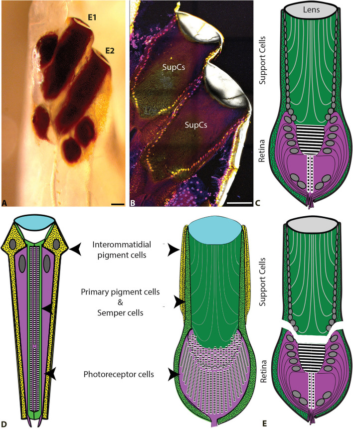
Animals typically have either compound eyes, or camera-type eyes, both of which have evolved repeatedly in the animal kingdom. Both eye types include two important kinds of cells: photoreceptor cells, which can be excited by light, and non-neuronal support cells (SupCs), which provide essential support to photoreceptors. At the molecular level deeply conserved genes that relate to the differentiation of photoreceptor cells have fueled a discussion on whether or not a shared evolutionary origin might be considered for this cell type. In contrast, only a handful of studies, primarily on the compound eyes of Drosophila melanogaster, have demonstrated molecular similarities in SupCs. D. melanogaster SupCs (Semper cells and primary pigment cells) are specialized eye glia that share several molecular similarities with certain vertebrate eye glia, including Müller glia. This led us to question if there could be conserved molecular signatures of SupCs, even in functionally different eyes such as the image-forming larval camera eyes of the sunburst diving beetle Thermonectus marmoratus. To investigate this possibility, we used an in-depth comparative whole-tissue transcriptomics approach. Specifically, we dissected the larval principal camera eyes into SupC- and retina-containing regions and generated the respective transcriptomes. Our analysis revealed several common features of SupCs including enrichment of genes that are important for glial function (e.g. gap junction proteins such as innexin 3), glycogen production (glycogenin), and energy metabolism (glutamine synthetase 1 and 2). To evaluate similarities, we compared our transcriptomes with those of fly (Semper cells) and vertebrate (Müller glia) eye glia as well as respective retinas. T. marmoratus SupCs were found to have distinct genetic overlap with both fly and vertebrate eye glia. These results suggest that T. marmoratus SupCs are a form of glia, and like photoreceptors, may be deeply conserved.

Keywords: Eye evolution; Gene regulatory network; Glia; Insects; Support cells; Transcriptomics.


Conflict of interest statement

The authors declare no competing interests.

Figures

Fig. 1
Fig. 1
T. marmoratus larvae have two high-resolution image-forming principal camera eyes on each side of the head (E1 and E2). A Both E1 and E2 are highly pigmented and tubular in shape, as illustrated by a freshly emerged larva in which the head cuticle is still transparent, scale bar = 100 µm. B DAPI staining highlights the nuclei of the support cells (SupCs) that form the distal region of the eye tubes, scale bar = 100 µm. C Schematic of a principal eye, illustrating its division into distally situated SupCs (green) and a proximal tiered retina (purple). D These camera eyes and D. melanogaster compound eyes share similar developmental plans [29]. Based on their organization, it has been hypothesized that the outer SupCs in T. marmoratus camera eyes are related to D. melanogaster interommatidial pigment cells (yellow) and the inner SupCs to D. melanogaster primary pigment and Semper cells (green). E As SupCs and photoreceptor cells are anatomically distinct, they can be dissected into separate regions for tissue-specific transcriptomics
Fig. 2
Fig. 2
Validation of SupC- and retina-specific transcriptomes. A and B Treemaps illustrating gene ontology (GO) terms for biological processes. A SupCs are enriched in genes from three major functional categories with multiple subclasses: anatomical structure development, tube size regulation, and cytosolic initiation complex formation. Additional categories include (1) cell developmental processes, (2) multicellular organismal processes, (3) molting, (4) response to external stimuli, (5) small molecule metabolic processes, (6) cellular localization, (7) carbohydrate metabolic processes, (8) cellular processes, and (9) oxoacid metabolic processes. B The functional categories with multiple subclasses in the retina are transport regulation, export from cells, response to light stimulus, neuron system processes, cell–cell signaling, and cell junction organization. Other categories in this tissue include (1) cell communication, (2) cell localization, (3) cell signaling, (4) response to stimuli, (5) cellular lipid metabolic processes, (6) cell processes, (7) cell locomotion, (8)ell homeostatic processes, (9) rhodopsin metabolic processes, (10) multicellular organismal process, (11) biological regulation, (12) circadian rhythm, (13) rhythmic processes, (14) lipid metabolic process, (15) developmental processes and (16) organic hydroxy compound metabolic process
Fig. 3
Fig. 3
Heat maps represent gene expression, where darker colors indicate higher expression levels. A As expected for this cell class, SupC transcriptomes are enriched in key lens (Ln) proteins (Ln 1–3, 5, 7, 9, and 10), with only one of six Ln 7 contigs being enriched in the retina transcriptomes. B As expected, the retina transcriptomes are enriched in genes related to phototransduction and reception. These genes include arrestins 1 (arr1 and 2 (arr2), retinal degeneration enzymes a (rdgA) and b (rdgB), neither inactivation nor afterpotential (ninaA, ninaB, and ninaC), and opsins rh3, 4, and 6. In contrast, nonvisual rh7 is enriched in the SupCs
Fig. 4
Fig. 4
Expression of important transcription factors in the principal camera eyes of T. marmoratus third instars. A Relative expression of transcription factors cut (ct) and bar homolog2 (bh2) shows a tendency towards but no significant enrichment in the SupCs. Conversely, prospero (pros) shows a tendency towards but no significant enrichment in the retina. In contrast, transcription factors eyes absent (eya) and sine oculis (so) as well as the sine oculis binding protein (sobp) are significantly enriched in the SupCs. B A Cut antibody (green), which is known to mark Semper cells in the compound eyes of T. marmoratus adults stains a subset of SupCs (cyan, arrow) in a section that is counter-stained with DAPI (blue) [8], stained a specific subset of proximally placed SupCs (teal, arrows), scale bar = 100 µm. See SupFig1 for separate channels of the staining. C As illustrated by the schematic, the staining pattern supports the deep conservation of this transcription factor and is consistent with our model, in which a portion of the SupCs in T. marmoratus larval eye tubes (green) are homologous to D. melanogaster Semper cells
Fig. 5
Fig. 5
Homeostasis-related glia-typical functions in T. marmoratus camera eyes. A Insect glia-typical genes such as stretchin-mick (strn-Mlck), myogialnin (myo), axotactin (axo), and tramtrack (ttk) are enriched in the SupCs, but the general insect glia marker reversed polarity (repo) is enriched in the retina. B The SupCs are enriched in immune response genes, including gram-negative bacteria binding protein 3 (gnbp3), limpet (lmpt), immune deficiency (imd), Toll-like receptor (tollo), defense repressor 1 (dnr1), and dorsal (dl). C SupC-enriched genes associated with blood–brain barrier (BBB) formation include pasiflora 2 (pasi2), fasciclin 3 isoform B (fas3), sinuous (sinu), and kune. D and E Genes required for potassium transport (inwardly rectifying potassium channel 2 (irk2) and acid-sensitive potassium channel 7 (task7)), sodium symport (rumpel/CG9657), chloride transport (chloride channels (clic and clc-c) and bestrophin (bes2)), amine transport (pathetic (path)), osmoregulation (serotonin receptor (5-ht2a), vacuolar H + ATPase (vha100-2), and osmotic stress response related gene inebriated (ine)) are enriched in the SupCs. The expression of aquaporin genes such as aqp, eglp4, prip, drip, and bib is not significantly different in the SupCs and retina
Fig. 6
Fig. 6
Metabolic and structurally related glia-typical support functions in T. marmoratus camera eyes. A Genes associated with the pentose phosphate pathway (PPP), including zwischenferment (zwi), phosphogluconate dehyrogenase (pgd), and glycogen binding subunit 76A (gbs76A), and with glycogenesis, including 1,4-alpha-glucan branching enzyme (abge), glycogenin (gyg) isoforms I and B, and ATP citrate lyase (atpcl). For glucose homeostasis, we found glucose transmembrane transporter pippin and three isoforms of glucose transporter 1 (glut1); isoforms S and P are enriched in the retina region, whereas isoform W is enriched in the SupCs. B A glutamate receptor and an associated protein (eye-enriched kainate receptor (Ekar)) as well as CG11155 are enriched in the retina, whereas kainate-type ionotropic glutamate receptor subunit 1D (Kair1D) and a glutamate receptor activator clumsy are enriched in the SupCs. Glutamine synthetase enzymes gs1 and gs2 are also enriched in the SupCs. C SupCs are enriched in several genes related to fatty acid metabolism including CG31683, CG31522, CG4020, CG6847, CG1441, waterproof (wat), desaturase 2 (desat2), saccherophin dehydrogenase 2 (sccpdh2), and fatty acid transporter protein 2 (fatp2). Structural support mediated by cell adhesion molecules is another important glial function. We found SupC enrichment of the following cell adhesion molecules: innexin 3 (inx3), vinculin (vin), unzipped (uzip), smallish (smash), and fat (ft)
Fig. 7
Fig. 7
Four-way analysis to identify genes that overlap between the specific eye tissues of T. marmoratus (beetle) and those of fly (D. melanogaster), fish (D. rerio), and mouse (M. musculus) [17, 51]. A Comparison of T. marmoratus SupCs with D. melanogaster Semper cells and mouse and zebrafish Müller glia, revealing six common genes. B The names and putative functions of overlapping genes are based on Flybase [53]. C Comparison of the T. marmoratus retina with D. melanogaster photoreceptors cells and mouse and zebrafish retinal neurons, revealing seven common genes. D The names and putative functions of these genes are based on Flybase

Update of

Similar articles

Cited by

References

    1. Meece M, Rathore S, Buschbeck EK. Stark trade-offs and elegant solutions in arthropod visual systems. J Exp Biol. 2021;224(Pt 4):jeb215541. doi: 10.1242/jeb.215541. - DOI - PubMed
    1. Land MF, Nilsson D-E. Animal Eyes. USA: Oxford University Press; 2012.
    1. Nilsson D-E. The Diversity of Eyes and Vision. Annu Rev Vis Sci. 2021;7:19–41. doi: 10.1146/annurev-vision-121820-074736. - DOI - PubMed
    1. Gehring WJ. The genetic control of eye development and its implications for the evolution of the various eye-types. Zoology. 2001;104:171–183. doi: 10.1078/0944-2006-00022. - DOI - PubMed
    1. Arendt D. Evolution of eyes and photoreceptor cell types. Int J Dev Biol. 2003;47:563–571. - PubMed

LinkOut - more resources