. 2023 Nov 22;23(1):1136.
doi: 10.1186/s12885-023-11631-6.

Lactate receptor GPR81 drives breast cancer growth and invasiveness through regulation of ECM properties and Notch ligand DLL4

Affiliations

Lactate receptor GPR81 drives breast cancer growth and invasiveness through regulation of ECM properties and Notch ligand DLL4

Kathrine Lundø et al. BMC Cancer. .

Abstract

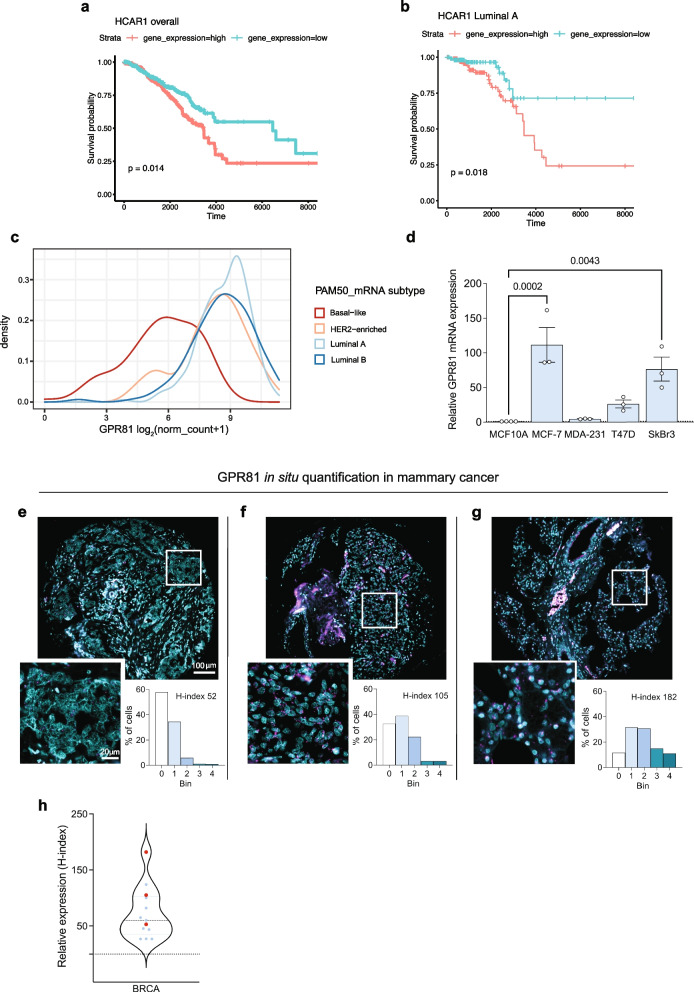
Background: The lactate receptor GPR81 contributes to cancer development through unclear mechanisms. Here, we investigate the roles of GPR81 in three-dimensional (3D) and in vivo growth of breast cancer cells and study the molecular mechanisms involved.

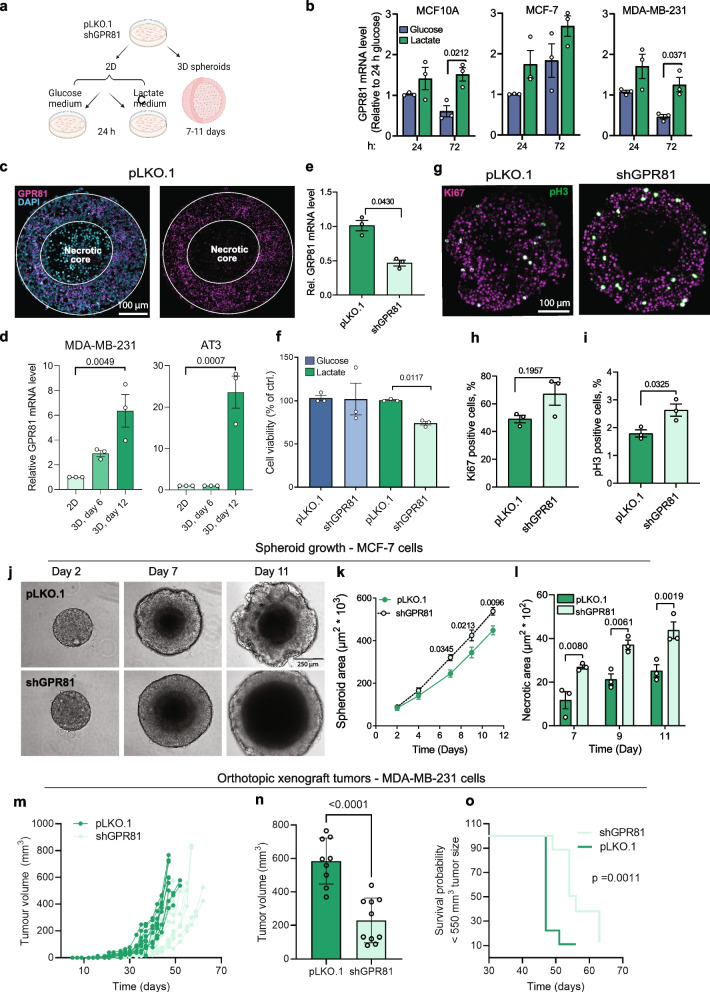
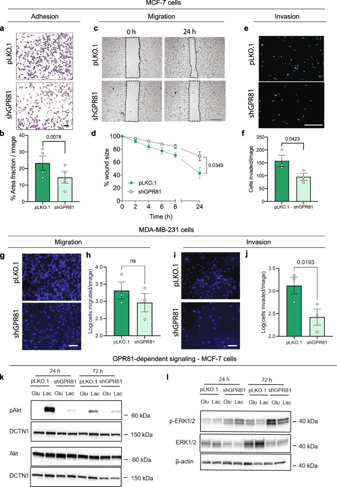
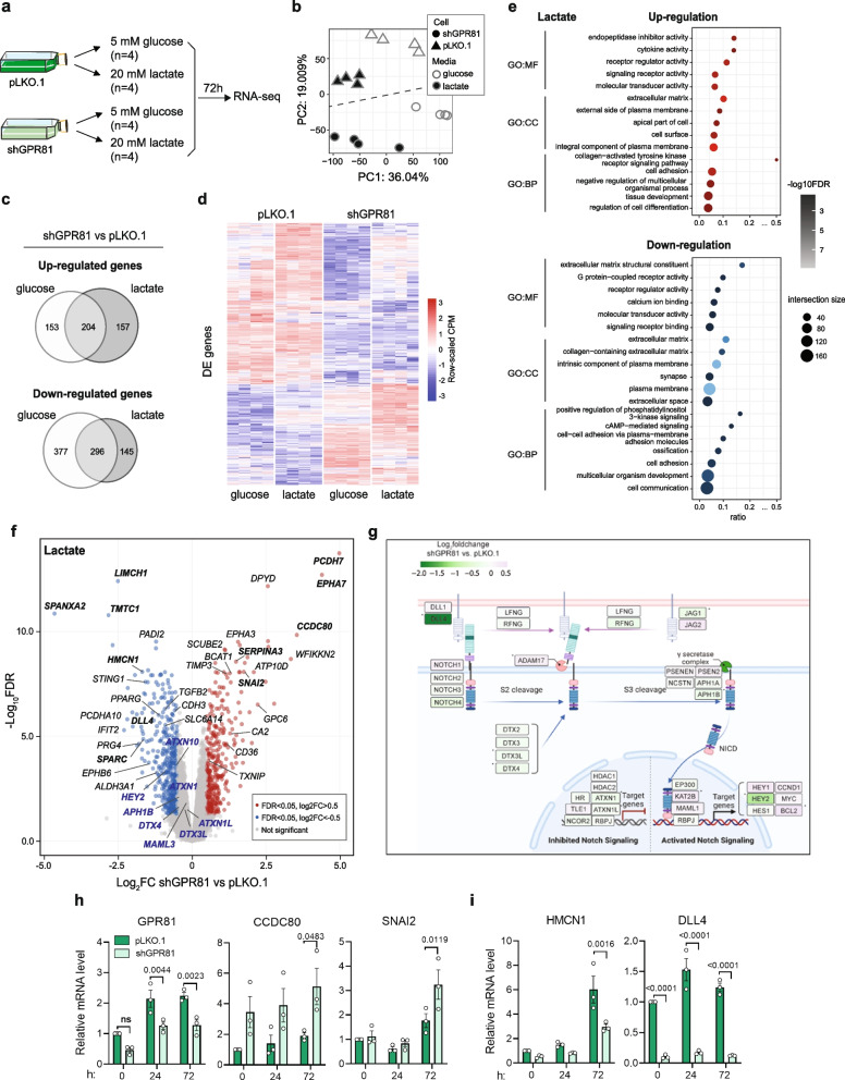
Methods: GPR81 was stably knocked down (KD) in MCF-7 human breast cancer cells which were subjected to RNA-seq analysis, 3D growth, in situ- and immunofluorescence analyses, and cell viability- and motility assays, combined with KD of key GPR81-regulated genes. Key findings were additionally studied in other breast cancer cell lines and in mammary epithelial cells.

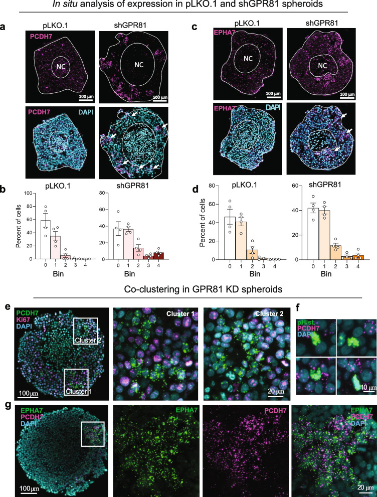
Results: GPR81 was upregulated in multiple human cancer types and further upregulated by extracellular lactate and 3D growth in breast cancer spheroids. GPR81 KD increased spheroid necrosis, reduced invasion and in vivo tumor growth, and altered expression of genes related to GO/KEGG terms extracellular matrix, cell adhesion, and Notch signaling. Single cell in situ analysis of MCF-7 cells revealed that several GPR81-regulated genes were upregulated in the same cell clusters. Notch signaling, particularly the Notch ligand Delta-like-4 (DLL4), was strikingly downregulated upon GPR81 KD, and DLL4 KD elicited spheroid necrosis and inhibited invasion in a manner similar to GPR81 KD.

Conclusions: GPR81 supports breast cancer aggressiveness, and in MCF-7 cells, this occurs at least in part via DLL4. Our findings reveal a new GPR81-driven mechanism in breast cancer and substantiate GPR81 as a promising treatment target.

Keywords: EPHA7; Extracellular matrix; HCAR1; Metabolite GPCR; Notch; PCDH7; Spheroid; Tumor microenvironment.


Conflict of interest statement

MT, TWS and SFP are co-founders of SOLID Therapeutics. All other authors declare no conflicts of interest.

Figures

Fig. 1
Fig. 1
GPR81 mRNA expression is upregulated in tumor tissues and cell lines. a-b Kaplan–Meier analysis of patient overall survival as a function of GPR81 (HCAR1) expression. a All 1194 breast cancer patient data sets in TCGA. b Luminal A subtype (222 patients) after stratification by PAM50 subtype. The corresponding data for Luminal B, Basal and HER2 enriched subtypes are found in Suppl. Figure 1. c Density plot shows GPR81 (HCAR1) expression in breast cancer patients of PAM50 subtypes from TCGA data. Luminal A subtype breast cancer tissue has the highest, and triple-negative breast cancer (TNBC) tissue has the lowest GPR81 expression. d qPCR analysis of relative GPR81 mRNA expression in human normal mammary epithelial cells (MCF10) and human mammary cancer cell lines (n = 3 independent replicates per cell line, indicated by dots). eg In situ RNAscope analysis of GPR81 expression, quantified as the H-index, a measure of GPR81 expression and heterogeneity. The figure shows examples and corresponding bin quantifications for three breast cancer cores with low, medium and high overall GPR81 H-index. The H-index was calculated by totalling % cells in each bin, according to a weighted equation where bin 0 corresponds to 0, bin 1 to 1, etc., after grouping of cells into 5 bins (groups) based on the number of dots per cell. Each sample was evaluated for the % cells in each bin (see Materials and Methods). The Y-axis shows the % of cells with a given bin distribution. h All RNAscope analyses of GPR81 expression in human patient breast cancer tissue. Each dot corresponds to a biopsy from single patient, with the red dots corresponding to the analyses shown in panel eg. In Suppl. Figure 1 h, these data are compared with similar analyses from other tumor types to illustrate the heterogeneity of GPR81 expression
Fig. 2
Fig. 2
GPR81 is upregulated by extracellular lactate and by 3D spheroid growth and GPR81 KD inhibits 2D growth and increases 3D necrosis of breast cancer cells. a Experiment overview. b qPCR analysis of relative GPR81 mRNA levels in 2D cultures of breast epithelial (MCF10A) and breast cancer cells (MDA-MB-231, MCF-7) cultured for 24 and 72 h with glucose (5 mM glucose/0 mM lactate) or lactate (20 mM lactate/0 mM glucose) medium. n = 3 independent replicates per cell line, two-way-ANOVA with Sidak Post Hoc test. c Representative in situ analyses of GPR81 expression in MCF-7 control (pLKO.1) and GPR81 knockdown (shGPR81) spheroids. n = 3. d qPCR analysis of GPR81 mRNA levels in 2D vs day 6 and 12 3D cultures of MDA-MB-231 and AT3 cells (n = 3, paired, two-sided Student’s t-test). e Relative GPR81 expression in MCF-7 control (pLKO.1) and GPR81 KD (shGPR81) cells, n = 3, paired, two-sided Students t-test. f Viability of MCF-7 GPR81 KD cells. Cells were incubated in glucose or lactate medium for 72 h before assessing cell viability. Data is presented as % cell viability compared to control (pLKO.1) (n = 3, paired, two-sided Students t-test). g-i Representative images (g) and corresponding quantification (h-i) of MCF-7 pLKO.1 and GPR81 KD (shGPR81) spheroids stained for Ki67 and phospho-Histone H3 (pH3). Graphs show % live cell positive for Ki67 (H) or pH3 (I). (n = 3, paired, two-sided Student’s t-test). j Spheroid growth of MCF-7 cells. Representative brightfield images taken on day 2, 7 and 11. Scalebar: 250 µm. k Spheroid area. l Necrotic core area (µm2 * 103) relative to spheroid size on day 7, 9 and 11. (n = 5). Two-way ANOVA with Tukey post-test. mo Orthotopic xenograft model using MDA-MB-231 GPR81 KD cells. Eight-week-old female NOD.Cg-Prkdcg Il2rgtm1Wjl/SzJ mice were inoculated with 0.25 × 106 pLKO.1 or shGPR81 cells (n = 10 per condition). Tumor volume was evaluated three times per week after cell inoculation. m Individual tumor growth curves for pLKO.1 and shGPR81 xenografts. n Tumor volume (mm3) on day 47. o Kaplan–Meier overall survival of mice bearing pLKO.1 or shGPR81 xenografts. Mice were sacrificed when they reached a tumor volume of 550–600 mm3 or had tumor ulcers. Log-rank statistics was used to compare statistical significances between groups (p = 0.0014)
Fig. 3
Fig. 3
GPR81 stimulates breast cancer cell adhesion, migration, invasion, and Akt activity. a-b Adhesion of MCF-7 cells to matrigel is reduced by GPR81 KD. Cells were incubated for 24 h in lactate medium prior to being seeded on matrigel. After 1 h, nonadherent cells were washed of, and cells were fixed and stained. a Representative images (n = 4). b %Area-fraction/image, i.e. the relative area covered by cells. c-d Migration of MCF-7 cells is reduced by GPR81 KD. c Cells were seeded in lactate medium in Ibidi insert plates and treated with aphidicolin to block proliferation. Inserts were removed after 24 h, and images were acquired at the time points shown (n = 3). Scalebar: 500 µm. d Average wound size as % of size at t=0, normalized to pLKO.1 cells. ef Invasion of MCF-7 cells through matrigel is reduced by GPR81 KD. Cells incubated in lactate medium for 24 h were seeded in the upper chamber of matrigel-coated Boyden chambers. The lower chamber contained the same medium plus 10% FBS. After 24 h, membranes were processed for imaging. e Representative images (n = 3). Scalebar: 500 µm. f Cells invaded/image. g-j Boyden chamber analysis of migration (g-h) and invasion (i-j) of MDA-MB-231 pLKO.1 and shGPR81 cells. g,i Representative images of migration (g) and invasion (i), h,j Migrated/invaded cells per image. Scale bars: 100 µm. k-l Representative Western Blots of active, Ser473-phosphorylated Akt (p-Akt) and total Akt (k), and active, Thr202/Tyr204-phosphorylated ERK1/2 and total ERK1/2 (l) in pLKO.1 and shGPR81 MCF-7 cells, following incubation in glucose- or lactate medium for 24 or 72 h. DCTN1 and β-actin serve as a loading controls. Statistics (b, d, f, h, j): Paired, two-sided Students t-test
Fig. 4
Fig. 4
GPR81 KD alters expression of genes regulating cell adhesion and ECM organization. a Experimental setup. b Principal component (PC) analysis of RNA-seq data. PCs 1 and 2 are shown as X- and Y-axes with % explained variance indicated. Triangles and dots represent RNA-seq libraries, coloured by condition. The dotted line separates shGPR81 and pLKO.1 libraries. c Venn diagrams showing overlap of significantly differentially expressed (DE) genes (FDR < 0.05 and abs logfold change) > 0.5, by limma analysis) following shGPR81 and pLKO.1 treatment in lactate and glucose conditions. d Heatmap of pLKO.1 and shGPR81 cell gene expression after incubation in glucose- or lactate conditions as indicated. Rows correspond to DE genes. Columns correspond to RNA-seq libraries, grouped first by cell type (pLKO.1 or shGPR81) and then by condition (glucose or lactate). Colours correspond to normalized RNA-seq read counts (CPM) that were subsequently row-scaled. e GO analysis of DE genes in the lactate condition, split by up- (upper panel) and down-regulated genes (lower panel). Colour intensity indicates over-representation significance (-log10-scaled FDR), while dot size indicates intersection size (number of DE genes with a given GO term). GO terms are ordered by class: molecular function (MF), cell compartment (CC) and biological process (BP). f Volcano plot based on RNA-seq data analysis of GPR81 KD in lactate environment. Dots correspond to genes, coloured by DE status as defined above. Genes of particular interest are labelled by name. Gene names in bold are discussed specifically in main text. Gene names in blue are Notch pathway genes with FDR < 0.05 but absolute logfold change < 0.5, by limma analysis. See text for details. g Expression changes of expressed genes involved in the Notch signaling pathway, color-coded by log2foldchange obtained from differential expression analysis between shGPR81 and pLKO.1 samples in lactate condition. Included genes based on KEGG map04330 [46]. Asterisk indicates FDR < 0.05. h-i qPCR validation of selected upregulated (h) and downregulated (i) DE genes, shown as mean ± SEM of relative mRNA level normalized to pLKO.1 per time point. Statistics: Two-way ANOVA
Fig. 5
Fig. 5
PCDH7 and EPHA7 exhibit clustered upregulation in spheroids upon GPR81 KD. a-d In situ hybridization of PCDH7 and EPHA7 in pLKO.1 and shGPR81 MCF-7 spheroids. Representative images and corresponding quantification of in situ hybridization of PCDH7 (a, b) and EPHA7 (l, m) in MCF-7 pLKO.1 and shGPR81 spheroids. b,d Quantification of the in situ signal. The Y-axis shows % cells with a given bin distribution (for a detailed description, see Materials and Methods). e-g High resolution in situ analysis of GPR81 KD (shGPR81) spheroids, detected by RNAscope.. In e and g, the middle and right panels represent higher magnifications of the boxed regions, illustrating PCDH7-positive cell clusters. e Ki67 positive cells (magenta) very rarely exhibit co-expression with PCDH7 (green). f Examples illustrating that p-H3 positive cells (green) do not stain for PCDH7 (magenta). g PCDH7 (magenta) co-localizes in cell clusters with EPHA7. Representative of 3 biological n per condition
Fig. 6
Fig. 6
DLL4, but not PCDH7 and EPHA7, contribute to the GPR81-mediated changes in cell survival, migration and invasion. a Representative Western blots of PCDH7 and EPHA7 protein levels in pLKO.1 and shGPR81 MCF-7 cells grown in glucose or lactate medium for the time indicated. α-tubulin serves as a loading control for PCDH7 and β-actin for EPHA7. b Immunofluorescence analysis of PCDH7 in pLKO.1 and shGPR81 MCF-7 cells grown in lactate medium for 24 h. Scalebar: 20 µm. c-e Spheroid growth of GPR81 KD MCF-7 cells with/without siRNA-mediated KD of PCDH7 or EPHA7. c Representative images, day 7 (n = 3). Scalebar: 200 µm. d Spheroid area (µm2 * 103). e Percent necrotic core relative to spheroid size, day 7. f-g Migration of GPR81 KD MCF-7 cells in lactate medium with/without KD of PCDH7 or EPHA7. f Representative images, n = 3. Scalebar: 500 µm. g Percent wound remaining after 48 h, normalized to control. h Boyden chamber invasion assay, quantification, invaded cells/image (n = 3). shGPR81 PCDH7 and EPHA7 cells were cultured in lactate medium for 24 h, seeded on matrigel in lactate medium in the upper chamber, and allowed to invade 24 h toward lactate medium with 10% FBS in the lower chamber. i DLL4 protein level is decreased by GPR81 KD. pLKO.1 and shGPR81 MCF-7 cells were cultured in glucose or lactate medium for 24 h. Representative Western blot, DCTN1 serves as a loading control (n = 3). j qPCR validation of siRNA mediated DLL4 KD in MCF-7 cells. k-m DLL4 KD increases necrotic core size in 3D spheroids of MCF-7 cells. k Representative day 7 images (n = 3). Scalebar: 200 µm. l Average spheroid area (µm2 * 103). m Percent necrotic core relative to spheroid size on day 7. no Adhesion of MCF-7 cells to matrigel is reduced by DLL4 KD. Cells were incubated for 24 h in lactate medium and seeded on matrigel. After 1 h, nonadherent cells were washed of and cells fixed and stained. n Representative images (n = 4). o Percent area-fraction/image. p-r DLL4 KD inhibits migration and invasion. Boyden chamber assays of MCF-7 cells with or without siRNA mediated KD of DLL4 or SPARC. MCF-7 cells with/without DLL4 KD were cultured in lactate medium for 24 h, seeded on matrigel in lactate medium in the upper chamber, and allowed to invade 24 h toward lactate medium with 10% FBS in the lower chamber (n = 3). p Representative images (n = 3). q-r Cells migrated/image (q) and cells invaded/image (r)

Similar articles

Cited by

References

    1. Boedtkjer E, Pedersen SF. The acidic tumor microenvironment as a driver of cancer. Annu Rev Physiol. 2020;82:103–126. doi: 10.1146/annurev-physiol-021119-034627. - DOI - PubMed
    1. Liberti MV, Locasale JW. The Warburg effect: how does it benefit cancer cells? Trends Biochem Sci. 2016;41(3):211–218. doi: 10.1016/j.tibs.2015.12.001. - DOI - PMC - PubMed
    1. Elingaard-Larsen LOR, M.G.; Sorensen, E. E.; Pedersen, S. F. : How reciprocal interactions between the tumor microenvironment and ion transport proteins drive cancer progression. Reviews of Physiology, Biochemistry, and Pharmacology 2020, In press. - PubMed
    1. DeBerardinis RJ, Mancuso A, Daikhin E, Nissim I, Yudkoff M, Wehrli S, Thompson CB. Beyond aerobic glycolysis: transformed cells can engage in glutamine metabolism that exceeds the requirement for protein and nucleotide synthesis. Proc Natl Acad Sci U S A. 2007;104(49):19345–19350. doi: 10.1073/pnas.0709747104. - DOI - PMC - PubMed
    1. Wu Q, Li B, Li Z, Li J, Sun S, Sun S. Cancer-associated adipocytes: key players in breast cancer progression. J Hematol Oncol. 2019;12(1):95. doi: 10.1186/s13045-019-0778-6. - DOI - PMC - PubMed