. 2023 Nov 22;20(1):44.
doi: 10.1186/s12989-023-00556-4.

Polystyrene nanoplastic exposure induces excessive mitophagy by activating AMPK/ULK1 pathway in differentiated SH-SY5Y cells and dopaminergic neurons in vivo

Affiliations

Polystyrene nanoplastic exposure induces excessive mitophagy by activating AMPK/ULK1 pathway in differentiated SH-SY5Y cells and dopaminergic neurons in vivo

Yuji Huang et al. Part Fibre Toxicol. .

Abstract

Background: Microplastics and nanoplastics (MNPs) are emerging environmental contaminants detected in human samples, and have raised concerns regarding their potential risks to human health, particularly neurotoxicity. This study aimed to investigate the deleterious effects of polystyrene nanoplastics (PS-NPs, 50 nm) and understand their mechanisms in inducing Parkinson's disease (PD)-like neurodegeneration, along with exploring preventive strategies.

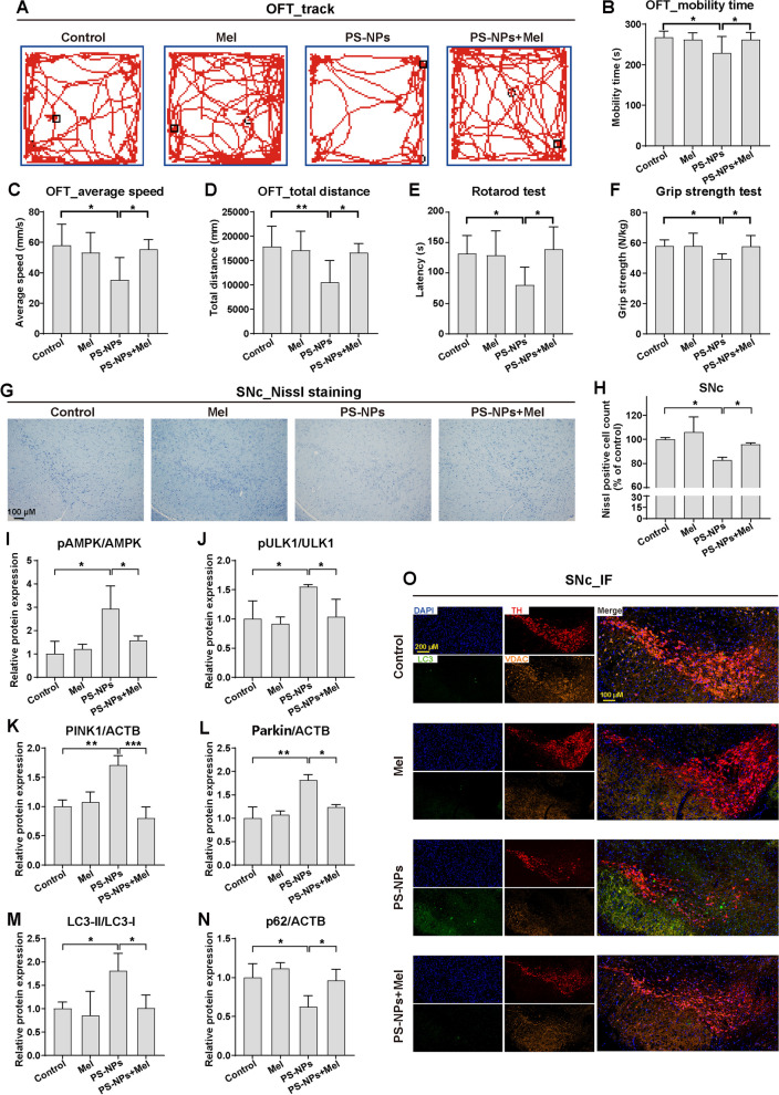
Methods: Following exposure to PS-NPs (0.5-500 μg/mL), we assessed cytotoxicity, mitochondrial integrity, ATP levels, and mitochondrial respiration in dopaminergic-differentiated SH-SY5Y cells. Molecular docking and dynamic simulations explored PS-NPs' interactions with mitochondrial complexes. We further probed mitophagy's pivotal role in PS-NP-induced mitochondrial damage and examined melatonin's ameliorative potential in vitro. We validated melatonin's intervention (intraperitoneal, 10 mg/kg/d) in C57BL/6 J mice exposed to 250 mg/kg/d of PS-NPs for 28 days.

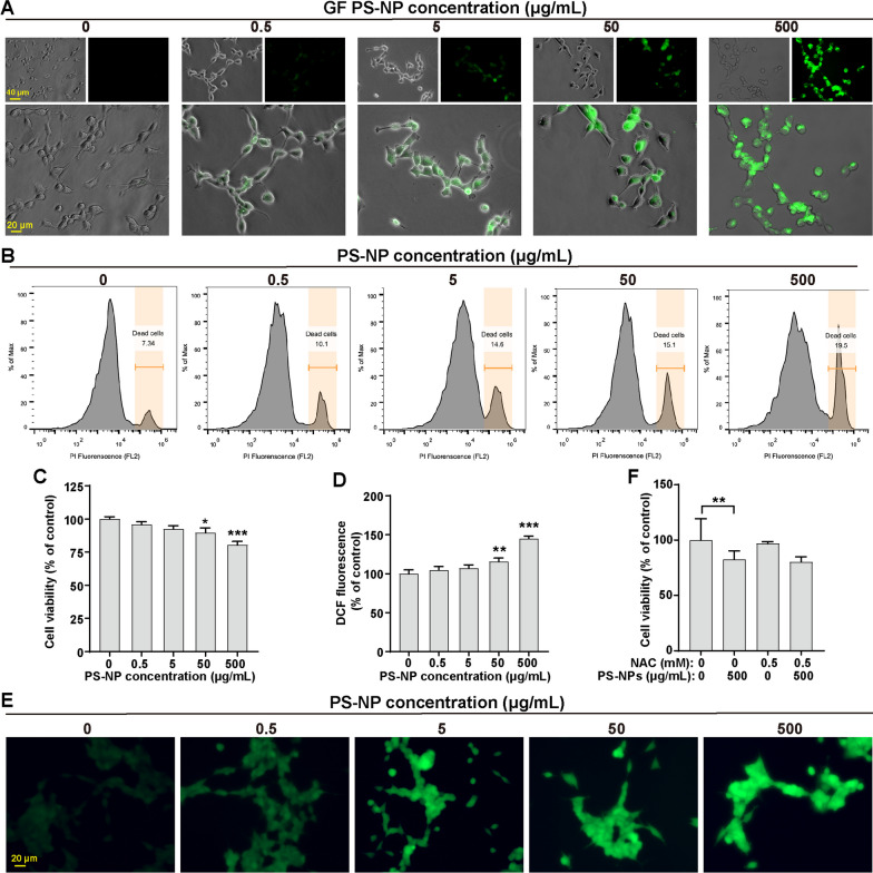
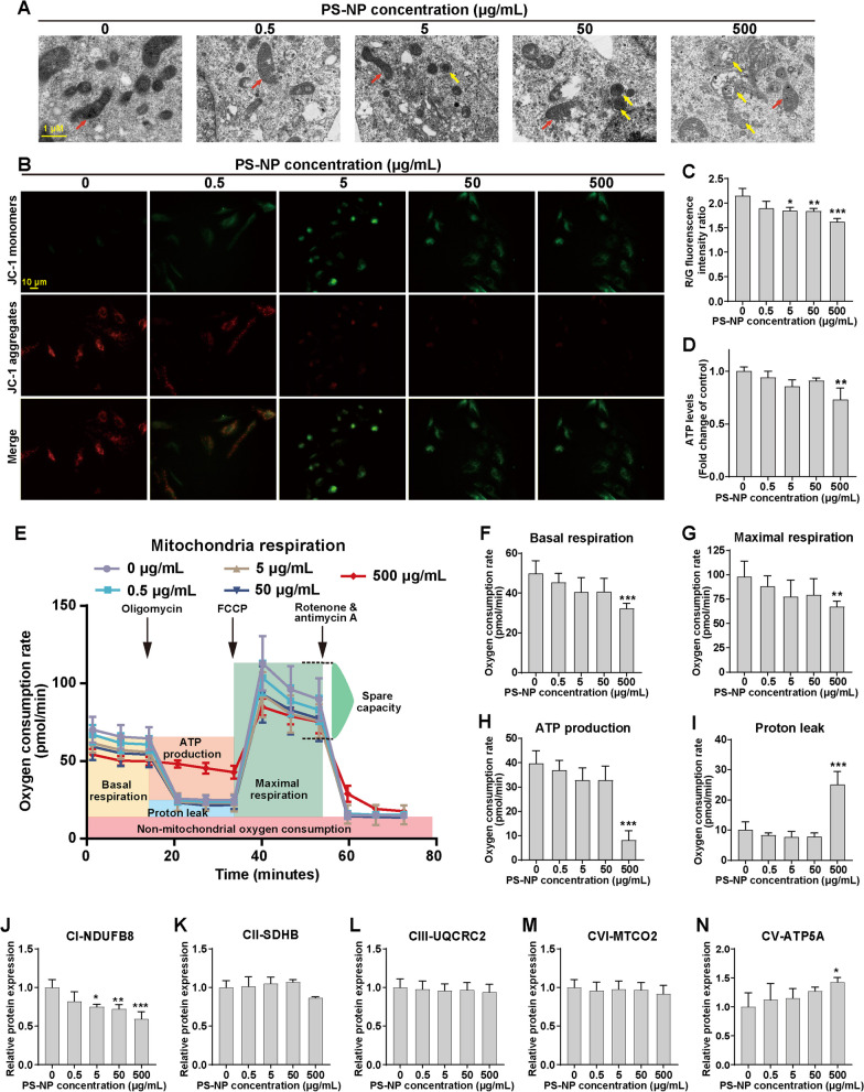
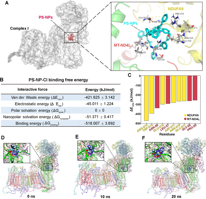
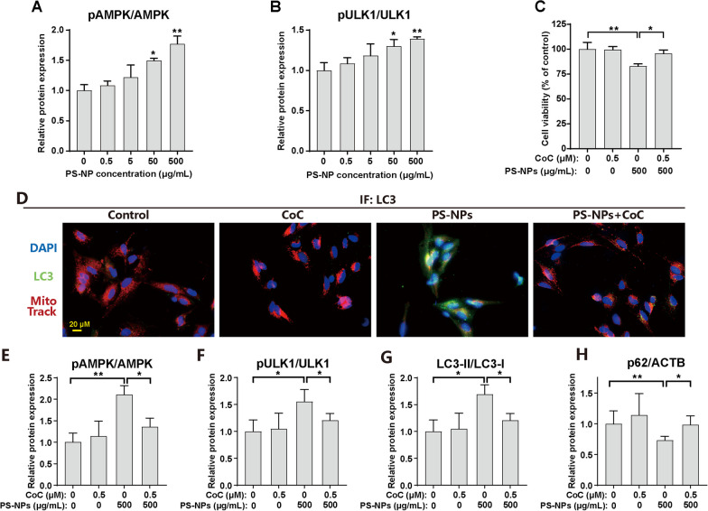
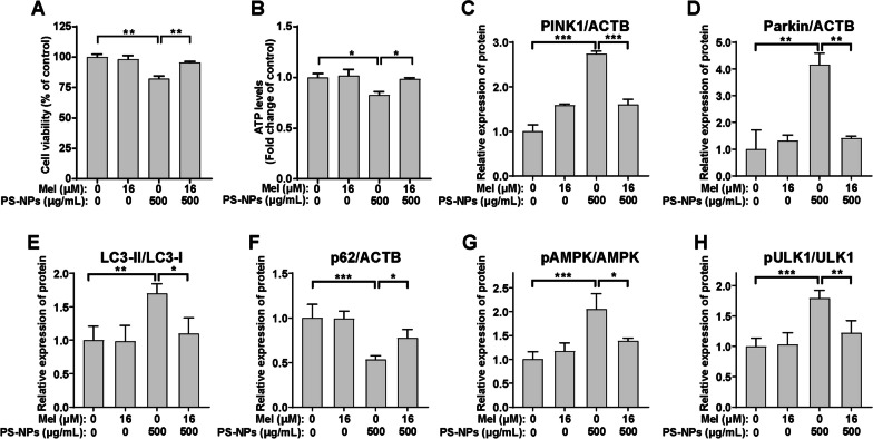
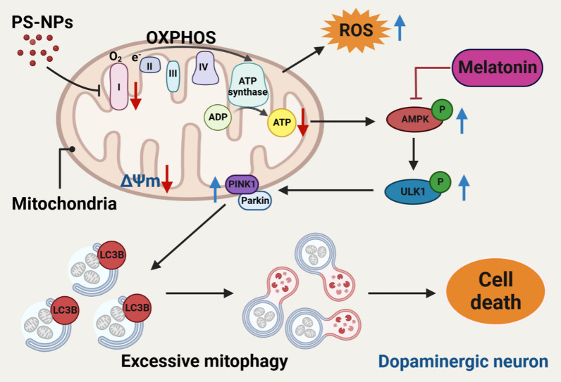
Results: In our in vitro experiments, we observed PS-NP accumulation in cells, including mitochondria, leading to cell toxicity and reduced viability. Notably, antioxidant treatment failed to fully rescue viability, suggesting reactive oxygen species (ROS)-independent cytotoxicity. PS-NPs caused significant mitochondrial damage, characterized by altered morphology, reduced mitochondrial membrane potential, and decreased ATP production. Subsequent investigations pointed to PS-NP-induced disruption of mitochondrial respiration, potentially through interference with complex I (CI), a concept supported by molecular docking studies highlighting the influence of PS-NPs on CI. Rescue experiments using an AMPK pathway inhibitor (compound C) and an autophagy inhibitor (3-methyladenine) revealed that excessive mitophagy was induced through AMPK/ULK1 pathway activation, worsening mitochondrial damage and subsequent cell death in differentiated SH-SY5Y cells. Notably, we identified melatonin as a potential protective agent, capable of alleviating PS-NP-induced mitochondrial dysfunction. Lastly, our in vivo experiments demonstrated that melatonin could mitigate dopaminergic neuron loss and motor impairments by restoring mitophagy regulation in mice.

Conclusions: Our study demonstrated that PS-NPs disrupt mitochondrial function by affecting CI, leading to excessive mitophagy through the AMPK/ULK1 pathway, causing dopaminergic neuron death. Melatonin can counteract PS-NP-induced mitochondrial dysfunction and motor impairments by regulating mitochondrial autophagy. These findings offer novel insights into the MNP-induced PD-like neurodegenerative mechanisms, and highlight melatonin's protective potential in mitigating the MNP's environmental risk.

Keywords: Complex I; Melatonin; Microplastics and nanoplastics; Mitochondrial dysfunction; Mitophagy; Neurotoxicity.


Conflict of interest statement

The authors declare no competing interests.

Figures

Fig. 1
Fig. 1
Cellular internalization and cytotoxicity of PS-NPs in differentiated SH-SY5Y cells. A GF PS-NPs distribution. B Flow cytometry analysis of PS-NP-induced cell death. C Cell viability after PS-NP exposure. D-E ROS levels post PS-NP exposure. F NAC effect on PS-NP-inhibited cell viability. Results are presented as mean ± SD (n = 3). *P < 0.05, **P < 0.01, ***P < 0.001, compared to the 0 μg/mL group
Fig. 2
Fig. 2
Impairment of mitochondria and mitochondrial stress by PS-NPs in differentiated SH-SY5Y cells. A Representative TEM images of cells after PS-NP exposure. Red arrows indicate mitochondria, and yellow arrows indicate mitophagosomes. Impact of PS-NPs on B, C ΔΨm and D cellular ATP levels. E Mitochondrial stress profiles. Relative changes in key parameters of mitochondrial function: F basal respiration, G maximal respiration, H mitochondrial ATP production, and I proton (H+) leakage. JN The quantification of protein expression levels of ETC CI-NDUFB8, CII-SDHB, CIII-UQCRC2, CVI-MTCO2, and CV-ATP5A. Results are presented as mean ± SD (n = 3). *P < 0.05, **P < 0.01, ***P < 0.001, compared to the 0 μg/mL group
Fig. 3
Fig. 3
Molecular docking and MD simulation showing interaction between PS-NPs and CI. A Molecular docking model illustrating the binding of PS-NPs to CI. Detailed interaction view (enlarged panel). B Binding free energy between PS-NPs and CI. C Van der Waals interactions between specific residues of CI and PS-NPs. D-F Conformational changes of the PS-NP- CI complex in MD simulation
Fig. 4
Fig. 4
PS-NP-induced aberrant mitophagy mediated by PINK1/Parkin in differentiated SH-SY5Y cells. A-D The quantification of mitophagy-related proteins, including LC3-II/LC3-I, p62, PINK1, and Parkin. E Representative IF images showing colocalization of LC3 (green) and Mito tracker (red). F-I Differentiated SH-SY5Y cells were exposed to 500 μg/mL PS-NPs with or without 3-MA (100 μM) for 48 h. The quantification of protein expression levels of (F) LC3-II/LC3-I and (G) p62. (H) Cell viability and (I) ATP levels. Results are presented as mean ± SD (n = 3). * P < 0.05, ** P < 0.01, *** P < 0.001, compared to the 0 μg/mL group or indicative group
Fig. 5
Fig. 5
Regulation of AMPK/ULK1 signaling pathway in PS-NP-activated mitophagy in differentiated SH-SY5Y cells. A, B The quantification of protein expression levels of the AMPK/ULK1 pathway, including phosphorylated and total AMPK and ULK1. C-H Differentiated SH-SY5Y cells were exposed to 500 μg/mL PS-NPs with or without CoC (0.5 μM) for 48 h. C Cell viability, and D representative IF images showing colocalization of LC3 (green) and Mito tracker (red). The quantification of protein expression levels of E pAMPK/AMPK, F pULK1/ULK1, G LC3-II/LC3-I, and H p62. Results are presented as mean ± SD (n = 3). * P < 0.05, ** P < 0.01, compared to the 0 μg/mL group or indicative group
Fig. 6
Fig. 6
Protective effects of melatonin on PS-NP-induced mitochondrial dysfunction and excessive mitophagy in differentiated SH-SY5Y cells. Differentiated SH-SY5Y cells were exposed to 500 μg/mL PS-NPs with or without Mel (16 μM) for 48 h. A Cell viability and B ATP levels. The quantification of protein expression levels of C PINK1, D Parkin, E LC3-II/LC3-I, F p62, G pAMPK/AMPK, and H pULK1/ULK1. Results are presented as mean ± SD (n = 3). * P < 0.05, ** P < 0.01, *** P < 0.001, compared to the 0 μg/mL group or indicative group. Note: Mel, melatonin
Fig. 7
Fig. 7
Protective effects of melatonin on PS-NP-induced motor and coordination impairments in mice by mitigating dopaminergic neurons' mitophagy. 250 mg/kg/day PS-NPs and 10 mg/kg/day melatonin were applied during 28-day exposure. A Activity trajectory, B mobility time, C average speed, and D total distance in the open field test (OFT). E Latency time in the rotarod test, and F grip strength in the grip strength test. G-H Representative images of Nissl staining and quantitative analyses in SNc neurons. Detection and quantification of protein expression levels of I pAMPK/AMPK, J pULK1/ULK1, K PINK1, L Parkin, M LC3-II/LC3-I, and N p62 in mouse midbrain. O Representative image of triple IF for LC3 (green, labeling autophagosomes), TH (red, labeling dopaminergic neurons), VDAC (orange, labeling mitochondria) and their merged images with DAPI (blue, labeling cell nucleus) in the SNc. n = 10 per group for neurobehavioral tests, and n = 5 per group for other experiments. Results are presented as mean ± SD. *P < 0.05, **P < 0.01, ***P < 0.001, compared to the indicative group. Note: Mel, melatonin
Fig. 8
Fig. 8
Graphical abstract. Dopaminergic neurons exposure to PS-NPs induces mitochondrial dysfunction and triggers excessive mitophagy, which can be restored by melatonin treatment

References

    1. Amato-Lourenco LF, Galvao LD, Wiebeck H, Carvalho-Oliveira R, Mauad T. Atmospheric microplastic fallout in outdoor and indoor environments in Sao Paulo megacity. Sci Total Environ. 2022;821:153450. doi: 10.1016/j.scitotenv.2022.153450. - DOI - PubMed
    1. Chen H, Chen YH, Xu YB, Xiao CQ, Liu JC, Wu RR, et al. Different functional areas and human activities significantly affect the occurrence and characteristics of microplastics in soils of the Xi'an metropolitan area. Sci Total Environ. 2022;852:158581. doi: 10.1016/j.scitotenv.2022.158581. - DOI - PubMed
    1. Gao SK, Yan K, Liang BG, Shu RL, Wang N, Zhang S. The different ways microplastics from the water column and sediment accumulate in fish in Haizhou Bay. Sci Total Environ. 2023;854:158575. doi: 10.1016/j.scitotenv.2022.158575. - DOI - PubMed
    1. Yuan WK, Christie-Oleza JA, Xu EG, Li JW, Zhang HB, Wang WF, et al. Environmental fate of microplastics in the world's third-largest river: basin-wide investigation and microplastic community analysis. Water Res. 2022;210:118002. doi: 10.1016/j.watres.2021.118002. - DOI - PubMed
    1. Wen SY, Zhao Y, Wang MQ, Yuan HB, Xu HY. Micro(nano)plastics in food system: potential health impacts on human intestinal system. Crit Rev Food Sci. 2022:1–19. - PubMed

Publication types

MeSH terms