. 2023 Nov 22;20(1):85.
doi: 10.1186/s12987-023-00492-7.

Local perfusion of capillaries reveals disrupted beta-amyloid homeostasis at the blood-brain barrier in Tg2576 murine Alzheimer's model

Affiliations

Local perfusion of capillaries reveals disrupted beta-amyloid homeostasis at the blood-brain barrier in Tg2576 murine Alzheimer's model

Amira Sayed Hanafy et al. Fluids Barriers CNS. .

Abstract

Background: Parenchymal accumulation of beta-amyloid (Aβ) characterizes Alzheimer's disease (AD). Aβ homeostasis is maintained by two ATP-binding cassette (ABC) transporters (ABCC1 and ABCB1) mediating efflux, and the receptor for advanced glycation end products (RAGE) mediating influx across the blood-brain barrier (BBB). Altered transporter levels and disruption of tight junctions (TJ) were linked to AD. However, Aβ transport and the activity of ABCC1, ABCB1 and RAGE as well as the functionality of TJ in AD are unclear.

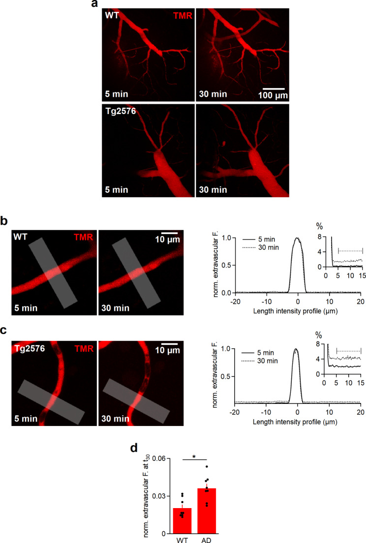
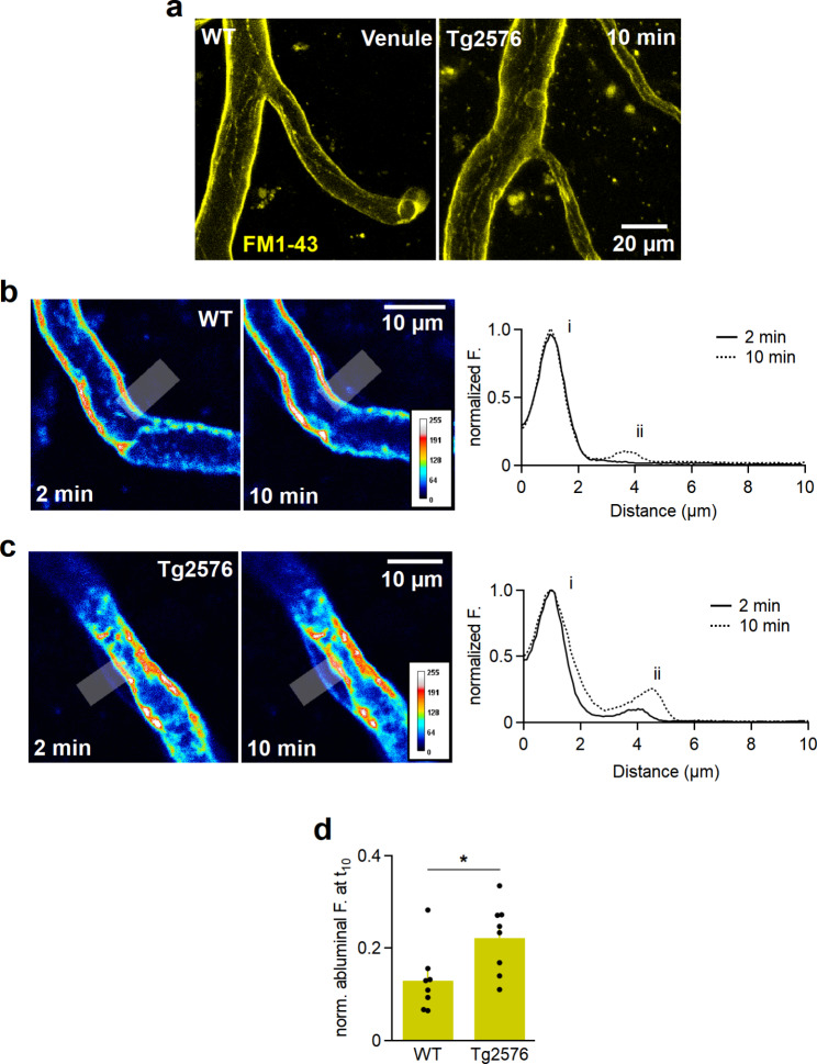
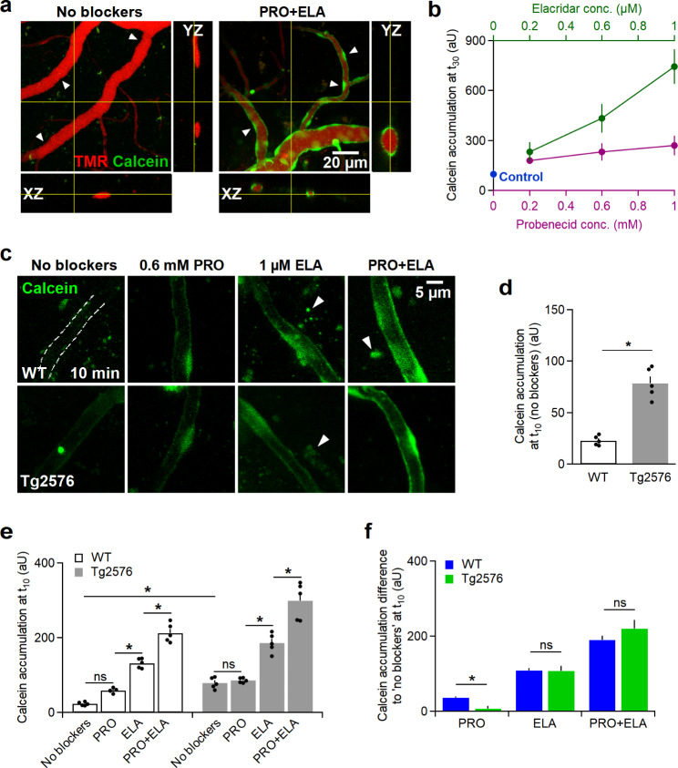
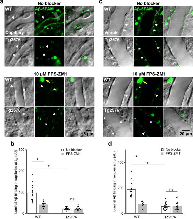
Methods: ISMICAP, a BBB model involving microperfusion of capillaries, was used to assess BBB properties in acute cortical brain slices from Tg2576 mice compared to wild-type (WT) controls using two-photon microscopy. TJ integrity was tested by vascularly perfusing biocytin-tetramethylrhodamine (TMR) and quantifying its extravascular diffusion as well as the diffusion of FM1-43 from luminal to abluminal membranes of endothelial cells (ECs). To assess ABCC1 and ABCB1 activity, calcein-AM was perfused, which is converted to fluorescent calcein in ECs and gets actively extruded by both transporters. To probe which transporter is involved, probenecid or Elacridar were applied, individually or combined, to block ABCC1 and ABCB1, respectively. To assess RAGE activity, the binding of 5-FAM-tagged Aβ by ECs was quantified with or without applying FPS-ZM1, a RAGE antagonist.

Results: In Tg2576 mouse brain, extravascular TMR was 1.8-fold that in WT mice, indicating increased paracellular leakage. FM1-43 staining of abluminal membranes in Tg2576 capillaries was 1.7-fold that in WT mice, indicating reduced TJ integrity in AD. While calcein was undetectable in WT mice, its accumulation was significant in Tg2576 mice, suggesting lower calcein extrusion in AD. Incubation with probenecid or Elacridar in WT mice resulted in a marked calcein accumulation, yet probenecid alone had no effect in Tg2576 mice, implying the absence of probenecid-sensitive ABC transporters. In WT mice, Aβ accumulated along the luminal membranes, which was undetectable after applying FPS-ZM1. In contrast, marginal Aβ fluorescence was observed in Tg2576 vessels, and FPS-ZM1 was without effect, suggesting reduced RAGE binding activity.

Conclusions: Disrupted TJ integrity, reduced ABCC1 functionality and decreased RAGE binding were identified as BBB alterations in Tg2576 mice, with the latter finding challenging the current concepts. Our results suggest to manage AD by including modulation of TJ proteins and Aβ-RAGE binding.

Keywords: ABC transporter; Alzheimer’s Disease; Blood-brain barrier; RAGE; Tight junctions.


Conflict of interest statement

The authors declare no competing interests.

Figures

Fig. 1
Fig. 1
The extracellular diffusion of TMR is significantly increased in Tg2576 than wild-type mice. a, Maximum intensity projections (MIPs) of TMR-perfused vascular tree in the cortices of WT mice (top) and Tg2576 mice (bottom) mice 5 and 30 min after impalement. b, A single frame of a capillary in a wild-type cortical slice at 5 and 30 min (left) and the corresponding spatial fluorescence profiles (right) of a line profile normalized to the intravascular fluorescence. The analyzed line profiles (grey, 10-µm thick) were within 10 μm in a 3-µm distance to the sides. The extravascular fluorescence was 1.3 ± 0.03% at 30 min (the dashed bar region was averaged for quantification, see inset). c, A single frame of a capillary in Tg2576 cortex recorded at t5 and t30 (left) and the corresponding spatial fluorescence profiles (right) of a line profile. After 30 min, the extravascular fluorescence rose to 4.2 ± 0.04% (the dashed bar region was averaged for quantification, see inset). d, Quantification of extravascular fluorescence intensities within the specified ROIs across capillaries, 100–120 μm away from the impalement point, in WT and Tg2576 mice at 30 min. Data are represented as mean ± SEM. All intensities were background corrected and normalized to the intravascular fluorescence. The asterisk denotes a statistically significant difference (Student’s t-test, *P < 0.05). Data were obtained from 10 injections/4 animals (nROI WT mice = 8, nROI Tg2576 mice = 8)
Fig. 2
Fig. 2
FM1-43 probe diffuses at a faster rate from the luminal to abluminal walls of endothelial cells in Tg2576 mouse brain tissue. a, MIPs of venules perfused with FM1-43 for 10 min in WT and Tg2576 mouse brains demonstrating the outlined ECs across the walls. b, A capillary in a WT cortical slice perfused with FM1-43 at 2 and 10 min (left) and the corresponding spatial fluorescence profiles (right) of a line profile (grey line, 5-µm thick) across the capillary wall normalized to the intravascular fluorescence. After 2 min, only the luminal membrane is stained (i), while the labeling of the abluminal membrane (ii) can be detected after 10 min. The LUT ‘royal’ is a color map that codes low-intensity pixels ‘blue’ and high-intensity pixels as ‘white’. c, A capillary in a Tg2576 cortical slice recorded at t2 and t10 (left) and the corresponding spatial fluorescence profiles (right) of a line profile normalized to the intravascular fluorescence. The labelling of the abluminal membrane can be detected at as early as 2 min after starting the FM1-43 perfusion, which intensifies over time (ii). d, Quantified fluorescence intensities within the abluminal membranes of capillaries in WT (nROI=8) and Tg2576 (nROI=8) mice. After 10 min, the diffused FM1-43 to the abluminal membranes in Tg2576 mice was 1.7-fold higher than in WT mice (Student’s t-test, *P < 0.05). The measured intensities were background-corrected and normalized to the corresponding luminal fluorescence intensities. Data are represented as mean ± SEM, obtained from 17 injections/5 animals
Fig. 3
Fig. 3
The endothelial efflux systems in Tg2576 mice are significantly hindered. a, Cross-sectional views of cortical vessels perfused with calcein-AM for 10 min under control conditions (left) and simultaneous inhibition of ABCC1 and ABCB1 transporters with 0.6 mM PRO and 1 µM ELA, respectively (right). Without blockers, no calcein accumulation was detected in endothelial cells (arrow heads, left), while it was observed after applying PEO and ELA (arrow heads, right). b, Dose-response curve showing the concentration-dependent accumulation of calcein 30 min after applying PRO or ELA. The ‘control’ datapoint denotes calcein accumulation in absence of both blockers. c, MIPs of capillaries recorded at 10 min demonstrating calcein accumulation under control conditions as well as inhibition of ABCC1, ABCB1 or both by 0.6 mM PRO, 1 µM ELA or a mixture of both, respectively, in WT mice (top row) and Tg2576 mice (bottom row). In WT mice capillaries, no calcein accumulation could be observed under control conditions, therefore the capillary’s lumen was manually outlined (dashed ROI) using simultaneously perfused TMR. Arrowheads denote extravascular autofluorescent aggregates in aged mice. d, Calcein accumulation in ECs at 10 min in absence of blockers in Tg2576 mice was significantly higher than that in WT mice (nROI WT mice = 5, nROI Tg2576 mice = 5, *P < 0.05). e, Quantification of calcein accumulating at 10 min in WT and Tg2576 mice in absence and presence of ABCC1 and ABCB1 blockers. A similar pattern of calcein accumulation was observed within both WT and Tg2576 mice groups. Compared to control conditions, applying PRO led to insignificant calcein accumulation, while applying ELA or ELA + PRO mixture resulted in significantly increased calcein accumulation. Data, presented as mean ± SEM, were obtained from 24 injections/6 animals (nROI = 5 for all groups, two-way ANOVA, *P < 0.05, ns; insignificant difference, see supplementary information for details). f, Inhibitor-induced increases in calcein fluorescence with respect to control (no blockers) conditions. PRO-sensitive ABC transporters were almost absent in Tg2576 mouse brains, while ELA-sensitive extrusion remained unchanged (multiple t-tests, *P < 0.05, ns; insignificant difference).
Fig. 4
Fig. 4
Beta-amyloid uptake in capillaries and venules of Tg2576 mice is significantly decreased. a, Single frames of capillaries 30 min after perfusing 5-FAM-tagged Aβ monomers in WT and Tg2576 murine vasculature with/without inhibiting RAGE transporter by 10 µM FPS-ZM1. All images are displayed in parallel as fluorescence scan, laser DIC scan and an overlay of both. In WT mice, Aβ fluorescence is localized at the luminal walls (arrow heads) under control conditions, which significantly decreased after RAGE inhibition by FPS-ZM1 (see b). In Tg2576 mice, negligible Aβ uptake was noted under both experimental conditions (see b). An example of line profiles used for analysis is shown (grey line, 3-µm thick). Asterisks signify ECs, while yellow arrows refer to autofluorescence commonly found in aged tissue. b, Quantification of luminal fluorescence intensities in capillaries under control conditions (nROI WT mice = 14, nROI Tg2576 mice = 16) and RAGE inhibition (nROI WT mice = 11, nROI Tg2576 mice = 12) represented as mean ± SEM (two-way ANOVA, *P < 0.05; ns, insignificant, see supplementary information). c, Single frames of venules after perfusing 5-FAM-tagged Aβ monomers for 30 min under control conditions and after preincubation with 10 µM FPS-ZM1 in WT and Tg2576 murine vasculature. In WT mouse brain tissue under no-blocking condition, Aβ fluorescence is localized within the luminal walls (arrow heads), which significantly decreased upon inhibiting RAGE with FPS-ZM1 (see d). Significantly lower Aβ uptake was recorded in Tg2576 venules, which remained unaltered after applying FPS-ZM1 to block RAGE (see d). An example of the analyzed line profile is indicated (grey line, 3-µm thick). Similarly, ECs are marked by asterisks, while yellow arrows indicate autofluorescence. d, Quantification of luminal fluorescence in venules under no-blocking condition (nROI WT mice = 13, nROI Tg2576 mice = 19) and RAGE inhibition (nROI WT mice = 7, nROI Tg2576 mice = 14) represented as mean ± SEM. (two-way ANOVA, *P < 0.05; ns, insignificant, see supplementary information). Data were collected from 10 injections/4 animals
Fig. 5
Fig. 5
Schematic representation of the healthy blood-brain barrier (top) compared to pathological blood-brain barrier alterations identified in Alzheimer’s Tg2576 mouse brain (bottom) in the current study along with the associated experimental observations

Similar articles

Cited by

References

    1. Liu P-P, Xie Y, Meng X-Y, Kang J-S. History and progress of hypotheses and clinical trials for Alzheimer’s Disease. Signal Transduct Target Therapy. 2019;4:1–22. - PMC - PubMed
    1. Hoogmartens J, Cacace R, Van Broeckhoven C. Insight into the genetic etiology of Alzheimer’s Disease: a comprehensive review of the role of rare variants. Alzheimer’s & Dementia: Diagnosis Assessment & Disease Monitoring. 2021;13:e12155. - PMC - PubMed
    1. Hampel H, Hardy J, Blennow K et al. The Amyloid-β pathway in Alzheimer’s Disease. Mol Psychiatry. 2021: 1–23. - PMC - PubMed
    1. Sturchio A, Dwivedi AK, Young CB, et al. High cerebrospinal amyloid-β 42 is associated with normal cognition in individuals with brain amyloidosis. EClinicalMedicine. 2021;38:100988. doi: 10.1016/j.eclinm.2021.100988. - DOI - PMC - PubMed
    1. Storck SE, Hartz AM, Bernard J, et al. The concerted amyloid-beta clearance of LRP1 and ABCB1/P-gp across the blood-brain barrier is linked by PICALM. Brain Behav Immun. 2018;73:21–33. doi: 10.1016/j.bbi.2018.07.017. - DOI - PMC - PubMed