Review
. 2024 Feb 1;136(2):283-297.
doi: 10.1152/japplphysiol.00723.2022. Epub 2023 Nov 23.

Idiosyncratic bone responses to blood flow restriction exercise: new insights and future directions

Affiliations
Review

Idiosyncratic bone responses to blood flow restriction exercise: new insights and future directions

Luke Hughes et al. J Appl Physiol (1985). .

Abstract

Applying blood flow restriction (BFR) during low-load exercise induces beneficial adaptations of the myotendinous and neuromuscular systems. Despite the low mechanical tension, BFR exercise facilitates a localized hypoxic environment and increase in metabolic stress, widely regarded as the primary stimulus for tissue adaptations. First evidence indicates that low-load BFR exercise is effective in promoting an osteogenic response in bone, although this has previously been postulated to adapt primarily during high-impact weight-bearing exercise. Besides studies investigating the acute response of bone biomarkers following BFR exercise, first long-term trials demonstrate beneficial adaptations in bone in both healthy and clinical populations. Despite the increasing number of studies, the physiological mechanisms are largely unknown. Moreover, heterogeneity in methodological approaches such as biomarkers of bone metabolism measured, participant and study characteristics, and time course of measurement renders it difficult to formulate accurate conclusions. Furthermore, incongruity in the methods of BFR application (e.g., cuff pressure) limits the comparability of datasets and thus hinders generalizability of study findings. Appropriate use of biomarkers, effective BFR application, and befitting study design have the potential to progress knowledge on the acute and chronic response of bone to BFR exercise and contribute toward the development of a novel strategy to protect or enhance bone health. Therefore, the purpose of the present synthesis review is to 1) evaluate current mechanistic evidence; 2) discuss and offer explanations for similar and contrasting data findings; and 3) create a methodological framework for future mechanistic and applied research.

Keywords: blood flow restriction; bone remodeling; exercise; hypoxia.


Conflict of interest statement

No conflicts of interest, financial or otherwise, are declared by the authors.

Figures
None
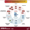
Graphical abstract
Figure 1.
Figure 1.
Overview of the bone remodeling process. Osteocytes initiate bone remodeling and limit bone resorption and formation processes to preserve bone mass. The RANKL/RANK/OPG and Wnt/β-catenin pathways are 2 key signaling pathways that regulate the differentiation and function of osteoclasts and osteoblasts. Osteocytes stimulate osteoclastogenesis via production of RANKL at the initiation of the bone remodeling cycle. RANKL then binds to osteoclastic precursor cells, stimulating osteoclast differentiation and facilitating fusion. Osteoblasts and osteocytes secrete OPG, which binds to RANKL, inhibiting osteoclastic bone resorption by preventing its binding to RANK. The Wnt/β-catenin pathway facilitates osteoblast differentiation. Sclerostin reduces osteoblastic bone formation by inhibiting canonical Wnt/β-catenin signaling. Created with BioRender with permission.
Figure 2.
Figure 2.
Hormonal and growth factors involved in the regulation of bone remodeling. Parathyroid hormone (PTH) stimulates proliferation and differentiation of mesenchymal progenitors to mature osteoblasts via insulin-like growth factor-I (IGF-I). PTH also induces differentiation of committed mesenchymal progenitors and induces receptor activator of nuclear factor kappa ligand (RANKL) from mature osteoblasts to promote osteoclastogenesis. Vitamin D3 stimulates osteoblastogenesis via differentiation of mesenchymal stem cells into osteoblasts. Calcitonin inhibits osteoclast activity to suppress increased bone resorption drive by PTH and also increases osteoblast proliferation and differentiation. Estrogen primarily attenuates osteoclastogenesis by stimulating osteoclast apoptosis. Growth hormone stimulates osteoblast proliferation directly and indirectly via increased IGF-I production. IGF-I stimulates osteoblast proliferation, function, and survival and promotes osteoclast differentiation. Bone morphogenetic proteins (BMPs) induce differentiation of mesenchymal cells to osteoblasts and also enhance the differentiated function of the osteoblast. Transforming growth factor-beta (TGF-β) can both stimulate early osteoblast differentiation and inhibit late osteoblast differentiation. IL-6 induces RANKL production in osteoblastic cells, which in turn stimulates differentiation of osteoclast precursors into mature osteoclasts. Created with BioRender with permission.
Figure 3.
Figure 3.
Hypothetical mechanisms of an effect of blood flow restriction (BFR) on bone remodeling processes. Partial or full occlusion of limb blood flow may decrease bone arterial blood perfusion in different areas of bone, causing hypoxia and withdrawal of nutrients. This may lead to a cascade of events including upregulation of several systemic hormones, transforming factors, and cytokines that impact bone remodeling processes. Deflation of the BFR cuff may trigger postexercise bone hyperemia, increasing blood flow to bone tissue. GH, growth hormone; HIF-1α, hypoxia-inducible factor 1-alpha; IGF-1, insulin-like growth factor-I; IL-6, interleukin-6; ROS, reactive oxygen species; VEGF, vascular endothelial growth factor. Created with BioRender with permission.

Similar articles

Cited by

References

    1. Rittweger J, Simunic B, Bilancio G, De Santo NG, Cirillo M, Biolo G, Pisot R, Eiken O, Mekjavic IB, Narici M. Bone loss in the lower leg during 35 days of bed rest is predominantly from the cortical compartment. Bone 44: 612–618, 2009. doi:10.1016/j.bone.2009.01.001. - DOI - PubMed
    1. Sowers MR, Jannausch M, McConnell D, Little R, Greendale GA, Finkelstein JS, Neer RM, Johnston J, Ettinger B. Hormone predictors of bone mineral density changes during the menopausal transition. J Clin Endocrinol Metab 91: 1261–1267, 2006. doi:10.1210/jc.2005-1836. - DOI - PubMed
    1. Daly RM, Rosengren BE, Alwis G, Ahlborg HG, Sernbo I, Karlsson MK. Gender specific age-related changes in bone density, muscle strength and functional performance in the elderly: a-10 year prospective population-based study. BMC Geriatr 13: 71, 2013. doi:10.1186/1471-2318-13-71. - DOI - PMC - PubMed
    1. Wolff J. The Law of Bone Remodelling. Heidelberg: Springer, 1986.
    1. Kohrt WM, Bloomfield SA, Little KD, Nelson ME, Yingling VR; American College of Sports Medicine. Physical activity and bone health. Med Sci Sports Exerc 36: 1985–1996, 2004. doi:10.1249/01.MSS.0000142662.21767.58. - DOI - PubMed

LinkOut - more resources