

Radiographic Anatomy of the Lateral Ankle Ligament Complex: A Cadaveric Study
- PMID: 37994643
- PMCID: PMC10860354
- DOI: 10.1177/10711007231213355
Radiographic Anatomy of the Lateral Ankle Ligament Complex: A Cadaveric Study
Abstract
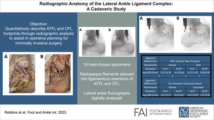
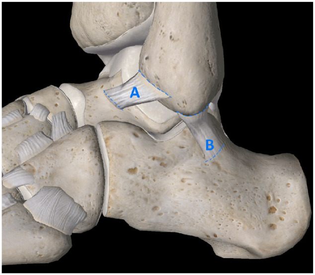
Background: When lateral ankle sprains progress into chronic lateral ankle instability (CLAI), restoring precise anatomic relationships of the lateral ankle ligament complex (LALC) surgically is complex. This study quantifies the radiographic relationships between the anterior talofibular ligament (ATFL), calcaneofibular ligament (CFL), and prominent osseous landmarks visible under fluoroscopy to assist in perioperative practices for minimally invasive surgery for CLAI.
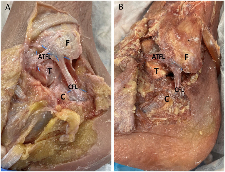
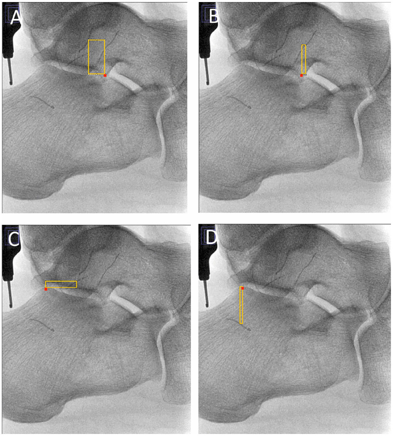
Methods: Ten fresh frozen ankle specimens were dissected to expose the LALC and prepared by threading a radiopaque filament through the ligamentous footprints of the ATFL and CFL. Fluoroscopic images were digitally analyzed to define dimensional characteristics of the ATFL and CFL. Directional measurements of the ligamentous footprints relative to the lateral process of the talus and the apex of the posterior facet of the calcaneus were calculated.
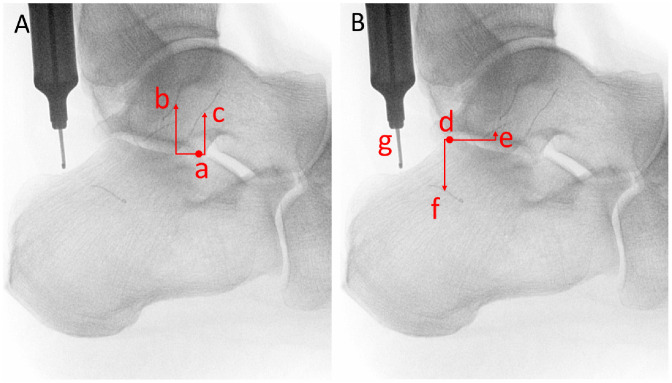
Results: Dimensional measurements of the ATFL were a mean length of 9.3 mm, fibular footprint of 9.4 mm, and talar footprint of 9.1 mm. Dimensional measurements of the CFL were a mean length of 19.4 mm, fibular footprint of 8.2 mm, and calcaneal footprint of 7.3 mm. From the radiographic apparent tip of the lateral process of the talus, the fibular attachment of the ATFL was found 13.3 mm superior and 4.4 mm posterior, whereas the talar attachment was found 11.5 mm superior and 4.8 mm anterior. From the radiographic apparent posterior apex of the posterior facet of the calcaneus, the fibular attachment of the CFL was found 0.2 mm inferior and 6.8 mm anterior, whereas the calcaneal attachment was found 14.3 mm inferior and 5.9 mm posterior.
Conclusion: The ATFL and CFL were radiographically analyzed using radiopaque filaments to outline the ligamentous footprints in their native locations. These ligaments were also localized with reference to 2 prominent osseous landmarks. These findings may assist in perioperative practices for keyhole incision placement and arthroscopic guidance. Perfect lateral ankle joint imaging with talar domes superimposed is required to be able to do this.
Clinical relevance: Radiographic evaluation of the ATFL and CFL with reference to prominent osseous landmarks identified under fluoroscopy may assist in perioperative practices for minimally invasive surgery to address CLAI for keyhole incision placement and arthroscopic guidance.
Keywords: anatomic landmarks; ankle instability; ankle lateral ligaments; anterior talofibular ligament; calcaneofibular ligament; fluoroscopy; minimally invasive surgery.
Conflict of interest statement
Declaration of Conflicting InterestsThe author(s) declared no potential conflicts of interest with respect to the research, authorship, and/or publication of this article. ICMJE forms for all authors are available online.
Figures

Comment on
-
Bony landmarks available for minimally invasive lateral ankle stabilization surgery: a cadaveric anatomical study.Knee Surg Sports Traumatol Arthrosc. 2017 Jun;25(6):1916-1924. doi: 10.1007/s00167-016-4218-7. Epub 2016 Jun 28. Knee Surg Sports Traumatol Arthrosc. 2017. PMID: 27351549
References
-
- Best R, Mauch F, Fischer KM, Rueth J, Brueggemann GP. Radiographic monitoring of the distal insertion of the calcaneofibular ligament in anatomical reconstructions of ankle instabilities: a preliminary cadaveric study. Foot Ankle Surg. 2015;21(4):245-249. doi: 10.1016/j.fas.2015.01.006 - DOI - PubMed
Publication types
MeSH terms
LinkOut - more resources
Full Text Sources
Miscellaneous
