Review
. 2023 Dec;25(12):1745-1758.
doi: 10.1007/s11886-023-01990-8. Epub 2023 Nov 23.

The Brain-Heart Axis: Neuroinflammatory Interactions in Cardiovascular Disease

Affiliations
Review

The Brain-Heart Axis: Neuroinflammatory Interactions in Cardiovascular Disease

Jiun-Ruey Hu et al. Curr Cardiol Rep. 2023 Dec.

Abstract

Purpose of review: The role of neuroimmune modulation and inflammation in cardiovascular disease has been historically underappreciated. Physiological connections between the heart and brain, termed the heart-brain axis (HBA), are bidirectional, occur through a complex network of autonomic nerves/hormones and cytokines, and play important roles in common disorders.

Recent findings: At the molecular level, advances in the past two decades reveal complex crosstalk mediated by the sympathetic and parasympathetic nervous systems, the renin-angiotensin aldosterone and hypothalamus-pituitary axes, microRNA, and cytokines. Afferent pathways amplify proinflammatory signals via the hypothalamus and brainstem to the periphery, promoting neurogenic inflammation. At the organ level, while stress-mediated cardiomyopathy is the prototypical disorder of the HBA, cardiac dysfunction can result from a myriad of neurologic insults including stroke and spinal injury. Atrial fibrillation is not necessarily a causative factor for cardioembolic stroke, but a manifestation of an abnormal atrial substrate, which can lead to the development of stroke independent of AF. Central and peripheral neurogenic proinflammatory factors have major roles in the HBA, manifesting as complex bi-directional relationships in common conditions such as stroke, arrhythmia, and cardiomyopathy.

Keywords: Arrhythmia-related stroke; Cardiometabolic syndrome; Cardiovascular neurogenic aging; Heart brain axis; Neuroinflammatory interactions; Stress cardiomyopathy.


Conflict of interest statement

Conflict of Interest: Michael G. Nanna reports personal fees from Merck and HeartFlow, Inc., outside the submitted work. The other authors declare that they have no conflict of interest.

Figures

Figure 1:
Figure 1:
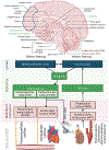
Neural pathways involved in the brain-heart axis [92]. Sagittal view of the brain, showing the cerebral cortex, the thalamus, the hypothalamus, and the brainstem, which includes the midbrain, pons, and medulla oblongata (bottom left). The afferent (“from bottom to top”) pathway begins with input from chemoreceptors and baroreceptors and culminates in the insular cortex (bottom right). The efferent (“from top to bottom”) pathway begins with the medial prefrontal cortex and insular cortex and culminates in cardiac myocytes and coronary epithelial cells. Abbreviations: ACTH: adrenocorticotropic hormone; Hypothal: hypothalamic. Original figure created with BioRender.com.
Figure 2:
Figure 2:
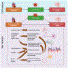
Relationship between AF and development of stroke. In the old paradigm, AF was considered a causative factor of stroke, with left atrial size being merely a surrogate marker for chronicity of AF. In the new paradigm, AF is considered to be a manifestation of abnormal atrial substrate, which can lead to development of stroke independently of AF. Cerebral vascular risk factors and central vascular risk factors predispose both to abnormal atrial substrate and development of stroke. Original figure created with BioRender.com.
Figure 3:
Figure 3:
Relationship between acute MI and development of stroke. In the old paradigm, acute MI predisposed to development of LV thrombus which led to stroke. In the new paradigm, it is recognized that stroke can develop post-MI regardless of whether an LV thrombus is present. This occurs through multiple mechanisms, including decreased cerebral blood flow from decreased cardiac output, neuronal injury from microRNA and neuroinflammation, altered synaptic transmission from microRNA and neuroinflammation, increased blood brain barrier permeability from upregulation of angiotensin II, DAMPs, and pro-inflammatory cytokines, and activation of astrocytes, microglia, choroid plexus, and endothelial cells. Original figure created in BioRender.com.

References

    1. Pavlov VA, Chavan SS, Tracey KJ. Molecular and Functional Neuroscience in Immunity. Annu Rev Immunol. 2018;36:783–812. doi: 10.1146/annurev-immunol-042617-053158. - DOI - PMC - PubMed
    1. Pereira VH, Cerqueira JJ, Palha JA, Sousa N. Stressed brain, diseased heart: A review on the pathophysiologic mechanisms of neurocardiology. International Journal of Cardiology. 2013;166(1):30–7. doi: 10.1016/j.ijcard.2012.03.165. - DOI - PubMed
    1. Rijkers K, Majoie HJ, Hoogland G, Kenis G, De Baets M, Vles JS. The role of interleukin-1 in seizures and epilepsy: a critical review. Exp Neurol. 2009;216(2):258–71. doi: 10.1016/j.expneurol.2008.12.014. - DOI - PubMed
    1. Liu T, Young PR, McDonnell PC, White RF, Barone FC, Feuerstein GZ. Cytokine-induced neutrophil chemoattractant mRNA expressed in cerebral ischemia. Neurosci Lett. 1993;164(1-2):125–8. doi: 10.1016/0304-3940(93)90873-j. - DOI - PubMed
    1. Yu Y, Zhang ZH, Wei SG, Chu Y, Weiss RM, Heistad DD, et al. Central gene transfer of interleukin-10 reduces hypothalamic inflammation and evidence of heart failure in rats after myocardial infarction. Circ Res. 2007;101(3):304–12. doi: 10.1161/CIRCRESAHA.107.148940. - DOI - PubMed

Publication types