. 2023 Nov 23;9(1):156.
doi: 10.1038/s41531-023-00595-w.

Accurate long-read sequencing identified GBA1 as major risk factor in the Luxembourgish Parkinson's study

Collaborators, Affiliations

Accurate long-read sequencing identified GBA1 as major risk factor in the Luxembourgish Parkinson's study

Sinthuja Pachchek et al. NPJ Parkinsons Dis. .

Erratum in

Abstract

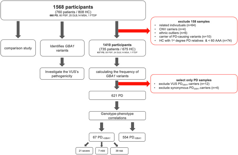
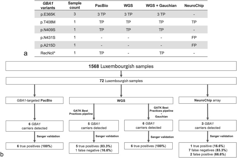
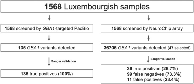
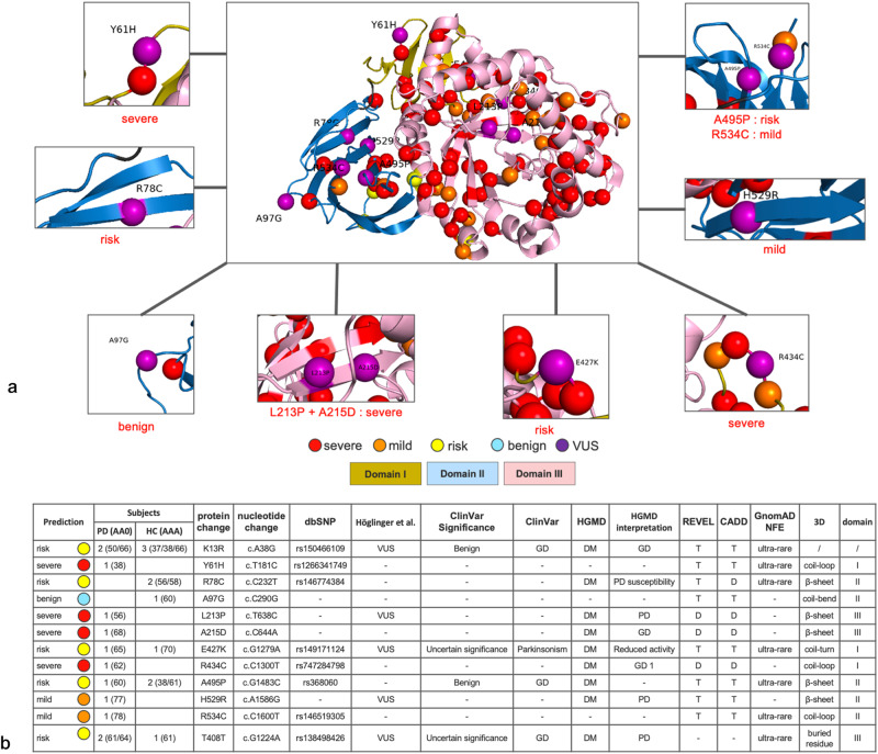
Heterozygous variants in the glucocerebrosidase GBA1 gene are an increasingly recognized risk factor for Parkinson's disease (PD). Due to the GBAP1 pseudogene, which shares 96% sequence homology with the GBA1 coding region, accurate variant calling by array-based or short-read sequencing methods remains a major challenge in understanding the genetic landscape of GBA1-associated PD. We analyzed 660 patients with PD, 100 patients with Parkinsonism and 808 healthy controls from the Luxembourg Parkinson's study, sequenced using amplicon-based long-read DNA sequencing technology. We found that 12.1% (77/637) of PD patients carried GBA1 variants, with 10.5% (67/637) of them carrying known pathogenic variants (including severe, mild, risk variants). In comparison, 5% (34/675) of the healthy controls carried GBA1 variants, and among them, 4.3% (29/675) were identified as pathogenic variant carriers. We found four GBA1 variants in patients with atypical parkinsonism. Pathogenic GBA1 variants were 2.6-fold more frequently observed in PD patients compared to controls (OR = 2.6; CI = [1.6,4.1]). Three novel variants of unknown significance (VUS) were identified. Using a structure-based approach, we defined a potential risk prediction method for VUS. This study describes the full landscape of GBA1-related parkinsonism in Luxembourg, showing a high prevalence of GBA1 variants as the major genetic risk for PD. Although the long-read DNA sequencing technique used in our study may be limited in its effectiveness to detect potential structural variants, our approach provides an important advancement for highly accurate GBA1 variant calling, which is essential for providing access to emerging causative therapies for GBA1 carriers.


Conflict of interest statement

The authors declare no competing interests.

Figures

Fig. 1
Fig. 1. Description of the study dataset and methodology.
HC Healthy controls, PD Parkinson’s Disease and Parkinson’s Disease with Dementia, PSP Progressive Supranuclear Palsy, DLB Dementia with Lewy Body, MSA Multiple System Atrophy, FTDP Fronto-temporal dementia with parkinsonism, GBA1 glucocerebrosidase gene, VUS Variants of unknown significance, PD+GBA1 PD patients with GBA1 pathogenic variant, PD-GBA1 PD patients without GBA1 pathogenic variant, CNV copy number variants, AAA age at assessment.
Fig. 2
Fig. 2. Comparison of variant calls from PacBio, WGS and NeuroChip genotyping data using 72 matched samples for the GBA1 gene and validated by Sanger sequencing.
a *RecNcil (p.L483P; p.A495P; p.V499V); Sanger sequencing results: TP, true positive; FP, false positive. Sample count gives the total number of samples carrying the variant found by each method. b Comparative study of GBA1 variants detection by the GBA1-targeted PacBio DNA sequencing method and NeuroChip array methods in the Luxembourg Parkinson’s study. Due to overrepresented variants with the NeuroChip array, we applied for the detected variants a study-wide threshold of 1% in our cohort.
Fig. 3
Fig. 3. Comparative study of GBA1 variants detection by the GBA1-targeted PacBio DNA sequencing and NeuroChip array methods in the Luxembourg Parkinson’s study.
Due to overrepresented variants with the NeuroChip array, we applied for the detected variants a study-wide threshold of 1% in our cohort.
Fig. 4
Fig. 4. Sub-classification of VUS found in the Luxembourg Parkinson’s study.
a GBA1 missense and stop gain variants mapped onto the three-dimensional structure of GCase. Domain I is shown in dark yellow, domain II in blue, and domain III in pink. Domain I begins at residue 40 after the signal peptide sequence. Variants classified as severe are colored red, mild are colored orange, risk in yellow and VUS are colored purple. The 3D structure of GCase (PDB code 1ogs) was generated using PYMOL (http://www.pymol.org). b Proposed sub-classification of identified VUSs with their score in a known database. GBA1 glucocerebrosidase gene, GD Gaucher’s disease, PD Parkinson’s disease, AAO age at onset, AAA age at assessment in visit1. HGMD The Human Gene Mutation Database, REVEL Rare Exome Variant Ensemble Learner, CADD Combined Annotation Dependent Depletion, gnomAD The Genome Aggregation Database. DM Disease causing mutation, D Deleterious, T Tolerate. Variants classified as severe are colored red, mild are colored orange, risk in yellow and VUS are colored purple.

References

    1. Hruska KS, Lamarca ME, Scott CR, Sidransky E. Gaucher disease: mutation and polymorphism spectrum in the glucocerebrosidase gene (GBA) Hum. Mutat. 2008;29:567–583. doi: 10.1002/humu.20676. - DOI - PubMed
    1. Vieira SRL, Schapira AHV. Glucocerebrosidase mutations and Parkinson disease. J. Neural Transm. 2022;129:1105–1117. doi: 10.1007/s00702-022-02531-3. - DOI - PMC - PubMed
    1. Horowitz M, et al. The human glucocerebrosidase gene and pseudogene: structure and evolution. Genomics. 1989;4:87–96. doi: 10.1016/0888-7543(89)90319-4. - DOI - PubMed
    1. Do J, Mckinney C, Sharma P, Sidransky E. Glucocerebrosidase and its relevance to Parkinson disease. Mol. Neurodegener. 2019;14:36. doi: 10.1186/s13024-019-0336-2. - DOI - PMC - PubMed
    1. Graham OEE, et al. Nanopore sequencing of the glucocerebrosidase (GBA) gene in a New Zealand Parkinson’s disease cohort. Parkinson. Relat. Disord. 2020;70:36–41. doi: 10.1016/j.parkreldis.2019.11.022. - DOI - PubMed