. 2023 Nov 23;14(1):7668.
doi: 10.1038/s41467-023-43349-1.

Nod1-dependent NF-kB activation initiates hematopoietic stem cell specification in response to small Rho GTPases

Affiliations

Nod1-dependent NF-kB activation initiates hematopoietic stem cell specification in response to small Rho GTPases

Xiaoyi Cheng et al. Nat Commun. .

Abstract

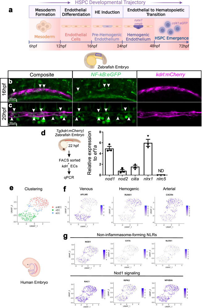
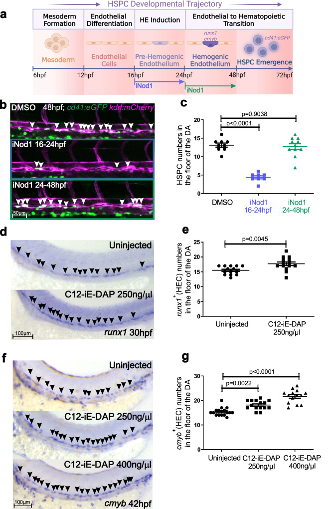
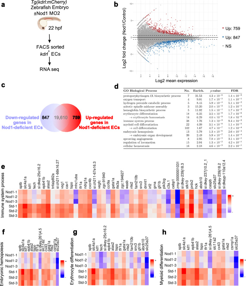
Uncovering the mechanisms regulating hematopoietic specification not only would overcome current limitations related to hematopoietic stem and progenitor cell (HSPC) transplantation, but also advance cellular immunotherapies. However, generating functional human induced pluripotent stem cell (hiPSC)-derived HSPCs and their derivatives has been elusive, necessitating a better understanding of the developmental mechanisms that trigger HSPC specification. Here, we reveal that early activation of the Nod1-Ripk2-NF-kB inflammatory pathway in endothelial cells (ECs) primes them to switch fate towards definitive hemogenic endothelium, a pre-requisite to specify HSPCs. Our genetic and chemical embryonic models show that HSPCs fail to specify in the absence of Nod1 and its downstream kinase Ripk2 due to a failure on hemogenic endothelial (HE) programming, and that small Rho GTPases coordinate the activation of this pathway. Manipulation of NOD1 in a human system of definitive hematopoietic differentiation indicates functional conservation. This work establishes the RAC1-NOD1-RIPK2-NF-kB axis as a critical intrinsic inductor that primes ECs prior to HE fate switch and HSPC specification. Manipulation of this pathway could help derive a competent HE amenable to specify functional patient specific HSPCs and their derivatives for the treatment of blood disorders.


Conflict of interest statement

The authors declare no competing interests.

Figures

Fig. 1
Fig. 1. NF-kB activation and NLRs expression during HE patterning.
a HSPC developmental trajectory in zebrafish embryos. Mesoderm is specified from 6 hpf, and endothelial cells from 12 hpf. Hemogenic endothelium (HE) can be visualized within the dorsal aorta by WISH for runx1 and cmyb from 24-48 hpf, being runx1 expressed prior to cmyb. From 48 hpf, emergent HSPCs can be traced within the dorsal aorta using the cd41:eGFP transgenic line. b, c Maximum projections from aorta-gonad-pronephros (AGP) of 16 hpf (b) and 20 hpf (c) NF-kB:eGFP; kdrl:mCherry double-transgenic embryos visualized by live confocal microscopy. Arrowheads denote NF-kB+ ECs. n = 3, 16hpf; n = 3, 20hpf. All views are lateral, with anterior to the left. Images are representative of two independent experiments. d kdrl:mCherry+ ECs were purified by FACS at 22hpf for gene expression analysis by qPCR (n = 3 from ~40 pooled embryos each). Levels of indicated transcripts along x-axis are shown relative to ef1a and multiplied by 10,000. eg UMAP visualization of the expression of indicated genes from CS 12 human endothelial AGM regions. Each dot represents one cell. Gray denotes minimal expression, purple intermediate, and blue high. aHEC, Arterial Hemogenic Endothelial cell; vEC, Venous Endothelial cell; aEC, Arterial Endothelial cell. Illustrations created with BioRender.com. All quantifications are represented with mean ± SEM. Source data are provided as a Source Data file.
Fig. 2
Fig. 2. The non-inflammasome-forming NLR Nod1 is required for HSPC generation in vivo.
a Schematic representation of the experimental design of (b, c). Nod1 was inhibited with Nodonitib-1 from 12-48 hpf, and emergent HSPCs quantified by confocal microscopy at 48 hpf. Illustration created with BioRender.com. b Live confocal maximum projections from DA of cmyb:eGFP; kdrl:mCherry transgenic embryos at 48 hpf treated with DMSO control, 15 µM, or 30 µM of Nodinitib-1 (Nod1 inhibitor) from 12hpf. Arrowheads denote cmyb+, kdrl+ HSPCs along the vDA. c Enumeration of cmyb+; kdrl+ HSPCs shown in (b). Each dot is the number of cmyb+; kdrl+ cells per embryo. n = 5, 6, 4 biological replicates from left to right. d, f Single z-plane live confocal images from DA of cd41:eGFP; kdrl:mCherry transgenic embryos at 48 hpf injected with Std MO control and Nod1 MO1 (d); or cas9 mRNA control and cas9 mRNA plus nod1 gRNA (f). Arrowheads denote cd41+, kdrl+ HSPCs along the vDA. e, g Quantification of cd41+, kdrl+ HSPCs from (d) and (f), respectively. Each dot represents the number of HSPCs per embryo. n = 10, Std MO; n = 10, Nod1 MO1 (e); n = 10, cas9 control; n = 10, nod1 gRNA+cas9 (g). h nod1+/+ and nod1-/- embryos were examined by WISH for runx1 expression in DA at 28hpf. Arrowheads denote runx1+ HECs. All views are lateral, with anterior to the left. i Quantification of runx1 HECs from (h). n = 17, nod1+/+; n = 30, nod1-/-. All quantifications are represented with mean ± SEM. Data were analyzed by ordinary one-way ANOVA with Tukey’s multiple comparisons test (c), or unpaired two-tailed T-test (e, g, i). Source data are provided as a Source Data file.
Fig. 3
Fig. 3. Nod1 programs the endothelium to become hemogenic.
a Schematic representation of the experimental design of (b, c). Nod1 was inhibited with Nodonitib-1 (iNod1) from either 16-24 hpf (HE induction), or 24-48 hpf (EHT), and emergent HSPCs quantified by confocal microscopy at 48 hpf in cd41:eGFP; kdrl:mCherry double transgenic embryos. Illustration created with BioRender.com. b 16-24 hpf, or 24-48 hpf cd41:eGFP; kdrl:mCherry double-transgenic embryos were treated with 15 µM of the Nod1 inhibitor Nodinitib-1 (iNod1) and lived imaged by confocal microscopy at 48hpf. Arrowheads denote cd41+, kdrl+ HSPCs along the DA within the maximum projections. c Enumeration of cd41+, kdrl+ HSPCs shown in (b). Each dot represents total cd41+; kdrl+ cells per embryo. n = 9, 8, 11 biological replicates from left to right. d, f Wildtype (wt) embryos injected with the Nod1 agonist C12-iE-DAP at one-cell stage were subjected to WISH for the HEC markers runx1 at 30 hpf (d), or cmyb at 42 hpf (f). e, g Quantification of runx1+ (e), or cmyb+ (g) HECs from (d) and (f), respectively. Each dot represents total HECs per embryo. Arrowheads denote HECs. n = 14 uninjected, n = 14 injected (e); n = 16, 15, 14 biological replicates from left to right. All views are lateral, with anterior to the left. All quantifications are represented with mean ± SEM. Data were analyzed by ordinary one-way ANOVA with Tukey’s multiple comparisons test (c, g), or unpaired two-tailed T-test (e). Source data are provided as a Source Data file.
Fig. 4
Fig. 4. Ripk2 hyperactivation restored the phenotypic lack of HSPCs in Nod1-deficient embryos.
a Single z-plane confocal images from DA of cd41:eGFP; kdrl:mCherry transgenic embryos at 48 hpf injected with Std-control and Ripk2 MOs. Arrowheads denote cd41+, kdrl+ HSPCs along the DA. b Enumeration of cd41+, kdrl+ HSPCs shown in (a). Each dot represents total cd41+; kdrl+ cells per embryo. n = 9, Std MO; n = 10, Ripk2 MO. c Wildtype (wt) and ripk2-/- embryos were examined by WISH for runx1 or cmyb expression at 30hpf and 42hpf, respectively. Arrowheads denote runx1+ or cmyb+ HECs. d Quantification of runx1+ or cmyb+ HECs from (c). Each dot represents total runx1+ or cmyb+ HECs per embryo. n = 10, 10, 10, 10 biological replicates from left to right. e) One-cell stage embryos were injected with Std-control or Nod1 MOs in the absence or presence of ripk2104ASP mRNA, and analyzed for runx1 expression by WISH at 30 hpf. Arrowheads denote runx1+ HECs. f Quantification of runx1+ HECs from (e). Each dot represents total HECs per embryo. n = 9, 9, 9, 9, biological replicates from left to right. g Fluorescence microscopy images from 5 dpf rag2:eGFP embryos injected with Std MO, Nod1 MO2 or Ripk2 MO (n = 10 embryos per condition). Arrowheads denote eGFP+ T-lymphocytes. All views are lateral, with anterior to the left. Images are representative of two independent experiments. Illustration created with BioRender.com. All quantifications are represented with mean ± SEM. All quantifications are represented with mean ± SEM. Data were analyzed by ordinary one-way ANOVA with Tukey’s multiple comparisons test (f), or unpaired two-tailed T-test (b, d). Source data are provided as a Source Data file.
Fig. 5
Fig. 5. RNA-seq transcriptomic analysis of FACS purified ECs identified deregulated hematopoietic and immune programs.
a Schematic representation of experimental design. Tg(kdrl:mCherry) zebrafish embryos were injected with Std MO or Nod1 MO2 and kdrl+ ECs were FACS purified at 22hpf for subsequent RNA-seq analysis. 40 embryos per condition (n = 3). Illustration created with BioRender.com. b MA plot displaying the log fold-change between Nod1-deficient ECs vs. Std-control ECs. Blue dots are significantly downregulated genes; red dots are significantly upregulated genes (adjusted p-value < 0.05). c 847 differentially downregulated and 759 upregulated genes were identified from (a, b). d Enriched GO processes for significantly down-regulated genes in Nod1-deficient versus Std-control ECs. eh Heatmaps displaying the log_2 gene expression of significantly downregulated genes between 22 hpf Nod1-deficient ECs versus Std-control ECs with log_2 foldchange < −1 under the GO terms ‘immune system process’ (e), ‘embryonic hemopoiesis’ (f), ‘erythrocyte differentiation’ (g), and ‘myeloid differentiation’ (h). Source data are provided as a Source Data file.
Fig. 6
Fig. 6. Nod1/Ripk2 signaling activates NF-kB within ECs.
a, c Maximum confocal projections of trunk regions of Std MO, Nod1 MO1- (a), or Ripk2-MO-injected NF-kB:eGFP; kdrl:mCherry (c), double-transgenic zebrafish embryos at 22 hpf. Arrowheads denote ECs with active NF-kB. b, d Quantification of NF-kB:eGFP activity from (a), or (c), respectively. Each dot represents eGFP+ mean pixel intensity within the kdrl+ EC area delimited by two intersegmental vessels. 3-4 areas were quantified per embryo. a, b n = 3, Std MO; n = 4, Nod1 MO1. c, d n = 3, Std MO; n = 3, Ripk2 MO. e Partial alignment between human (hIKKB) and zebrafish (zIkkb) proteins, showing the high conservation among Ser-177/181 (red) and their surrounding amino acids (top panel). Fluorescence microscopy images of NF-kB:eGFP embryos injected with wt ikkb mRNA (n = 12) or constitutively active ikkb (IkkbCA) (n = 10) (bottom panel). Notice that the eGFP expression pattern in embryos injected with control wt ikkb mRNA was identical to previously described uninjected NF-kB:eGFP embryos. Images are representative of two independent experiments. f One-cell stage embryos were injected with Std MO or Ripk2 MO in the absence or presence of IkkbCA mRNA, or Ikkb wt control and analyzed for cmyb expression by WISH at 42 hpf. Arrowheads denote cmyb+ HECs. g Quantification of cmyb+ HECs from (f). Each dot represents total cmyb+ HECs per embryo. n = 10, 10, 10, 10 biological replicates from left to right. h fli1b:Gal4+; UAS:ikkbCA- control, or fli1b:Gal4+; UAS:ikkbCA+ embryos analyzed for runx1 expression by WISH at 26hpf. To identify the UAS:ikkbCA transgene, PCR from genomic DNA was performed using specific primers spanning ikkbCA and mRFP sequences. Arrowheads denote runx1+ HECs. All views are lateral, with anterior to the left. i Quantification of runx1+ HECs from (h). Each dot represents total runx1+ HECs per embryo. n = 38, fli1b:Gal4+, UAS:ikkbCA-; n = 45, fli1b:Gal4+, UAS:ikkbCA+. All quantifications are represented with mean ± SEM. Data were analyzed by ordinary one-way ANOVA with Tukey’s multiple comparisons test (g), or unpaired two-tailed T-test (b, d, i). Source data are provided as a Source Data file.
Fig. 7
Fig. 7. Rac1 activates Nod1 signaling to specify HSPCs.
a kdrl:mCherry+ ECs were purified by FACS at 22hpf for gene expression analysis by qPCR (n = 3, 40 pooled embryos per biological replicate). Levels of indicated transcripts are shown relative to ef1a and multiplied by 10,000. b Schematic representation of the experimental design of (c). Zebrafish embryos were incubated with specific inhibitors for small Rho GTPases from 16-48 hpf, and emergent HSPCs quantified by confocal microscopy at 48 hpf. c 16hpf cd41:eGFP; kdrl:mCherry embryos were treated with DMSO, hydrochloride (Rac1 inhibitor), rho kinase inhibitor III (Rho inhibitor), or ML141 (Cdc42 inhibitor) and imaged by confocal microscopy at 48hpf. Maximum projection images are shown. n = 10 per condition. Arrowheads denote cd41+, kdrl+ HSPCs along the DA. Images are representative of two independent experiments. d cd41:eGFP; kdrl:mCherry embryos were injected with ikkb wt or ikkbCA mRNA, treated from 16 hpf with hydrochloride, rho kinase inhibitor III, or ML141 and quantified by confocal microscopy at 48hpf for cd41+, kdrl+ HSPCs. Each dot represents total HSPCs per embryo. n = 9, 10, 9, 11, 9, 9, 9, 9, biological replicates from left to right. e Embryos were injected with Cas9 mRNA, or Cas9 mRNA+rac1a/b gRNA in the absence or presence of ripk2104ASP mRNA, and analyzed by WISH for runx1 at 30 hpf, or cmyb at 42 hpf. Arrowheads denote HECs. f Quantification of HECs from (e). Each dot represents total runx1+ or cmyb+ HECs per embryo. n = 10, 8, 8, 9, 9, 10, 12, 7 biological replicates from left to right. g One-cell stage embryos were injected with transposase or transposase plus kdrl:rac1CA Tol2 vector and analyzed for cmyb expression at 38hpf by WISH. Arrowheads denote HECs. All views are lateral, with anterior to the left. h Quantification of HECs from (g). Each dot represents total cmyb+ HECs per embryo. n = 37 control; n = 33 kdrl:rac1CA. Horizontal lines indicate mean ± SEM. Illustrations created with BioRender.com. All quantifications are represented with mean ± SEM. Data were analyzed by ordinary one-way ANOVA with Tukey’s multiple comparisons test (d), (f) or unpaired two-tailed T-test (h). Source data are provided as a Source Data file.
Fig. 8
Fig. 8. NOD1 inactivation impairs the formation of definitive human PSC-derived HPCs.
a Schematic representation of the experimental design of (b, c). NOD1 was inhibited (iNOD1) with Nodinitib-1 from day 2-15, or 8-15 of definitive hematopoietic differentiation. At day 8, CD34+ cells were purified, and 20 K plated on OP9 cultures. At day 15, human definitive hematopoietic progenitors in suspension were quantified. b Representative bright-field microscopy images from cells in suspension at day 15 from 20 K CD34+ cells cultured on OP9 treated with DMSO control (left panel), or NOD1 inhibition (middle and right panels). c Enumeration of live cells in suspension at day 15 shown in (b). n = 3, 6, 3 biological replicates from left to right. d Schematic representation of the experimental design of (e, f). NOD1 was inhibited (iNOD1) with Nodinitib-1 at day 4, or at day 4 and refreshed at day 6. At day 9, CD34+ cells were isolated and analyzed for arterial-like, venous-like, and hemogenic-like markers. e Quantification at day 9 of CD34 frequencies of CD34+/CD184+/CD73- arterial-like, CD34+/CD184-/CD73+ venous-like, and CD34+/CD184-/CD73- hemogenic-like endothelium fractions after NOD1 inhibition at day 4, or days 4 + 6, compared to DMSO-treated control. f Percentage of RUNX1+ HE-like cells within the CD34+ fraction at day 9 of differentiation. n = 3, 3, 3 biological replicates from left to right. g Schematic representation of Nod1 signaling occurring within ECs to drive hemogenic endothelial fate. Briefly, Nod1 senses Rac1 activation, which leads to Nod1 oligomerization and recruitment of multiple Ripk2 units, forming a molecular platform that activates NF-kB to prime ECs to become hemogenic and their subsequent specification to HSPCs. Illustrations created with BioRender.com. All quantifications are represented with mean ± SEM. Data were analyzed by one-Way ANOVA with Kruskal-Wallis test and Dunnett’s multiple comparisons test (c), two-way ANOVA with Dunnett’s multiple comparisons test (e), or RM one-way ANOVA with Dunnett’s multiple comparisons test (f). Source data are provided as a Source Data file.

References

    1. Tie R, et al. Interleukin-6 signaling regulates hematopoietic stem cell emergence. Exp. Mol. Med. 2019;51:1–12. doi: 10.1038/s12276-019-0320-5. - DOI - PMC - PubMed
    1. Sawamiphak S, Kontarakis Z, Stainier DY. Interferon gamma signaling positively regulates hematopoietic stem cell emergence. Dev. Cell. 2014;31:640–653. doi: 10.1016/j.devcel.2014.11.007. - DOI - PMC - PubMed
    1. Li Y, et al. Inflammatory signaling regulates embryonic hematopoietic stem and progenitor cell production. Genes Dev. 2014;28:2597–2612. doi: 10.1101/gad.253302.114. - DOI - PMC - PubMed
    1. Lefkopoulos S, et al. Repetitive elements trigger RIG-I-like receptor signaling that regulates the emergence of hematopoietic stem and progenitor cells. Immunity. 2020;53:934–951.e939. doi: 10.1016/j.immuni.2020.10.007. - DOI - PubMed
    1. He Q, et al. Inflammatory signaling regulates hematopoietic stem and progenitor cell emergence in vertebrates. Blood. 2015;125:1098–1106. doi: 10.1182/blood-2014-09-601542. - DOI - PubMed

Publication types

MeSH terms

Associated data