. 2024 Jan;31(1):40-52.
doi: 10.1038/s41418-023-01237-7. Epub 2023 Nov 23.

MLKL polymerization-induced lysosomal membrane permeabilization promotes necroptosis

Affiliations

MLKL polymerization-induced lysosomal membrane permeabilization promotes necroptosis

Shuzhen Liu et al. Cell Death Differ. 2024 Jan.

Abstract

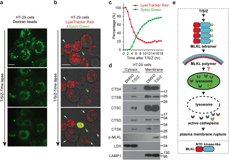
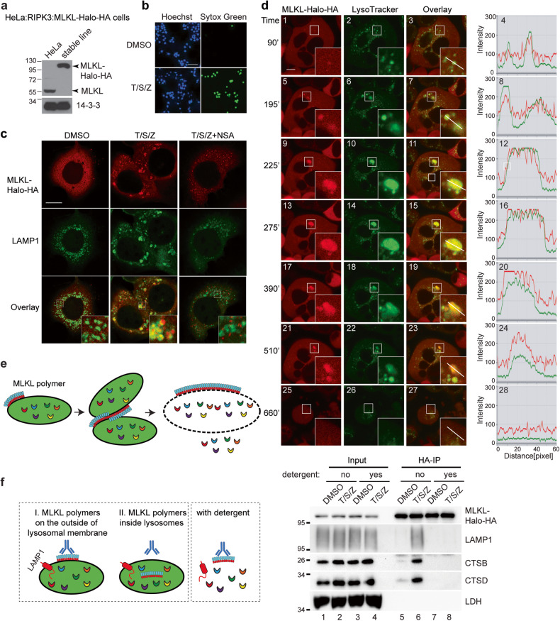
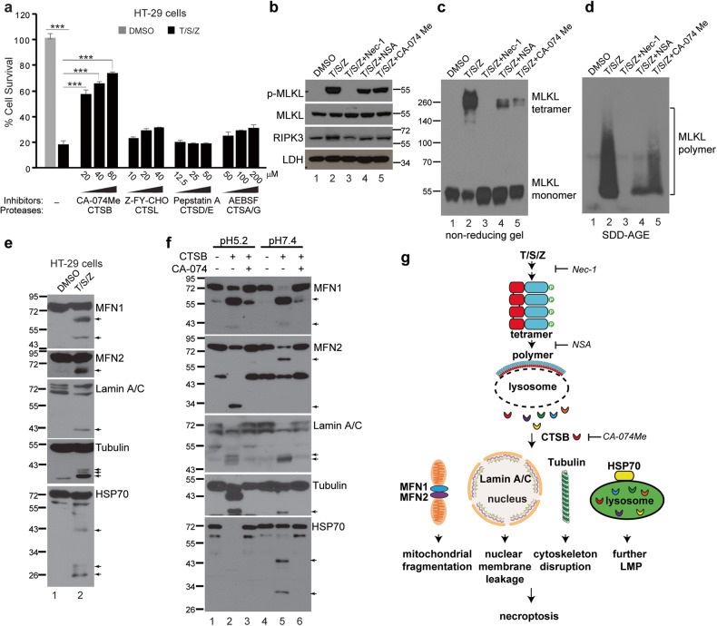
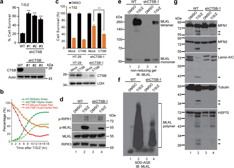
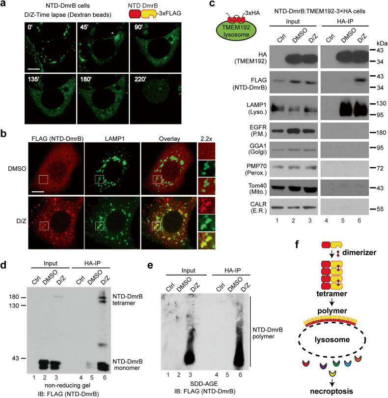
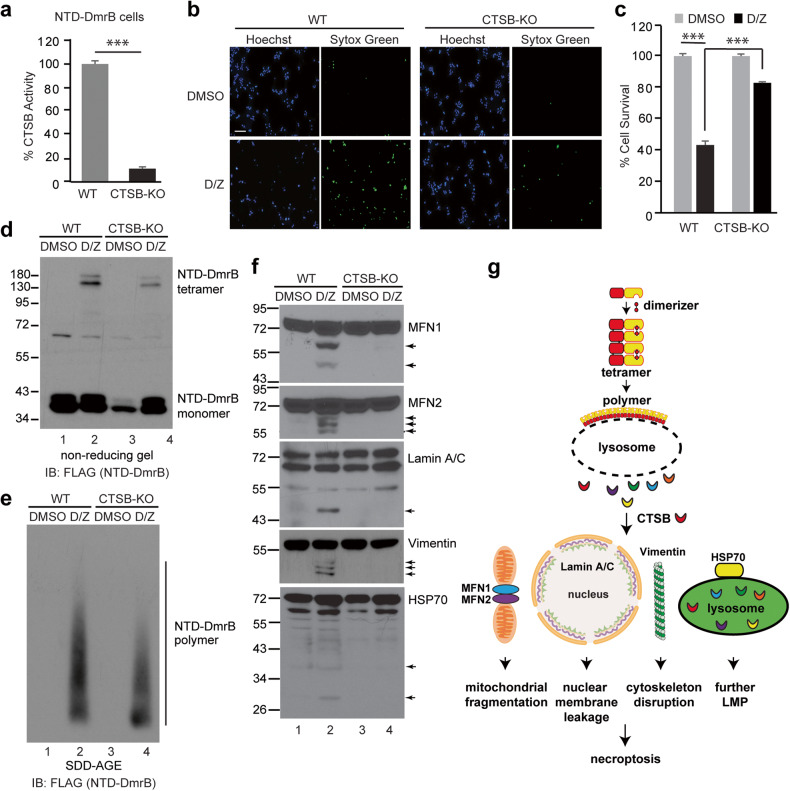
Mixed lineage kinase-like protein (MLKL) forms amyloid-like polymers to promote necroptosis; however, the mechanism through which these polymers trigger cell death is not clear. We have determined that activated MLKL translocates to the lysosomal membrane during necroptosis induction. The subsequent polymerization of MLKL induces lysosome clustering and fusion and eventual lysosomal membrane permeabilization (LMP). This LMP leads to the rapid release of lysosomal contents into the cytosol, resulting in a massive surge in cathepsin levels, with Cathepsin B (CTSB) as a significant contributor to the ensuing cell death as it cleaves many proteins essential for cell survival. Importantly, chemical inhibition or knockdown of CTSB protects cells from necroptosis. Furthermore, induced polymerization of the MLKL N-terminal domain (NTD) also triggers LMP, leading to CTSB release and subsequent cell death. These findings clearly establish the critical role of MLKL polymerization induced lysosomal membrane permeabilization (MPI-LMP) in the process of necroptosis.


Conflict of interest statement

The authors declare no competing interests.

Figures

Fig. 1
Fig. 1. Lysosomal membrane permeabilization precedes plasma membrane rupture and active cathepsins are released into cytosol during necroptosis.
a HT-29 cells were preloaded with 10 kDa Green Dextran beads overnight. Live cell imaging was recorded after treatment with TNF (T), Smac-mimetic (S) and Z-VAD-FMK (Z). Scale bar, 10 μm. b HT-29 cells were stained with 1 μM LysoTracker Red DND-99 for 2 h followed by 3 washes with PBS. Cells were then treated with 1 μM Sytox Green and T/S/Z, followed by live cell imaging. Scale bar, 10 μm. The green arrowhead identifies a cell undergoing necroptosis and the white arrowhead marks a relatively normal neighboring cell. c HT-29 cells were seeded at a density of 2000 cells/well and stained with LysoTracker Red and Sytox Green. The cells were then treated with DMSO or T/S/Z and florescent images were captured every hour for 18 h. Percentage of red and green signal intensity at each time point was calculated as described in the methods. d HT-29 cells were fractionated into cytosol and membrane fractions after DMSO or T/S/Z treatment for 4 h, followed by Western blotting with the indicated antibodies. The mature active cathepsins were shown. LDH (lactate dehydrogenase) and LAMP1 served as cytosol and membrane markers respectively. Antibody p-MLKL recognizes phospho-S358 of MLKL. e Working model. Upon activation, phosphorylated MLKL forms tetramers and later polymers, somehow leading to LMP and the release of active cathepsins into cytosol, followed by plasma membrane rupture. Diagram on the bottom shows two domains of MLKL, the N-terminal domain (NTD) and the C-terminal kinase-like domain.
Fig. 2
Fig. 2. Activated MLKL translocates to the lysosomal membrane after necroptotic induction.
a HT-29 cells were treated with DMSO or T/S/Z for 4 h, followed by staining with antibodies against p-MLKL and LAMP2. Arrowheads mark cytosolic puncta and arrows denote plasma membrane puncta. b Lysosomal membrane protein TMEM192 with 3×HA tags at C-terminus was stably expressed in HT-29 cells to establish HT-29:TMEM192-3xHA cell line. Cells were treated with DMSO or T/S/Z for 4 h and lysosomes were precipitated with anti-HA magnetic beads as described in methods. Western blotting was performed with the indicated antibodies. * denotes non-specific signals from the IgG heavy chain. For tetramer detection, non-reducing SDS-PAGE was performed. LAMP1, lysosome marker; GAPDH, cytosol marker; EGFR, plasma membrane marker; GRP78, ER marker; and Tom20, mitochondria marker. Antibodies against phospho-S166 of RIPK1 (p-RIPK1) and phospho-S227 of RIPK3 (p-RIPK3) were also used. c Immunoprecipitated lysosomes were subjected to semi-denaturing detergent agarose gel electrophoresis (SDD-AGE) and Western blotting was performed with an MLKL antibody. d Working model. Upon activation, MLKL translocates to the lysosome membrane, leading to LMP and release of active cathepsins into cytosol, and eventual plasma membrane rupture.
Fig. 3
Fig. 3. Activated MLKL polymerizes on the lysosomal membrane to promote lysosome fusion and lysosomal membrane permeabilization.
a Characterization of the HeLa:RIPK3:MLKL-Halo-HA cell line. After CRISPR/Cas9-mediated knockout of endogenous MLKL in HeLa cells, MLKL fused with C-terminal Halo-Tag and HA-tag as well as FLAG-RIPK3 was engineered to stably express in these cells. Western blotting was performed with an MLKL antibody. b HeLa:RIPK3:MLKL-Halo-HA cells were treated with DMSO or T/S/Z overnight and then stained with Hoechst and a cell-impermeable DNA dye Sytox Green. Hoechst stains all cells and Sytox Green stains dead cells with compromised cell membranes. Scale bar, 100 μm. c Cells were treated with the indicated inducers for 4 h, and the fluorescent dye TMR was added to stain MLKL-Halo. Cells were then fixed and stained with an anti-LAMP1 antibody. Insets were shown at a 4 × magnification. Scale bar: 10 μm. d HeLa:RIPK3:MLKL-Halo-HA cells were stained with TMR for MLKL-Halo and LysoTracker Green DND-26 for lysosomes. Live cell imaging was recorded after T/S/Z treatment. Insets were shown at a 3 × magnification. A line intensity profile of the inset was analyzed with Zeiss software ZEN and shown on the right. Scale bar: 10 μm. e Working model. Upon activation, MLKL polymerizes on the lysosomal membrane, and MLKL polymers on different lysosomes further polymerize to promote lysosome clustering and fusion, eventually leading to LMP. f The left panel depicts a diagram illustrating the predicted outcomes of MLKL-Halo-HA-IP based on its localization, either outside or inside lysosomes. HeLa:RIPK3:MLKL-Halo-HA cells were treated with DMSO or T/S/Z for 4 h and harvested as described for lysosome-IP. MLKL-Halo-HA fusion protein was precipitated with anti-HA magnetic beads with or without 1% Triton X-100. Western blotting was performed with the indicated antibodies.
Fig. 4
Fig. 4. Inhibition of lysosomal cysteine protease CTSB attenuates necroptosis and CTSB cleaves vital proteins at neutral pH.
a HT-29 cells were treated with DMSO or T/S/Z for 16 h in the presence of various concentrations of different protease inhibitors and cell survival was assayed by CellTiter-Glo. ***p < 0.001, mean ± SD are shown. b HT-29 cells were treated with the indicated inducers for 4 h and cell lysates were subjected to Western blotting with the indicated antibodies. 20 μM of CA-074Me, 10 μM of Nec-1 (RIPK1 inhibitor) and 5 μM of NSA (MLKL inhibitor) were used. Cell lysates were analyzed by non-reducing SDS-PAGE (c) or SDD-AGE (d) and probed with an MLKL antibody. e HT-29 cells were treated with DMSO or T/S/Z for 4 h and cell lysates were subjected to Western blotting with the indicated antibodies. Arrows denote the cleaved bands. f In vitro CTSB cleavage assay. Recombinant Tubulin or HSP70, or membrane fractions from HT-29 cells which were used as starting material for MFN1, MFN2, and Lamin A/C, were incubated with 100 ng of recombinant CTSB under conditions of pH5.2 or pH7.4, followed by Western blotting. CA-074 (20 μΜ) is a CTSB inhibitor. Arrows denote the cleaved bands. g Working model. Upon activation, MLKL tetramers form polymers on the lysosome membrane, leading to LMP and the release of active cathepsins into cytosol. Released CTSB cleaves MFN1, MFN2, Lamin A/C, Tubulin and HSP70 to promote mitochondrial fragmentation, nuclear membrane leakage, cytoskeleton disruption and further lysosome permeabilization, eventually resulting in cell death.
Fig. 5
Fig. 5. Loss of CTSB suppresses protein cleavage and necroptosis in HT-29 cells.
a Upper panel, parental HT-29 (WT) and CTSB knockdown (shCTSB) cells were treated with DMSO or T/S/Z for 16 h. Cell survival was measured by CellTiter-Glo assay. ***p < 0.001. Lower panel, Western blotting with antibodies against CTSB and Actin. b LysoTracker Red and Sytox Green staining images for HT-29 or shCTSB-1 cells were recorded and analyzed as in Fig. 1c. c Upper panel, HT-29 or shCTSB-1 cells were transfected with an empty vector or a CTSB expressing plasmid that is resistant to shRNA. Thirty-six hours later, the cells were treated with T/S/Z for 16 h and CellTiter-Glo was performed to assay cell survival. Lower panel, Western blotting with antibodies against CTSB and LDH. d Cells were treated with DMSO or T/S/Z for 4 h and cell lysates were subjected to Western blotting with the indicated antibodies. Cell lysates were analyzed with non-reducing SDS-PAGE (e) or SDD-AGE (f) and probed with an MLKL antibody. g Cell lysates were subjected to Western blotting with the indicated antibodies. Arrows denote cleaved bands.
Fig. 6
Fig. 6. NTD-DmrB forms polymers on the lysosomal membrane and induces LMP during necroptosis.
a NTD-DmrB cells were preloaded with green Dextran beads and live cell imaging was recorded following dimerizer (D) and Z-VAD-FMK (Z) treatment. Scale bar, 10 μm. b NTD-DmrB cells were treated with DMSO or D/Z for 2 h and stained with antibodies against FLAG and LAMP1. Scale bar, 10 μm. c Lysosomal membrane protein TMEM192 with 3×HA tags at C-terminus was stably expressed in NTD-DmrB cells. Cells were treated with DMSO or D/Z for 2 h and lysosomes were precipitated with anti-HA magnetic beads and Western blotting was performed with the indicated antibodies. Control sample was from DMSO-treated parental NTD-DmrB cells. LAMP1, lysosome marker; EGFR, plasma membrane marker; GGA1, Golgi marker; PMP70, peroxisome marker; Tom40, mitochondria marker; CALR (Calreticulin), ER marker. Immunoprecitated lysosomes were analyzed with non-reducing SDS-PAGE (d) or SDD-AGE (e) and probed with an anti-FLAG antibody. f Working model. Upon D/Z treatment, NTD-DmrB tetramers form polymers on lysosomal membrane and induces LMP.
Fig. 7
Fig. 7. Loss of CTSB prevents proteins cleavage and suppresses necroptosis in NTD-DmrB cells.
a CTSB was inactivated by CRISPR/Cas9-mediated knockout in NTD-DmrB cells and CTSB enzyme activity was analyzed in lysates from WT and CTSB-KO cells. ***p < 0.001. b Cells were treated with DMSO or D/Z for 8 h and stained with Hoechst and Sytox Green. Scale bar, 100 μm. c Cells were treated with DMSO or D/Z for 8 h and cell survival was analyzed by CellTiter-Glo assay. ***p < 0.001. Cells were treated for 2 h and cell lysates were analyzed by non-reducing SDS-PAGE (d) or SDD-AGE (e) and probed with a FLAG antibody. f Cell lysates were analyzed by Western blotting with the indicated antibodies. Arrows denote cleaved bands. g Working model. Upon D/Z treatment, NTD-DmrB tetramers form polymers on the lysosomal membrane, leading to LMP and release of active cathepsins into cytosol. Released CTSB cleaves MFN1, MFN2, Lamin A/C, Vimentin and HSP70 to promote mitochondrial fragmentation, nuclear membrane leakage, cytoskeleton disruption and further lysosome permeabilization, eventually resulting in cell death.

References

    1. Galluzzi L, Vitale I, Aaronson SA, Abrams JM, Adam D, Agostinis P, et al. Molecular mechanisms of cell death: recommendations of the nomenclature committee on cell death 2018. Cell Death Differ. 2018;25:486–541. doi: 10.1038/s41418-017-0012-4. - DOI - PMC - PubMed
    1. Mocarski ES, Guo H, Kaiser WJ. Necroptosis: the trojan horse in cell autonomous antiviral host defense. Virology. 2015;479-480:160–6. doi: 10.1016/j.virol.2015.03.016. - DOI - PMC - PubMed
    1. Chan FK, Luz NF, Moriwaki K. Programmed necrosis in the cross talk of cell death and inflammation. Annu Rev Immunol. 2015;33:79–106. doi: 10.1146/annurev-immunol-032414-112248. - DOI - PMC - PubMed
    1. He S, Wang X. RIP kinases as modulators of inflammation and immunity. Nat Immunol. 2018;19:912–22. doi: 10.1038/s41590-018-0188-x. - DOI - PubMed
    1. Shan B, Pan H, Najafov A, Yuan J. Necroptosis in development and diseases. Genes Dev. 2018;32:327–40. doi: 10.1101/gad.312561.118. - DOI - PMC - PubMed

Publication types

MeSH terms

Substances