Review
. 2023 Dec;26(12):2052-2062.
doi: 10.1038/s41593-023-01487-1. Epub 2023 Nov 23.

Skull bone marrow channels as immune gateways to the central nervous system

Affiliations
Review

Skull bone marrow channels as immune gateways to the central nervous system

Jose A Mazzitelli et al. Nat Neurosci. 2023 Dec.

Abstract

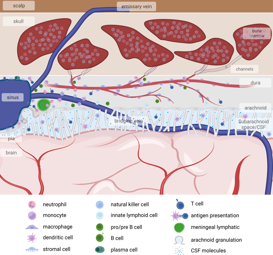
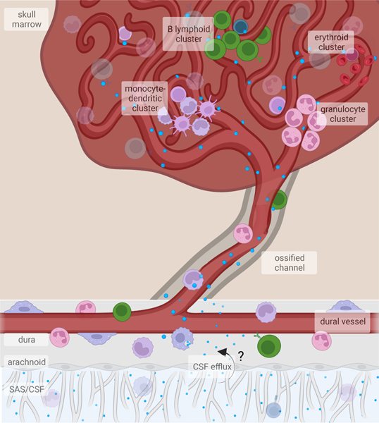
Decades of research have characterized diverse immune cells surveilling the CNS. More recently, the discovery of osseous channels (so-called 'skull channels') connecting the meninges with the skull and vertebral bone marrow has revealed a new layer of complexity in our understanding of neuroimmune interactions. Here we discuss our current understanding of skull and vertebral bone marrow anatomy, its contribution of leukocytes to the meninges, and its surveillance of the CNS. We explore the role of this hematopoietic output on CNS health, focusing on the supply of immune cells during health and disease.


Conflict of interest statement

Competing interests

J.K. is a scientific advisor for Sana Biotechnology. M.N. has received funds or material research support from Alnylam, Biotronik, CSL Behring, GlycoMimetics, GSK, Medtronic, Novartis and Pfizer, as well as consulting fees from Biogen, Gimv, IFM Therapeutics, Molecular Imaging, Sigilon, Verseau Therapeutics and Bitterroot. The other authors declare no competing interests.

Figures

Fig. 1.
Fig. 1.. Immune landscape at the outer borders of the CNS
The CNS contains three membranous coverings, termed the meninges. The innermost layer, the pia mater, is in direct contact with the brain parenchyma. Above the pia is the arachnoid mater, and the subarachnoid space between these two membranes is filled with CSF. The subarachnoid space also contains arteries and veins that extend into the brain parenchyma. Above the arachnoid lies the dura mater, which contains the venous sinuses, draining cerebral veins, as well as meningeal lymphatic vessels, draining CSF. The dura mater is connected to the overlying skull bone marrow. These meningeal layers and the skull bone marrow harbor diverse immune cell subsets, many of which play important roles in brain development, social and cognitive behaviors, and CNS antigen presentation to the rest of the immune system.
Fig. 2.
Fig. 2.. CNS-associated bone marrow channels are bidirectional conduits
CNS-associated bone marrow is anatomically connected to the underlying dural meninges through skull channels that cross the inner bone cortex into the marrow cavity. These channels contain a vessel that extends from the dura into the marrow and integrates into the sinusoidal vasculature. Within the marrow, HSPCs reside in perisinusoidal niches, where the processes of myelopoiesis, lymphopoiesis, and erythropoiesis are spatially segregated. Immune cells produced in the bone marrow traffic into the underlying meninges through channels. In addition to permitting cellular trafficking, the perivascular space allows CSF to flow into the bone marrow. Despite strong evidence supporting CSF efflux to the dura as well as the surrounding bone marrow, the precise route by which CSF is able to bypass the arachnoid mater remains unknown.
Fig. 3.
Fig. 3.. Skull bone marrow responds to CNS perturbations
Due to its proximity to the underlying CNS, the skull bone marrow can sense CNS impairment and mobilize immune supply to the underlying tissue. Alternatively, bone marrow niche perturbations can alter hematopoietic output to the CNS. a) Following CNS injury, such as spinal cord injury or stroke, HSPCs rapidly proliferate. This expands the monocyte and neutrophil supply into the underlying dura and injured CNS parenchyma. Such rapid response is due, in part, to CSF-contained cues entering the bone marrow and is likely a response to damage-associated molecular patterns (DAMP) resulting from tissue injury and cell death. Additionally, local retention cues such as Cxcl12 are downregulated, resulting in the mobilization of HSPCs and egress of monocytes and neutrophils. b) In bacterial meningitis, pathogens exploit the skull channel anatomy to invade the bone marrow. The rapid expansion of HSPCs as well as monocytes and neutrophils results from innate pathogen sensing, as well as local production of pro-inflammatory cytokines. c) In CNS autoimmune disease, autoreactive T cells home to the bone marrow and signal to hematopoietic stem cells to promote preferential over-production of monocytes and neutrophils. While this T cell homing and skewing of myelopoiesis seems to occur in both skull/vertebral bone marrow as well as femoral bone marrow, recent evidence suggests that skull/verterbal bone marrow-derived myeloid cells may be functionally distinct from their circulating counterparts during EAE. While there appears to be a conserved response in favor of myelopoiesis, whether these myeloid cells acquire phenotypes that are context-specific remains to be seen.

References

    1. Vajkoczy P, Laschinger M. & Engelhardt B. α4-integrin-VCAM-1 binding mediates G protein–independent capture of encephalitogenic T cell blasts to CNS white matter microvessels. J Clin Invest 108, 557–565 (2001). - PMC - PubMed
    1. Steinman L. Blocking adhesion molecules as therapy for multiple sclerosis: natalizumab. Nat Rev Drug Discov 4, 510–518 (2005). - PubMed
    1. Mrdjen D. et al. High-Dimensional Single-Cell Mapping of Central Nervous System Immune Cells Reveals Distinct Myeloid Subsets in Health, Aging, and Disease. Immunity 48, 380–395.e6 (2018). - PubMed
    1. Hove HV et al. A single-cell atlas of mouse brain macrophages reveals unique transcriptional identities shaped by ontogeny and tissue environment. Nat Neurosci 22, 1021–1035 (2019). - PubMed
    1. Fitzpatrick Z. et al. Gut-educated IgA plasma cells defend the meningeal venous sinuses. Nature 587, 472–476 (2020). - PMC - PubMed