. 2023 Nov 23;13(1):20541.
doi: 10.1038/s41598-023-47942-8.

Long-term HIF-1α stabilization reduces respiration, promotes mitophagy, and results in retinal cell death

Affiliations

Long-term HIF-1α stabilization reduces respiration, promotes mitophagy, and results in retinal cell death

Nana Yaa Nsiah et al. Sci Rep. .

Abstract

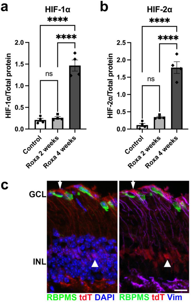
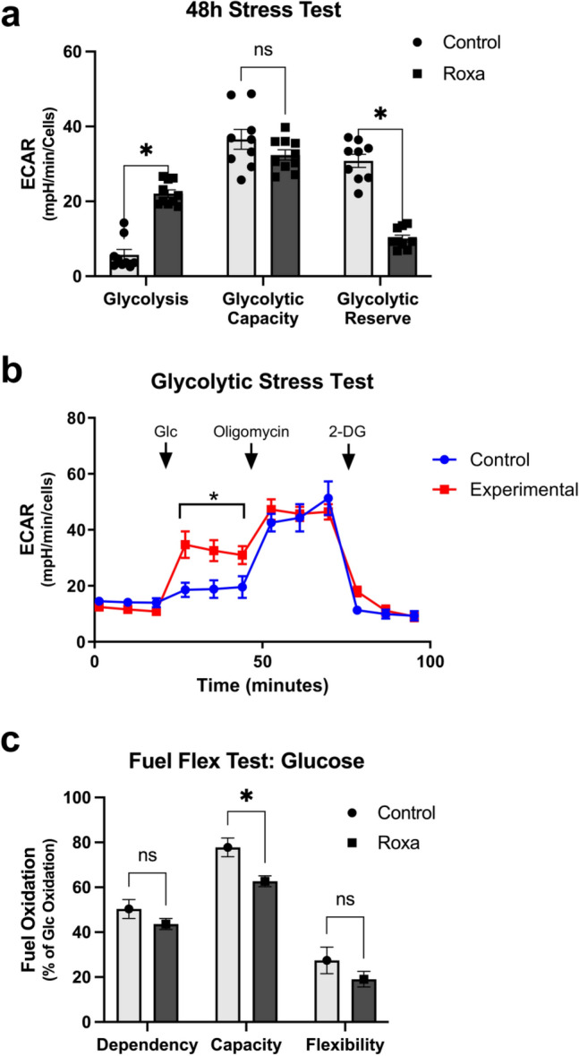
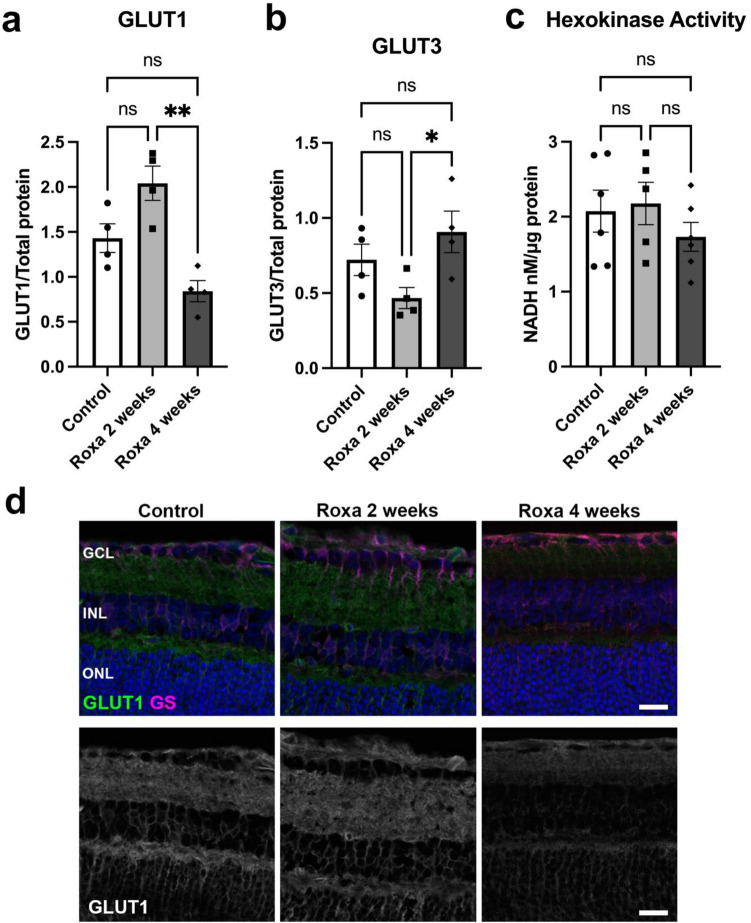
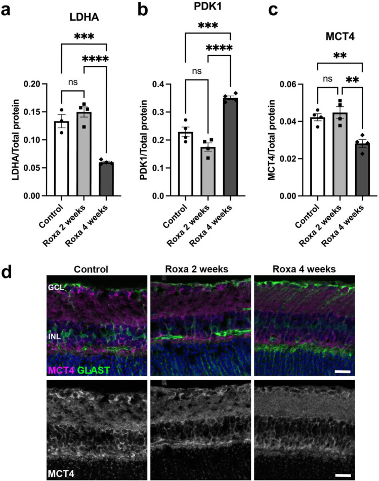
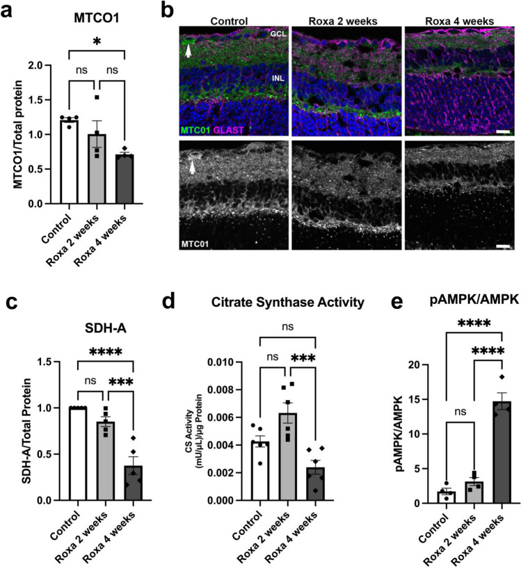
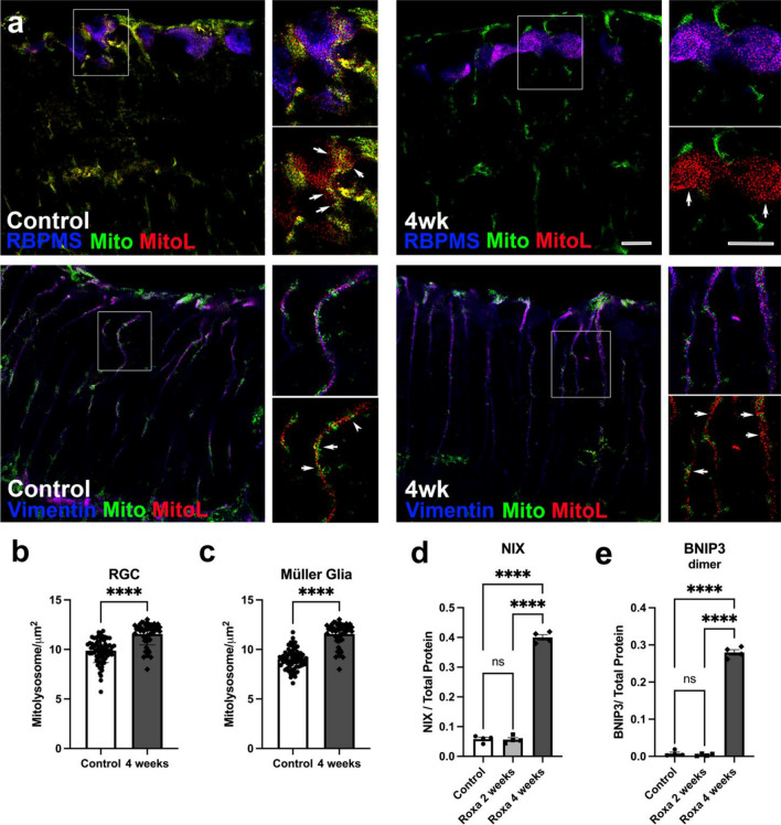
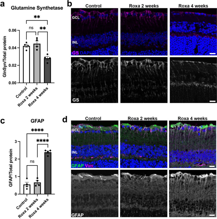
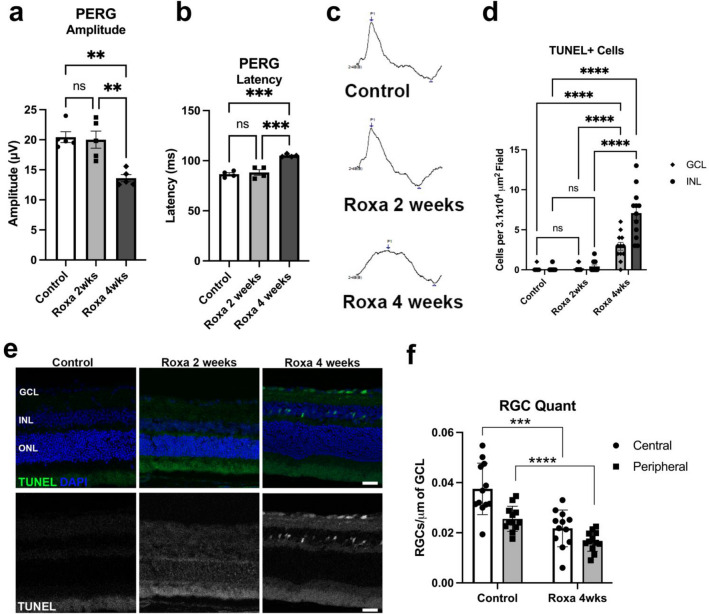
Ocular hypertension during glaucoma can lead to hypoxia, activation of the HIF transcription factors, and a metabolic shift toward glycolysis. This study aims to test whether chronic HIF activation and the attendant metabolic reprogramming can initiate glaucoma-associated pathology independently of ocular hypertension. HIF-1α stabilization was induced in mice for 2 and 4 weeks by inhibiting prolyl hydroxylases using the small molecule Roxadustat. HIF-1α stabilization and the expression of its downstream bioenergetic targets were investigated in the retina by immunofluorescence, capillary electrophoresis, and biochemical enzyme activity assays. Roxadustat dosing resulted in significant stabilization of HIF-1α in the retina by 4 weeks, and upregulation in glycolysis-associated proteins (GLUT3, PDK-1) and enzyme activity in both neurons and glia. Accordingly, succinate dehydrogenase, mitochondrial marker MTCO1, and citrate synthase activity were significantly decreased at 4 weeks, while mitophagy was significantly increased. TUNEL assay showed significant apoptosis of cells in the retina, and PERG amplitude was significantly decreased with 4 weeks of HIF-1α stabilization. A significant increase in AMPK activation and glial hypertrophy, concomitant with decreases in retinal ganglion cell function and inner retina cell death suggests that chronic HIF-1α stabilization alone is detrimental to retina metabolic homeostasis and cellular survival.


Conflict of interest statement

The authors declare no competing interests.

Figures

Figure 1
Figure 1
Verification that Roxadustat stabilizes HIF1-α in the retina. (a, b) Protein analysis of retinal lysates shows that I.P. injection of Roxadustat increased HIF-1α and HIF-2α protein in the retina. HIF-1α was significantly higher (F2,9 = 80, ****p < 0.0001) in 4-week Roxa retinas compared to 2-week Roxa and control retinas. HIF-2α was significantly higher (F2,9 = 77.23, ****p < 0.0001) in 4-week Roxa retinas compared to 2-week Roxa and control retinas. Tukey's multiple comparisons test showed a significant increase in HIF-1α and HIF-2α after 4 weeks of Roxa treatment (****p < 0.0001) compared to 2-week Roxa (****p < 0.0001) and control (****p < 0.0001) retinas. There was no difference in HIF-1α and HIF-2α in 2-week Roxa and control retinas (p = 0.8989, p = 0.7366, respectively). Values shown are mean ± SEM of n = 4 per group. C) Retinal cross-section from CAG-CreERT2-ODD x ROSA26 flox’d STOP tdTomato mice 3 days after injection with Roxa and no OHT showing tdTomato (tdT, red) expression in cells with stabilized HIF-1α. RGCs (green) and Müller glia (blue, right panel) indicate that the primary tdTomato immunolabel is in Müller glial cell somata (arrowhead), and Müller glial basal processes (arrow). The right panel of (c) is the same view as the left, though showing Vimentin immunolabel in lieu of DAPI. GCL: ganglion cell layer; INL: inner nuclear layer. Scale bar: 25 μm.
Figure 2
Figure 2
Roxadustat increases glycolysis in primary Müller glia. (a) Glycolysis, glycolytic capacity, and glycolytic reserve were calculated from the extracellular acidification rate (ECAR) in primary Müller glia exposed for 48 h to Roxadustat, as shown in (b). Müller glia exposed to Roxadustat engaged in significantly higher glycolysis as compared to control Müller glia (*p = 0.015); glycolytic capacity did not vary across the control and Roxa-treated cells. The Roxa-treated cells did exhibit significantly lower glycolytic reserve (*p = 0.02) than the control cells. (b) The moment-by-moment ECAR measures taken during the glycolytic stress test as glucose (Glc), then oligomycin, and finally, 2-deoxyglucose (2-DG) were sequentially added to the wells containing either control or Roxa-treated Müller glia. Glycolysis in the Roxa-treated cells was significantly higher than control cells (*p = 0.015). Graph shows example trace from a biological replicate. (c) Primary Müller glia subjected to a fuel flex test using glucose showed that the Roxa-treated Müller glia were no different from control in terms of their dependency on glucose or their flexibility towards glucose utilization, but they had significantly lower capacity than the control cells (*p = 0.028). Seahorse Bioanalyzer experiments were run with three separate primary Muller glial cell isolations, with 4–9 wells assessed per run for the data shown in a and c.
Figure 3
Figure 3
Glycolytic protein expression in the retina of control and Roxa-treated mice. (a) GLUT1 protein was significantly reduced (F(2,9) = 8.719, p = 0.0078) in 4-week Roxa retinas compared to 2-week and control retinas. Tukey's multiple comparisons test showed a significant reduction in GLUT1 in 4-week Roxa retina compared to 2-week Roxa retina (p = 0.0062). There was no difference in GLUT1 protein in control retina compared to 2-week Roxa (p = 0.1034) and 4-week (p = 0.2103) retinas. Values shown are mean ± SEM, n = 4 samples per group. (b) GLUT3 protein was not different between control and Roxa-treatment groups (F(2,9) = 4.204, p = 0.0514). Tukey's multiple comparisons test showed GLUT3 in control retina was comparable to 2-week Roxa (p = 0.2688) and 4-week Roxa (p = 0.4714) retinas. 4-week Roxa GLUT3 protein was significantly higher than in 2-week Roxa retinas (p = 0.0430). Values shown are mean ± SEM of n = 6 samples per group. (c) Hexokinase activity did not vary across groups (F(2, 14) = 0.8535, p = 0.4469), n = 6 samples per group. Tukey's multiple comparisons test showed no difference in hexokinase activity between groups; control vs. 2-week Roxa (p = 0.9567), control vs. 4-week Roxa (p = 0.5974), and 2-week vs. 4-week Roxa (p = 0.4593). (D) Representative immunofluorescence images showing GLUT1 (green) and glutamine synthetase (magenta) protein levels in Control, Roxa 2-week, and Roxa 4-week retina cross-sections, Scale bar = 20 µm. GCL = ganglion cell layer; INL = inner nuclear layer; ONL = outer nuclear layer. Proteins were analyzed by capillary electrophoresis normalized with total protein levels in each capillary. Bar graph data presented as mean ± SEM, n = 4–6 per group.
Figure 4
Figure 4
Effect of HIF-1α stabilization on LDHA, PDK1, and MCT4 protein expression. (a) LDHA protein was significantly reduced (F(2,8) = 38.55, p < 0.0001) in 4-week Roxa retina compared to control and 2-week retinas. Tukey's multiple comparisons test showed a significant reduction in LDHA in 2- week (p = 0.0006) and 4-week Roxa (p < 0.0001) retinas compared to control retinas. (b) PDK1 protein was significantly higher (F(2,9) = 42.73, p < 0.0001) in 4-week Roxa retina compared to control and 2-week retinas. Tukey's multiple comparisons test showed a significant PDK1 increase in 4 weeks compared to control (p = 0.0004) and 2-week Roxa (p < 0.0001) retinas. (c) MCT4 protein was significantly lower (F(2,9) = 13.64, p = 0.0019) in 4-week Roxa retina compared to control and 2-week retinas. Tukey's multiple comparisons test showed MCT4 decreased in 4 weeks compared to control (p = 0.0064) and 2-week Roxa (p = 0.0023) retinas. (d) Representative immunofluorescence images showing MCT-4 (magenta) and GLAST (green) expression in retina cross-sections for the Control, Roxa 2-week, and Roxa 4-week groups. Scale bar = 20 µm. GCL = ganglion cell layer; INL = inner nuclear layer. Proteins were analyzed by capillary electrophoresis and normalized to total protein levels. Values shown are mean ± SEM of n = 3–4 per group.
Figure 5
Figure 5
Chronic HIF-1α stabilization impairs mitochondrial function. (a) MTCO1 protein was significantly different between groups (F(2,9) = 4.814, p = 0.0379). Tukey's multiple comparisons test showed MTCO1 was significantly lower in 4-week Roxa retinas compared to control retinas (p = 0.0316). MTCO1 protein levels were not significantly different between control vs. 2-week Roxa (p = 0.4497) and 2-week vs. 4-week retinas; n = 4 per group. (b) Mitochondrial cytochrome c subunit 1 (MTCO1, green) labels cytochrome c oxidase (COX) and GLAST (magenta) labels Müller glia. Arrow points to an RGC in the GCL with MTCO1 immunolabel. GCL = ganglion cell layer; INL = inner nuclear layer. Scale bar: 20 μm, (c) SDH-A protein was significantly lower (F(2,12) = 26.24, p < 0.0001) in Roxa-treated retinas compared to control. Tukey's multiple comparisons test showed SDH-A decreased in 4-week Roxa retinas (n = 6) compared to control (n = 6, p < 0.0001) and 2-week Roxa (n = 6, p = 0.0005) retinas. There were no differences in SDHA protein levels between control and 2-week Roxa retinas (p = 0.264). (d) Citrate synthase activity was significantly reduced in 4-week Roxa retinas (F(2, 15) = 12.26, p = 0.0007), n = 6 samples per group. Tukey's multiple comparisons test showed citrate synthase activity decreased in 4-week Roxa compared to 2-week Roxa (p = 0.0005). There was no significant difference in citrate synthase activity in control vs. 2-week Roxa (p = 0.05) and control vs. 4-week Roxa (p = 0.0788). (e) The ratio of pAMPK to AMPK was significantly higher (F(2,9) = 76.26, p < 0.0001) in 4-week Roxa retina compared to control and 2-week retinas. Tukey's multiple comparisons test showed pAMPK to AMPK ratio increased in 4-week retinas compared to control (p < 0.0001) and 2-week Roxa (p < 0.0001) retinas; n = 4 samples per group.
Figure 6
Figure 6
Roxa treatment upregulates mitophagy in RGCs. (a) Sections of control retina (left) and retina after 4 weeks of Roxa treatment (right) depict mitochondria (green and red, ‘Mito’) and mitolysosomes (red, ‘MitoL’) in RGCs immunolabeled for RBPMS (blue, top panels), and in Müller glia immunolabeled for Vimentin (blue, lower panels). Mitolysosomes in RGCs (b) and Müller glia (c) were significantly increased after 4 weeks of Roxa treatment; p < 0.0001 by Student’s t-test for RGCs (b) and Müller glia (c). Values are mean ± SEM of n = 6 retinas per group. (d) NIX was significantly higher (F(2,9) = 759.6, p < 0.0001) in 4-week Roxa retinas compared to control and 2-week retinas. Tukey's multiple comparisons test showed NIX levels were significantly higher in 4-week retinas compared to control (p < 0.0001) and 2-week Roxa (p < 0.0001) retinas. There was no significant difference between control vs. 2-week Roxa retinas (p = 0.9848); n = 4 per group. (e) BNIP3 dimer was significantly higher (F(2,9) = 1050, p < 0.0001) in 4-week Roxa retinas compared to control and 2-week retinas. Tukey's multiple comparisons test showed BNIP3 dimer levels were significantly higher in 4-week retinas compared to control (p < 0.0001) and 2-week Roxa (p < 0.0001) retinas. There was no significant difference between control vs. 2-week Roxa retinas (p = 0.8791). Values shown are mean ± SEM of n = 4–6 per group. Proteins were analyzed by capillary electrophoresis and normalized to total protein levels.
Figure 7
Figure 7
HIF-1α stabilization is associated with reactive gliosis. (a) GS protein was significantly lower (F(2,9) = 13.64, p = 0.0019) in 4-week Roxa retina compared to control and 2-week retinas. Tukey's multiple comparisons test showed GS decreased in 4-week Roxa compared to control (p = 0.0067) and 2-week Roxa (p = 0.0023) retinas. There were no significant differences in GS protein quantity in control vs. 2-week retinas (p = 0.7453). (b) Representative immunofluorescence image showing GS (magenta) on retinal cross-sections. (c) GFAP was significantly higher (F(2,9) = 1050, p < 0.0001) in 4-week Roxa retinas compared to control and 2-week retinas. Tukey's multiple comparisons test showed GFAP levels were significantly higher in 4-week retinas compared to control (p < 0.0001) and 2-week Roxa (p < 0.0001) retinas. There was no significant difference between control vs. 2-week Roxa retinas (p = 0.7241). (d) Immunofluorescence image showing GFAP (green) and vimentin (MG marker) on retinal cross-sections. GCL = ganglion cell layer; INL = inner nuclear layer. Scale bar: 20 μm. Values shown are mean ± SEM of n = 4 per group.
Figure 8
Figure 8
Chronic HIF-1α stabilization on RGC function. (a) PERG amplitude was significantly lower in 4-week Roxa mice compared to control and 2-week Roxa mice (F(2,12) = 13.64, p = 0.0008). Tukey's multiple comparisons test showed PERG amplitude decreased in 4-week Roxa compared to control (p = 0.0015) and 2-week Roxa (p = 0.0024) retinas. There were no significant differences in PERG amplitude between control vs. 2-week retinas (p = 0.9559). Values shown are mean ± SEM of n = 5 per group. (b) PERG latency was significantly higher in 4-week Roxa mice compared to control and 2-week Roxa mice (F(2,9) = 28.94, p = 0.0001). Tukey's multiple comparisons test showed PERG latency increased in 4-week Roxa compared to control (p = 0.0002) and 2-week Roxa (p = 0.0004) retinas. There were no significant differences in PERG latency between control vs. 2-week retinas (p = 0.7960). Values shown are mean ± SEM of n = 4 per group. (c) Representative sample PERG traces from the Control, Roxa 2-week, and Roxa 4-week groups. (d)TUNEL-positive cell quantification in the GCL and INL of control and Roxa 4-week retinal cross-sections. There was no difference between control and 2-week Roxa GCL and INL for TUNEL + cell number, while 4-week Roxa treatment resulted in significant increases in GCL and INL when compared to control retina (p < 0.0001) and 2-week Roxa (p < 0.0001); two-way ANOVA with multiple comparisons, n = 6 retinas per group, with two fields per retina. (e) TUNEL staining (green) of retinal cross-sections from control and 2 and 4-week Roxa-treated mice. Nuclei stained with DAPI. (f) Quantification of RGCs in central and peripheral regions of control and 4-week Roxa-treated mouse retinas; n = 6 retinas per group, with two fields per retina. Control retina had significantly greater RGC number than central (p < 0.003) and peripheral (p < 0.0001) 4-week Roxa-treated central and peripheral retina, respectively; unpaired t-test. GCL = ganglion cell layer; INL = inner nuclear layer; ONL = outer nuclear layer. Scale bar: 20 μm. Data are shown as mean ± SEM.

Similar articles

Cited by

References

    1. Casson RJ, Chidlow G, Wood JPM, Crowston JG, Goldberg I. Definition of glaucoma: Clinical and experimental concepts. Clin. Exp. Ophthalmol. 2012;40:341–349. doi: 10.1111/j.1442-9071.2012.02773.x. - DOI - PubMed
    1. Tezel G, Wax MB. Hypoxia-inducible factor 1alpha in the glaucomatous retina and optic nerve head. Arch. Ophthalmol. 2004;122:1348–1356. doi: 10.1001/archopht.122.9.1348. - DOI - PubMed
    1. Chidlow G, Wood JPM, Casson RJ. Investigations into hypoxia and oxidative stress at the optic nerve head in a rat model of glaucoma. Front. Neurosci. 2017;11:478. doi: 10.3389/fnins.2017.00478. - DOI - PMC - PubMed
    1. Jassim, A. H. & Inman, D. M. Evidence of hypoxic glial cells in a model of ocular hypertension. Invest Ophthalmol. Vis. Sci.60, (2019). - PMC - PubMed
    1. Ergorul C, et al. Hypoxia inducible factor-1α (HIF-1α) and some HIF-1 target genes are elevated in experimental glaucoma. J. Mol. Neurosci. 2010;42:183–191. doi: 10.1007/s12031-010-9343-z. - DOI - PMC - PubMed

Substances