

[18F]PSMA-1007 PET is comparable to [99mTc]Tc-DMSA SPECT for renal cortical imaging
- PMID: 37996712
- PMCID: PMC10667166
- DOI: 10.1186/s41824-023-00185-2
[18F]PSMA-1007 PET is comparable to [99mTc]Tc-DMSA SPECT for renal cortical imaging
Abstract
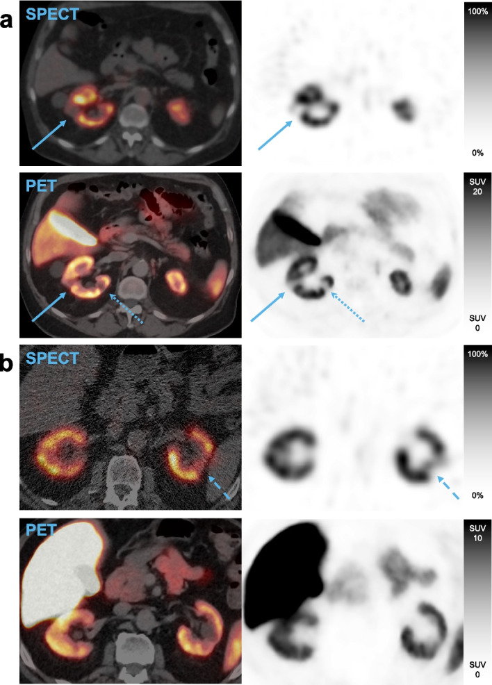
Background: Scintigraphy using technetium-99m labelled dimercaptosuccinic acid ([99mTc]Tc-DMSA), taken up in the proximal tubules, is the standard in functional imaging of the renal cortex. Recent guidelines recommend performing [99mTc]Tc-DMSA scintigraphy with single photon emission computed tomography (SPECT). Prostate-specific membrane antigen (PSMA) targeted positron emission tomography (PET) is used for staging and localization of recurrence in prostate cancer. A high renal uptake is often seen on PSMA PET, concordant with known PSMA expression in proximal tubules. This suggests PSMA PET could be used analogous to [99mTc]Tc-DMSA scintigraphy for renal cortical imaging. [18F]PSMA-1007 is a promising radiopharmaceutical for this purpose due to low urinary clearance. In this study, we aimed to compare [18F]PSMA-1007 PET to [99mTc]Tc-DMSA SPECT regarding split renal uptake and presence of renal uptake defects, in patients with prostate cancer. Three readers interpreted PET and SPECT images regarding presence of renal uptake defects, with each kidney split into cranial, mid and caudal segments. Kidneys were segmented in PET and SPECT images, and left renal uptake as a percentage of total renal uptake was measured.
Results: Twenty patients with prostate cancer were included. 2 participants had single kidneys; thus 38 kidneys were evaluated. A total of 29 defects were found on both [99mTc]Tc-DMSA SPECT and [18F]PSMA-1007 PET. Cohen's kappa for concordance regarding presence of any defect was 0.76 on a per-segment basis and 0.67 on a per-kidney basis. Spearman's r for left renal uptake percentage between [99mTc]Tc-DMSA SPECT and [18F]PSMA-1007 PET was 0.95.
Conclusions: [18F]PSMA-1007 PET is comparable to [99mTc]Tc-DMSA SPECT for detection of uptake defects in this setting. Measurements of split renal function made using [18F]PSMA-1007 PET are valid and strongly correlated to measurements made with [99mTc]Tc-DMSA SPECT.
Keywords: DMSA; PET; PSMA; Renal cortex; Renal function; SPECT.
© 2023. The Author(s).
Conflict of interest statement
The authors declare that they have no competing interests.
Figures


References
-
- Altman DG, Bland JM. Measurement in medicine: the analysis of method comparison studies. J R Stat Soc Series D (The Statistician) 1983;32:307–317. doi: 10.2307/2987937. - DOI
-
- Donswijk ML, Wondergem M, de Wit-van Veen L, Bruin NM, van Leeuwen PJ, van der Poel HG, et al. Effects of furosemide and tracer selection on urinary activity and peri-bladder artefacts in PSMA PET/CT: a single-centre retrospective study. EJNMMI Res. 2022;12:42. doi: 10.1186/s13550-022-00913-y. - DOI - PMC - PubMed
LinkOut - more resources
Full Text Sources
Miscellaneous