

Two-way adjustable double-knots intrascleral fixation and single sclerotomy looping technique: a novel minimal invasive adjustable intraocular lens fixation technique
- PMID: 37996816
- PMCID: PMC10668428
- DOI: 10.1186/s12886-023-03235-2
Two-way adjustable double-knots intrascleral fixation and single sclerotomy looping technique: a novel minimal invasive adjustable intraocular lens fixation technique
Abstract
Background: IOL fixation without capsular support presents challenges for surgeons. Although innovative techniques were developed to address subluxated IOLs, adjustable IOL fixation methods are seldom reported. We introduce a novel two-way adjustable double-knots intrascleral fixation combined with single sclerotomy looping technique for fixing intraocular lenses (IOL) or IOL-capsular bags.
Methods: A bent 30-gauge needle threaded with 8 - 0 polypropylene was introduced into the eye. A gripping forceps assisted the haptic looping. Two overhand knots were made with 8 - 0 polypropylene thread. The knots were incarcerated into a scleral tunnel made by a 30-gauge needle, with two ends of the thread left at each side of the tunnel. The IOL was adjusted to the premium position with adequate tension by pulling either end of the threads. The study included 19 eyes with aphakia, subluxated IOL-capsular bags, or subluxated crystalline lenses. The mean followed up period was 18.9 ± 7.1 months with evaluations of uncorrected visual acuity (UCVA), intraocular pressure, slit-lamp examination, and swept-source optical coherence tomography of the anterior segment.
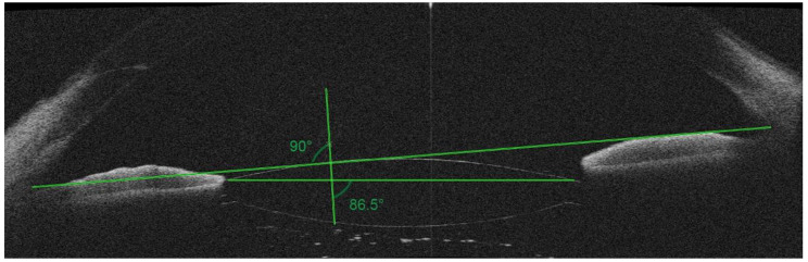
Results: UCVA increased from 1.28 ± 0.74 at baseline to 0.44 ± 0.51 (logMAR) at final visit (P < 0.001). All IOLs were fixed well-centered. The mean IOL tilt was 3.5°±1.1°. Postoperative complications included transient IOP elevation (15.8%), hypotony (10.5%), and cystoid edema (5.3%) which resolved within 4 weeks.
Conclusions: We presented a novel adjustable technique for IOL fixation, which stabilize IOLs by using an intrascleral double-knots structure. This technique minimized surgical manipulations by using a single sclerotomy looping technique without large conjunctival dissection and scleral flap creation. The technique offers a reliable and optimal IOL positioning and improved visual outcomes in patients undergoing scleral fixed IOL implantation.
Keywords: Adjustable; Intraocular lens; Surgical technique; Transscleral fixation.
© 2023. The Author(s).
Conflict of interest statement
The authors declare no competing interests.
Figures

Similar articles
-
Novel use of an adjustable single 8-0 polypropylene suture of scleral fixation without conjunctival dissection.BMC Ophthalmol. 2020 Jul 25;20(1):304. doi: 10.1186/s12886-020-01558-y. BMC Ophthalmol. 2020. PMID: 32711502 Free PMC article.
-
A modified intrascleral intraocular lens fixation technique with fewer anterior segment manipulations: 27-gauge needle-guided procedure with built-in 8-0 absorbable sutures.BMC Ophthalmol. 2019 Nov 21;19(1):234. doi: 10.1186/s12886-019-1239-2. BMC Ophthalmol. 2019. PMID: 31752875 Free PMC article.
-
Flanged Intrascleral Intraocular Lens Fixation with Double-Needle Technique.Ophthalmology. 2017 Aug;124(8):1136-1142. doi: 10.1016/j.ophtha.2017.03.036. Epub 2017 Apr 27. Ophthalmology. 2017. PMID: 28457613
-
[Sutureless scleral intraocular lens fixation: report of nine cases and literature review].J Fr Ophtalmol. 2013 Oct;36(8):658-68. doi: 10.1016/j.jfo.2012.09.009. Epub 2013 Jul 25. J Fr Ophtalmol. 2013. PMID: 23891322 Review. French.
-
Intrascleral IOL Fixation.Asia Pac J Ophthalmol (Phila). 2017 Jul-Aug;6(4):381-387. doi: 10.22608/APO.2017158. Epub 2017 Jun 26. Asia Pac J Ophthalmol (Phila). 2017. PMID: 28726356 Review.
Cited by
-
Re-fixation Using the Belt Loop Technique for Postoperative Subluxation of a Multifocal Intraocular Lens After Retinal Detachment Surgery: A Case Report.Cureus. 2025 Feb 24;17(2):e79585. doi: 10.7759/cureus.79585. eCollection 2025 Feb. Cureus. 2025. PMID: 40161112 Free PMC article.
References
MeSH terms
Substances
LinkOut - more resources
Full Text Sources
