. 2023 Nov 23;20(1):45.
doi: 10.1186/s12989-023-00555-5.

Perinatal foodborne titanium dioxide exposure-mediated dysbiosis predisposes mice to develop colitis through life

Affiliations

Perinatal foodborne titanium dioxide exposure-mediated dysbiosis predisposes mice to develop colitis through life

Caroline Carlé et al. Part Fibre Toxicol. .

Erratum in

Abstract

Background: Perinatal exposure to titanium dioxide (TiO2), as a foodborne particle, may influence the intestinal barrier function and the susceptibility to develop inflammatory bowel disease (IBD) later in life. Here, we investigate the impact of perinatal foodborne TiO2 exposure on the intestinal mucosal function and the susceptibility to develop IBD-associated colitis. Pregnant and lactating mother mice were exposed to TiO2 until pups weaning and the gut microbiota and intestinal barrier function of their offspring was assessed at day 30 post-birth (weaning) and at adult age (50 days). Epigenetic marks was studied by DNA methylation profile measuring the level of 5-methyl-2'-deoxycytosine (5-Me-dC) in DNA from colic epithelial cells. The susceptibility to develop IBD has been monitored using dextran-sulfate sodium (DSS)-induced colitis model. Germ-free mice were used to define whether microbial transfer influence the mucosal homeostasis and subsequent exacerbation of DSS-induced colitis.

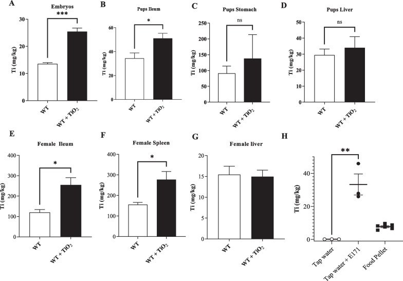
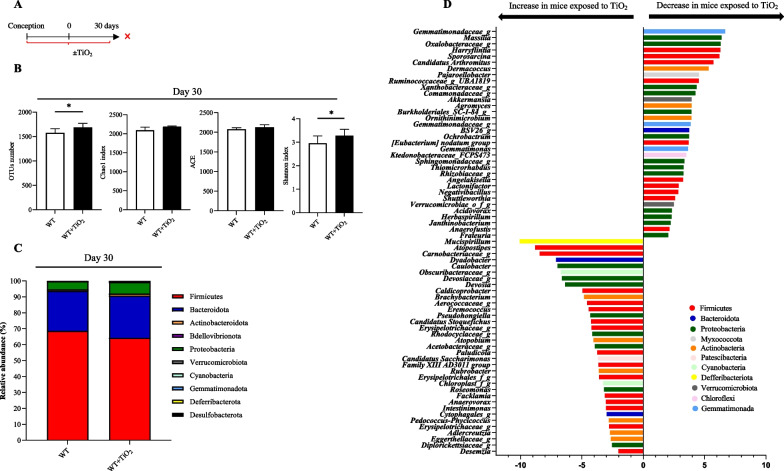
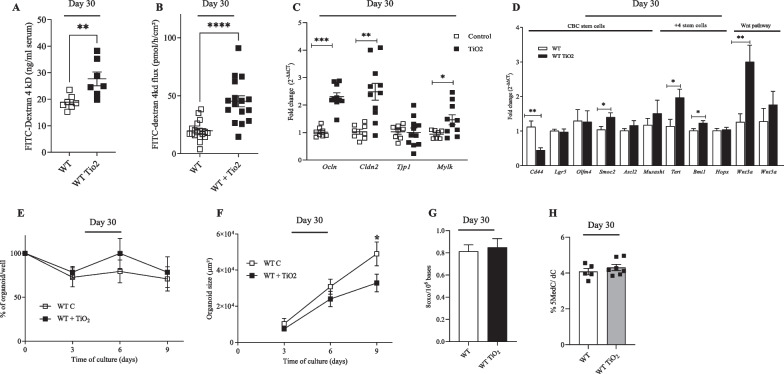
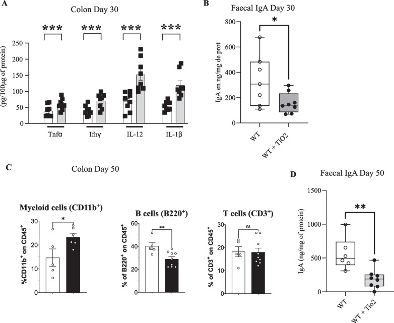
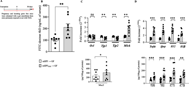
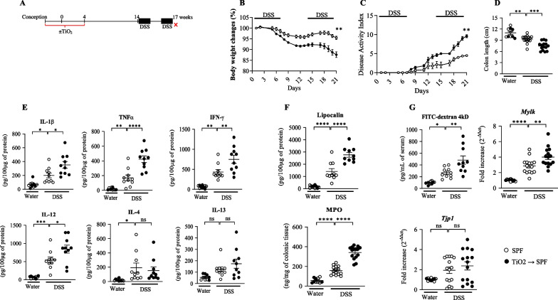
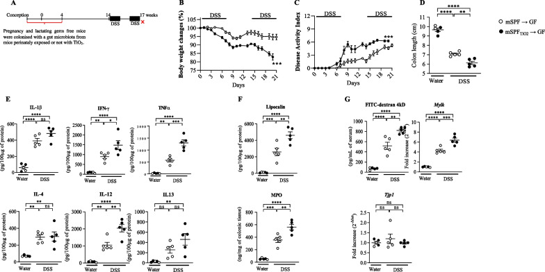
Results: In pregnant and lactating mice, foodborne TiO2 was able to translocate across the host barriers including gut, placenta and mammary gland to reach embryos and pups, respectively. This passage modified the chemical element composition of foetus, and spleen and liver of mothers and their offspring. We showed that perinatal exposure to TiO2 early in life alters the gut microbiota composition, increases the intestinal epithelial permeability and enhances the colonic cytokines and myosin light chain kinase expression. Moreover, perinatal exposure to TiO2 also modifies the abilities of intestinal stem cells to survive, grow and generate a functional epithelium. Maternal TiO2 exposure increases the susceptibility of offspring mice to develop severe DSS-induced colitis later in life. Finally, transfer of TiO2-induced microbiota dysbiosis to pregnant germ-free mice affects the homeostasis of the intestinal mucosal barrier early in life and confers an increased susceptibility to develop colitis in adult offspring.

Conclusions: Our findings indicate that foodborne TiO2 consumption during the perinatal period has negative long-lasting consequences on the development of the intestinal mucosal barrier toward higher colitis susceptibility. This demonstrates to which extent environmental factors influence the microbial-host interplay and impact the long-term mucosal homeostasis.

Keywords: Colitis; Foodborne TiO2; Intestinal barrier function; Intestinal stem cells; Microbiota; Perinatal period.


Conflict of interest statement

The authors declare no competing interests.

Figures

Fig. 1
Fig. 1
Abilities of foodborne TiO2 to translocate across the human barriers. AG Wild type female mice have been exposed to TiO2 (9 mg/Kg of BW/Day) during the perinatal period including gestational and lactating periods. Then pregnant and lactating females exposed to TiO2 or not, have been sacrificed and the concentrations of Ti in embryos (A; gestational day 20), pup’s organs (BD; postnatal day 12) and in female organs (EG; end of the weaning day 30), have been monitored by ICP-MS. H The concentrations of titanium have been determined by ICP-MS in regular food pellet and in tap water before and after the addition of TiO2 foodborne to reach the exposure dose: 9 mg of TiO2/kg of body weight/day. Data are expressed as mean ± SEM and were analysed by Student’s t-test. *p < 0.05, **p < 0.01 and ***p < 0.001 versus control group. At least n = 3 per group
Fig. 2
Fig. 2
Impact of perinatal exposure to foodborne TiO2 on colonic microbiota at day 30. AD Wild type female mice have been exposed to TiO2 (9 mg/Kg of BW/Day) during the perinatal period including gestational and lactating periods. Then at 30 days after birth, pups have been sacrificed and the structure of the colonic mucosa-associated microbiota has been monitored by 16S rRNA gene sequencing (BD). B Alpha diversity of colonic mucosal microbiota from exposed or non-exposed mice to foodborne TiO2 at day 30 after birth. C Composition of colonic microbiota at phyla level (C) and Fold changes 2 for bacterial genera significantly perturbed (D) from exposed or non-exposed mice to foodborne TiO2 at day 30 after birth. Data are expressed as median ± SEM and were analysed by Mann and Whitney test. *p < 0.05 versus control group. At least n = 8 per group
Fig. 3
Fig. 3
Impact of perinatal exposure to foodborne TiO2 on colonic epithelium at day 30. AD Wild type female mice have been exposed to TiO2 (9 mg/Kg of BW/Day) during the perinatal period including gestational and lactating periods. Then at days 30 after birth (weaning), pups have been sacrificed and several parameters including permeability (A and B), mRNA expression (C and D), abilities of intestinal stem cells to survive (E) and proliferate (F), oxidative stress (G) and DNA methylation (H) were assessed. A In vivo permeability was determined by measuring the level of plasmatic FITC-dextran 4 kDa, 3 h following oral administration. B Colonic permeability was monitored by measuring the flux of FITC-dextran 4 kDa across colonic biopsies mounted in Ussing chamber for 1 h. C mRNA expression of Occludin (Ocl), Claudin 2 (Cldn 2) Tight junction protein 1 (Tpj1) and myosin light chain kinase (Mlck) was studied at days 30 and 50 after birth. D Colonic mRNA expression of CD44, Leucine-rich repeat-containing G-protein coupled receptor 5 (Lgr5), Olfactome-din 4 (Olfm4), SPARC-related modular calcium-binding protein 2 (Smoc2), Achaete-scute complex homolog 2 (Ascl2), Musashi RNA-binding protein 1 (Musashi), Telomerase reverse transcriptase (Tert) and B lymphoma Mo-MLV insertion region 1 homolog (Bmi1), homeodomain-only protein homeobox (Hopx), canonical (Wnt3a) and non-canonical (Wnt5a). E The organoid survival has been monitored by measuring the percentage of viable organoids according to the time culture. F The organoid growth has been studied by measuring the organoid surface according to the time culture. G Oxidative stress has been monitored into the epithelial cells from the colonic based crypt of mice perinatally exposed or not to TiO2. H The quantity of cytosine methylated of DNA from the epithelial cells from the colonic based crypt of mice perinatally exposed or not to TiO2. Data are expressed as mean ± SEM and were analysed by Student’s t-test. *p < 0.05; **p < 0.01; ***p < 0.001 and ****p < 0.0001 vs control group. At least n = 8 per group
Fig. 4
Fig. 4
Impact of perinatal exposure to TiO2 foodborne on intestinal immune system. AD Wild type female mice have been exposed to TiO2 (9 mg/kg of BW/Day) during the perinatal period including gestational and lactating periods. A and B Then, at day 30 after birth, pups have been sacrificed and colonic protein expression of cytokines (A) and faecal levels of IgA (B) have been monitored. C and D At day 50 after birth, immune cells population of the colon (C) and faecal levels of IgA (D) have been measured. Data are expressed as mean ± SEM and were analysed by Student’s t-test. *p < 0.05 and **p < 0.01 versus control group. At least n = 8 per group
Fig. 5
Fig. 5
Impact of gut microbiota dysbiosis induced by perinatal exposure to foodborne TiO2 on the intestinal barrier function of offspring. AD Germ free female mice have been exposed to gut microbiota dysbiosis induced by perinatal foodborne until the weaning i.e. postnatal days 30. B Then, in vivo permeability was determined by measuring the level of plasmatic FITC-dextran 4 kDa, 3 h following oral administration. C, D On scrapped colonic epithelium, mRNA expression of Il1b, Il12, Tnfa, Ifng, Occludin (Ocl), Tight junction protein 1 (Tpj1) and Tight junction protein 2 (Tpj2) and myosin light chain kinase (Mlck) have been monitored at postnatal days 30. Data are expressed as mean ± SEM and were analysed by Student’s t-test. **p < 0.01 and ***p < 0.001 versus control group. At least n = 5 per group
Fig. 6
Fig. 6
Impact of perinatal exposure to foodborne TiO2 on susceptibility to develop colitis later in life. AG Wild type female mice have been exposed to TiO2 (9 mg/Kg of BW/Day) during the perinatal period including gestational and lactating periods (A). At 14 weeks of age, colitis has been orally induced by introducing Dextran Sulfate Sodium (DSS) into drinking water at 2% for 7 days followed by 7 days of regular water then 7 days of DSS (A). B and C, During the experimental DSS procedure (21 days), body weight and the disease activity index (DAI) have been monitored daily. DG Then, at the end of the DSS procedure, mice have been sacrificed and colonic length (D) and, cytokine expression (E) have been monitored. F and G Finally, lipocalin, MPO, permeability and mRNA expression of Mlck and Tpj1 have also been studied. Data are expressed as mean ± SEM and were analysed by Student’s t-test. *p < 0.05; **p < 0.01; ***p < 0.001 and ****p < 0.0001 versus control group. At least n = 5 per group
Fig. 7
Fig. 7
Impact of gut microbiota dysbiosis induced by perinatal exposure to TiO2 on the susceptibility to develop colitis later in life. AG Germ free mice female have been exposed to gut microbiota dysbiosis induced by perinatal foodborne until the weaning i.e. postnatal day 30 (A). At 14 weeks of age, colitis has been orally induced by introducing Dextran Sulfate Sodium (DSS) into drinking water at 2% for 7 days followed by 7 days of regular water then 7 days of DSS (A). B and C During the experimental DSS procedure (21 days), body weight and the disease activity index (DAI) have been monitored daily. DG Then, at the end of the DSS procedure, mice have been sacrificed and colonic length (D), cytokines expression (E) have been monitored. F and G Finally, lipocalin, MPO, permeability and mRNA expression of Mlck and Tpj1 have also been studied. Data are expressed as mean ± SEM and were analysed by Student’s t-test. *p < 0.05; **p < 0.01; ***p < 0.001 and ****p < 0.0001 versus control group. At least n = 5 per group

Similar articles

Cited by

References

    1. Turner JR. Intestinal mucosal barrier function in health and disease. Nat Rev Immunol. 2009;9(11):799–809. doi: 10.1038/nri2653. - DOI - PubMed
    1. Lievin-Le Moal V, Servin AL. The front line of enteric host defense against unwelcome intrusion of harmful microorganisms: mucins, antimicrobial peptides, and microbiota. Clin Microbiol Rev. 2006;19(2):315–337. doi: 10.1128/CMR.19.2.315-337.2006. - DOI - PMC - PubMed
    1. Hollingsworth MA, Swanson BJ. Mucins in cancer: protection and control of the cell surface. Nat Rev Cancer. 2004;4(1):45–60. doi: 10.1038/nrc1251. - DOI - PubMed
    1. Barreau F, Hugot J. Intestinal barrier dysfunction triggered by invasive bacteria. Curr Opin Microbiol. 2014;17C:91–98. doi: 10.1016/j.mib.2013.12.003. - DOI - PubMed
    1. Renz H, Brandtzaeg P, Hornef M. The impact of perinatal immune development on mucosal homeostasis and chronic inflammation. Nat Rev Immunol. 2012;12(1):9–23. doi: 10.1038/nri3112. - DOI - PubMed

Publication types

Substances