. 2023 Nov 24;21(1):444.
doi: 10.1186/s12951-023-02227-0.

Pathologically catalyzed physical coating restores the intestinal barrier for inflammatory bowel disease therapy

Affiliations

Pathologically catalyzed physical coating restores the intestinal barrier for inflammatory bowel disease therapy

Yuge Zhao et al. J Nanobiotechnology. .

Abstract

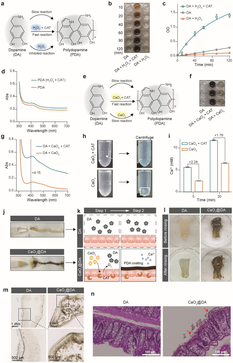
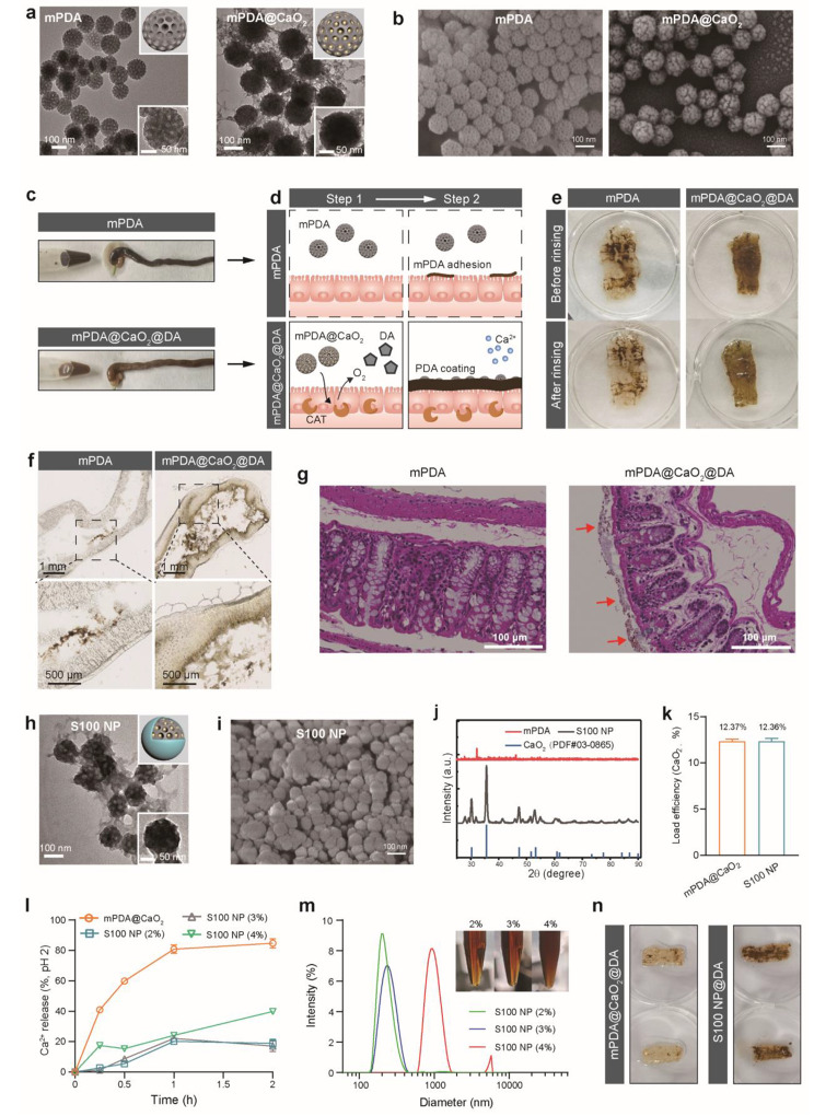
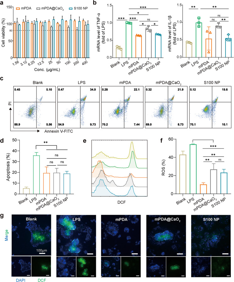
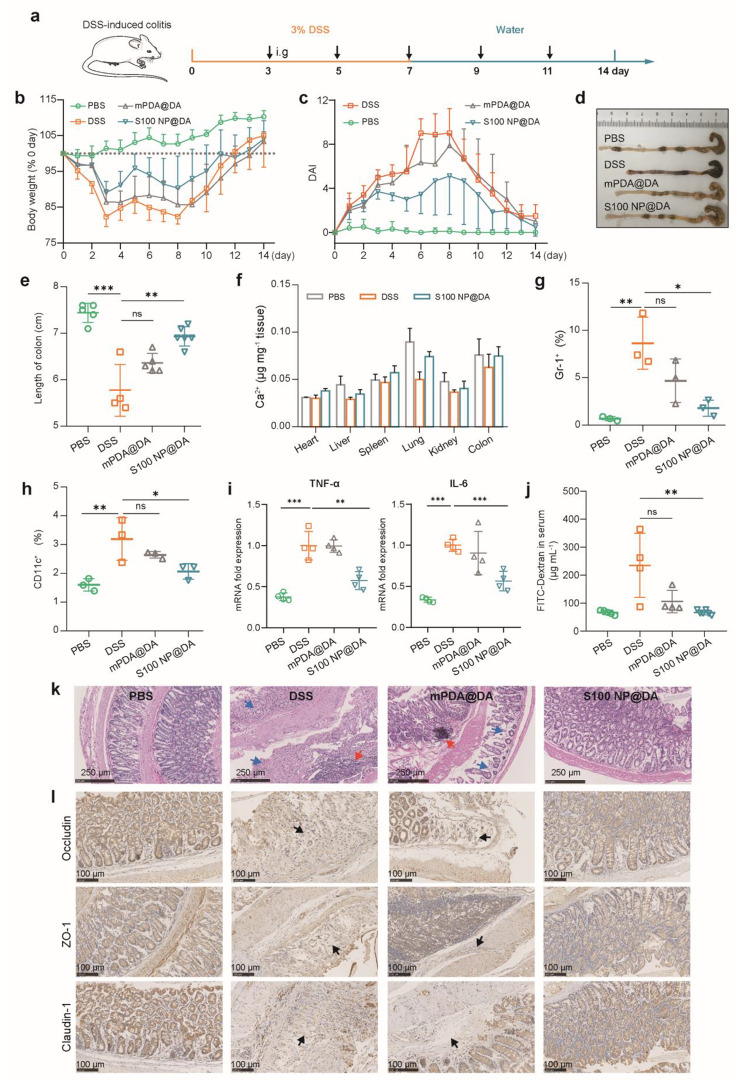
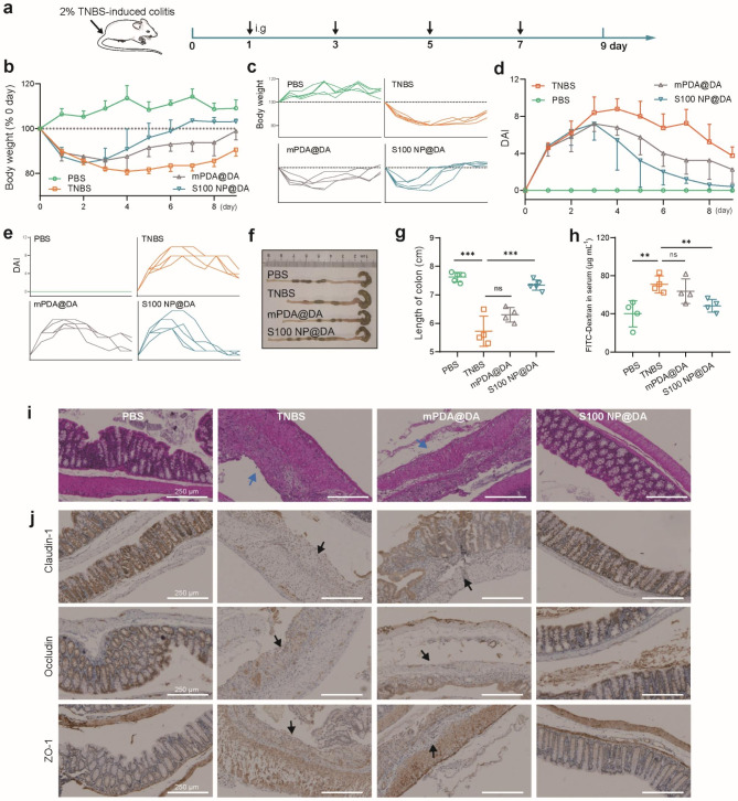
Intestinal epithelia impairment of inflammatory bowel disease (IBD) leads to the leakage of bacteria and antigens and the consequent persistent immune imbalance. Restoring the epithelial barrier is a promising therapeutic target but lacks effective and safe clinical interventions. By identifying the catalase (CAT) presence in the IBD pathological environment, we herein develop a CAT-catalyzed pathologically coating on the damaged epithelial barrier to inhibit intestinal leakage for IBD therapy. With the codelivery of CaO2 (a CAT substrate) and dopamine, the nanosystem can enable CAT-catalyzed oxygen (O2) production and in-situ polymerization of dopamine and then yield a thin and integrative polydopamine (PDA) coating on the intestinal barrier due to the highly adhesive property of PDA. In vivo study demonstrates that PDA coating provides not only a protective barrier by restricting intestinal leakage but also a favorable anti-inflammation effect. Beyond drug management, this work provides a physical repair strategy via catalyzed coating for IBD therapy.

Keywords: Inflammatory bowel Disease; Intestinal barrier; Pathological catalysis; Polydopamine coating.


Conflict of interest statement

The authors declare no competing interests.

Figures

Scheme 1
Scheme 1
Schematic illustration of S100 NP preparation and the in-situ growth mechanism of the catalase (CAT)-catalyzed polydopamine (PDA) coating to restore intestinal epithelia. (a) S100 NP synthesis and intragastric (i.g.) administration with dopamine (DA) solution. (b) Behavior in the gastrointestinal tract: (i) the Eudruagit S100 layer protects the S100 NP from premature release in the acidic environment (pH 2) but de-shells in the intestine due to pH change; (ii) calcium peroxide (CaO2) exposes and produces oxygen (O2) catalyzed by CAT; (iii) DA is oxidized into PDA coating on the diseased epithelia. (c) The PDA coating prevents inflammation caused by bacterial and antigen leakage and protects the tight junctions between epithelia
Fig. 1
Fig. 1
Ex vivo study of CAT-catalyzed PDA growth on the colon tissue. (a) Schematic illustration of DA polymerization under different conditions (with H2O2 and CAT, or H2O2 alone). (b) Visual results of DA polymerization under conditions shown in (a) at various time points (DA is colorless and PDA is dark brown). (c) OD value of the samples in (b) at 700 nm (n = 3). (d) UV-vis spectra of PDA standard and PDA with CAT catalysis. (e) Schematic illustration of DA polymerization under different conditions (with CaO2 and CAT, or CaO2 alone). (f) Visual results of DA polymerization under conditions shown in (e). (g) UV-vis spectra of PDA under conditions in (e). (h) Images showing the reaction of CaO2 and CAT, or CaO2 alone (Left column, the O2 is indicated by a white arrow). Image showing the centrifuged solution (Right column, the aggregated CaO2 is indicated by the white box). (i) [Ca2+] in the supernatant after centrifugation of solution in (h) (n = 3). (j) The colon was incubated with DA and CaO2@DA ex vivo. (k) Schematic illustration of the PDA coating on the epithelium ex vivo. (l) Images showing the PDA coating on the intestine. (m) Microscopic analysis of the PDA coating on the surface of the epithelium. (n) The histological examination (H&E staining) of the colon in DA and DA@CaO2 groups (The PDA coating is shown by the red arrows). Results were expressed as mean ± SD.
Fig. 2
Fig. 2
Preparation and functional study of mPDA@CaO2 and S100 NP. TEM (a) and SEM images (b) of mPDA and mPDA@CaO2. (c) The colon was incubated with mPDA@CaO2@DA and mPDA ex vivo. (d) Schematic illustration of the growth of PDA coating on the epithelium by mPDA@CaO2@DA. (e) Images showing the PDA coating on the intestine. (f) Microscopic analysis of the PDA coating on the intestine. (g) H&E staining of the colon in mPDA and mPDA@CaO2@DA groups (The PDA coating is shown by the red arrows). TEM (h) and SEM image (i) of S100 NP. (j) XRD of mPDA, S100 NP, and CaO2(PDF#03-0865) (k) Loading efficiency of CaO2 on mPDA@CaO2 and S100 NP (n = 3). (l) Ca2+ release of mPDA@CaO2 and S100 NP in the simulated gastric acid (pH 2) (n = 3). (m) The diameter of S100 NP with different concentrations of Eudragit S100. (n) After co-incubation with simulated gastric acid, the formation of PDA coating on the intestine via mPDA@CaO2@DA and S100 NP@DA. Results were expressed as mean ± SD.
Fig. 3
Fig. 3
The biocompatibility and anti-inflammatory effect of S100 NP on Caco2. (a) The biocompatibility of nanoparticles for Caco2 (n = 3). (b) The mRNA level of pro-inflammatory cytokines (TNF-α, IL-1β) in the LPS-induced Caco2 (n = 3). The flow cytometry (c) and quantitative results of apoptosis (d) of LPS-induced Caco2 (n = 3). (e) The flow cytometry analysis of ROS level (Blank, LPS, mPDA, mPDA@CaO2, and S100 NP, respectively). (f) Quantitative results of ROS level of LPS-induced Caco2 (n = 3). (g) Fluorescence of ROS in Caco2 (Scale bar: 100 μm). Results were expressed as mean ± SD. *p < 0.05, **p < 0.01, ***p < 0.001; ns represented not significant
Fig. 4
Fig. 4
In vivo growth of the pathologically catalyzed PDA coating. (a) Schematic illustration of DSS-induced colitis. S100 NP@DA was administered by i.g. (b) CAT in the intestine of normal mice and DSS-induced colitis mice detected by western blot. (c) Formation of PDA coating on the colon in normal and DSS-colitis mice in vivo. (d) Microscopic analysis of the PDA coating on the surface of the colon (Scale bar: 2.5 mm). (e) Schematic illustration of TNBS-induced colitis. S100 NP@DA was administered by i.g. (f) CAT in the intestine of normal mice and TNBS-induced colitis mice detected by western blot. (g) Formation of PDA coating on the colon in normal and TNBS-colitis mice in vivo. (h) Microscopic analysis of the PDA coating on the surface of the colon (Scale bar: 2.5 mm). (i) In vivo imaging of normal and colitis mice. (j) Quantitative analysis of in vivo radiant efficiency (n = 3, unit: (p sec– 1 cm– 2 sr– 1) (µW cm– 2)– 1). (k) Ex vivo radiant efficiency of the colon. (l) Quantitative analysis of ex vivo colonic radiant efficiency (n = 3, unit: (p sec–1 cm–2 sr–1) (µW cm–2)– 1). Results were expressed as mean ± SD. *p < 0.05, **p < 0.01
Fig. 5
Fig. 5
Therapeutic efficacy of pathologically catalyzed PDA coating against DSS-colitis. (a) Schematic illustration of colitis induction and treatment. S100 NP@DA was i.g. administered. (b) Changes in body weight (n = 4–6). (c) Disease activity index (DAI) record (n = 4–6). (d) Image showing the colon tissue of each group. (e) Statistical analysis of colon length (n = 4–6). (f) [Ca2+] in major organs and colons (n = 3). (g) The population of neutrophils (Gr-1+) in colon tissue (n = 3). (h) The population of dendritic cells (CD11c+) in colon tissue (n = 3). (i) The mRNA level of TNF-α and IL-6. (j) Intestinal permeability indicated by serum FITC-Dextran (n = 4–6). (k) The histopathological assessment of the colon with H&E staining. Blue arrow: the loss of the epithelial barrier; Red arrow: the infiltration of immune cells. (l) Immunohistochemical staining of Occludin, ZO-1, and Claudin-1 (scale bar: 100 μm). Results were expressed as mean ± SD. *p < 0.05, **p < 0.01, ***p < 0.001; ns represented not significant
Fig. 6
Fig. 6
Therapeutic efficacy of pathologically catalyzed PDA coating against TNBS-colitis. (a) Schematic illustration of colitis induction and treatment. S100 NP@DA was i.g. administered. (b) Changes in body weight (n = 4–5). (c) Individual body weight (%) of each group. (d) DAI record (n = 4–5). (e) Individual DAI of each group. (f) Image showing the colon tissue of each group. (g) Statistical analysis of colon length (n = 4–5). (h) Intestinal permeability indicated by serum FITC-Dextran (n = 4). (i) The histopathological assessment of the colon with H&E staining (scale bar: 250 μm). Blue arrow: the loss of the epithelial barrier. (i) Immunohistochemical staining of Claudin-1, Occludin, and ZO-1 (scale bar: 250 μm). Results were expressed as mean ± SD. *p < 0.05, **p < 0.01, ***p < 0.001; ns represented not significant

Similar articles

Cited by

References

    1. Parikh K, Antanaviciute A, Fawkner-Corbett D, Jagielowicz M, Aulicino A, Lagerholm C, Davis S, Kinchen J, Chen HH, Alham NK, Ashley N, Johnson E, Hublitz P, Bao L, Lukomska J, Andev RS, Bjorklund E, Kessler BM, Fischer R, Goldin R, Koohy H, Simmons A. Colonic epithelial cell diversity in health and inflammatory bowel Disease. Nature. 2019;567(7746):49–55. doi: 10.1038/s41586-019-0992-y. - DOI - PubMed
    1. Peterson LW, Artis D. Intestinal epithelial cells: regulators of barrier function and immune homeostasis. Nat Rev Immunol. 2014;14(3):141–53. doi: 10.1038/nri3608. - DOI - PubMed
    1. Podolsky DK. Inflammatory bowel Disease. N Engl J Med. 2002;347(6):417–29. doi: 10.1056/NEJMra020831. - DOI - PubMed
    1. Kaplan GG. The global burden of IBD: from 2015 to 2025. Nat Rev Gastroenterol Hepatol. 2015;12(12):720–7. doi: 10.1038/nrgastro.2015.150. - DOI - PubMed
    1. Kayama H, Okumura R, Takeda K. Interaction between the Microbiota, Epithelia, and Immune cells in the intestine. Annu Rev Immunol. 2020;38:23–48. doi: 10.1146/annurev-immunol-070119-115104. - DOI - PubMed

LinkOut - more resources