

Temporal changes in regulatory T cell subsets defined by the transcription factor Helios in stroke and their potential role in stroke-associated infection: a prospective case-control study
- PMID: 37996909
- PMCID: PMC10666369
- DOI: 10.1186/s12974-023-02957-w
Temporal changes in regulatory T cell subsets defined by the transcription factor Helios in stroke and their potential role in stroke-associated infection: a prospective case-control study
Abstract
Background: Regulatory T cells (Tregs) are involved in the systemic immune response after ischemic stroke. However, their role remains unclear, and the effect appears to be both neuroprotective and detrimental. Treg suppressor function may result in immunodepression and promote stroke-associated infection (SAI). Thus we assume that the bidirectional effects of Tregs may be in part attributed to the intracellular transcription factor Helios. Tregs with Helios expression (H+ Tregs) constitute 70-90% of all Treg cells and more frequently than Helios-negative Tregs (H- Tregs) express molecules recognized as markers of Tregs with suppressor abilities.
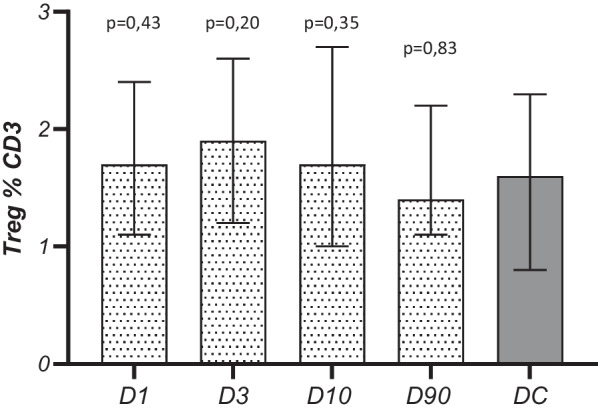
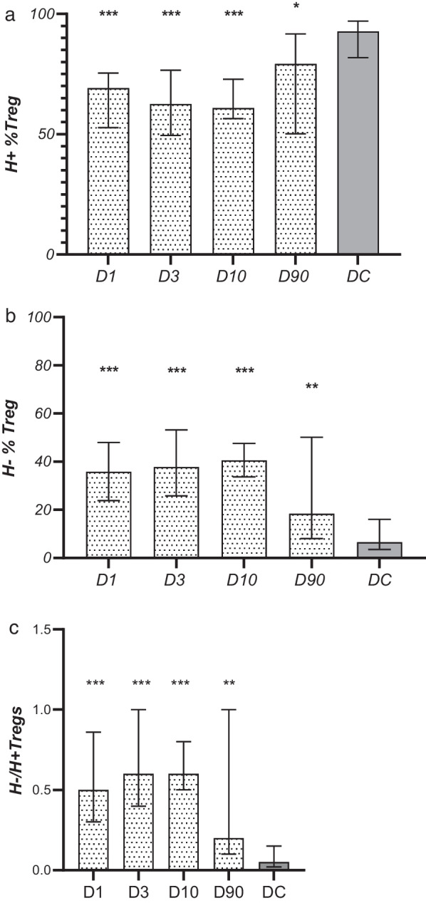
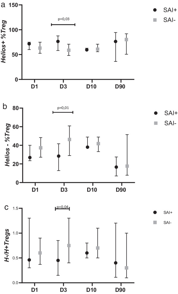
Methods and results: We prospectively assessed the circulating Treg population with flow cytometry in 52 subjects on days 1, 3, 10 and 90 after ischemic stroke and we compared the results with those obtained in concurrent age-, sex- and vascular risk factor-matched controls. At all studied time points the percentage of H+ Tregs decreased in stroke subjects-D1: 69.1% p < 0.0001; D3: 62.5% (49.6-76.6), p < 0.0001; D10: 60.9% (56.5-72.9), p < 0.0001; D90: 79.2% (50.2-91.7), p = 0.014 vs. controls: 92.7% (81.9-97.0) and the percentage of H- Tregs increased accordingly. In patients with SAI the percentage of pro-suppressor H+ Tregs on post-stroke day 3 was higher than in those without infection (p = 0.03). After adjustment for confounders, the percentage of H+ Tregs on day 3 independently correlated with SAI [OR 1.29; CI 95%: 1.08-1.27); p = 0.02]. Although the percentage of H+ Tregs on day 3 correlated positively with NIHSS score on day 90 (rS = 0.62; p < 0.01) and the infarct volume at day 90 (rS = 0.58; p < 0.05), in regression analysis it was not an independent risk factor.
Conclusions: On the first day after stroke the proportion of H+ vs. H- Tregs changes in favor of pro-inflammatory H- Tregs, and this shift continues toward normalization when assessed on day 90. A higher percentage of pro-suppressive H+ Tregs on day 3 independently correlates with SAI and is associated positively with NIHSS score, but it does not independently affect the outcome and stroke area in the convalescent phase of stroke.
Keywords: Regulatory T cells; Stroke; Stroke immunology; Stroke-associated infection; Transcription factor Helios.
© 2023. The Author(s).
Conflict of interest statement
The authors declare that they have no competing interests.
Figures
Similar articles
-
Differences in Maturation Status and Immune Phenotypes of Circulating Helios+ and Helios- Tregs and Their Disrupted Correlations With Monocyte Subsets in Autoantibody-Positive T1D Individuals.Front Immunol. 2021 May 12;12:628504. doi: 10.3389/fimmu.2021.628504. eCollection 2021. Front Immunol. 2021. PMID: 34054801 Free PMC article.
-
Upregulated TIGIT+ and Helios+ regulatory T cell levels in bronchoalveolar lavage fluid of NSCLC patients.Mol Immunol. 2022 Jul;147:40-49. doi: 10.1016/j.molimm.2022.04.005. Epub 2022 Apr 30. Mol Immunol. 2022. PMID: 35504057
-
FOXP3+Helios+ Regulatory T Cells, Immune Activation, and Advancing Disease in HIV-Infected Children.J Acquir Immune Defic Syndr. 2016 Aug 15;72(5):474-84. doi: 10.1097/QAI.0000000000001000. J Acquir Immune Defic Syndr. 2016. PMID: 27003495 Free PMC article.
-
Infection of Epstein-Barr Virus is Associated with the Decrease of Helios+FoxP3+Regulatory T Cells in Active Ulcerative Colitis Patients.Immunol Invest. 2021 Jan;50(1):23-36. doi: 10.1080/08820139.2020.1723021. Epub 2020 Feb 11. Immunol Invest. 2021. PMID: 32046542
-
Coexpression of Helios in Foxp3+ Regulatory T Cells and Its Role in Human Disease.Dis Markers. 2021 Jun 22;2021:5574472. doi: 10.1155/2021/5574472. eCollection 2021. Dis Markers. 2021. PMID: 34257746 Free PMC article. Review.
Cited by
-
The Alteration of Circulating Invariant Natural Killer T, γδT, and Natural Killer Cells after Ischemic Stroke in Relation to Clinical Outcomes: A Prospective Case-Control Study.Cells. 2024 Aug 22;13(16):1401. doi: 10.3390/cells13161401. Cells. 2024. PMID: 39195289 Free PMC article.
-
Prospective Quantitative and Phenotypic Analysis of Platelet-Derived Extracellular Vesicles and Its Clinical Relevance in Ischemic Stroke Patients.Int J Mol Sci. 2024 Oct 18;25(20):11219. doi: 10.3390/ijms252011219. Int J Mol Sci. 2024. PMID: 39457001 Free PMC article.
-
The Immunomodulatory Role of Regulatory T Cells in Preterm Birth and Associated Pregnancy Outcomes.Int J Mol Sci. 2024 Nov 5;25(22):11878. doi: 10.3390/ijms252211878. Int J Mol Sci. 2024. PMID: 39595948 Free PMC article. Review.
-
Neuroinflammation in Adaptive Immunodeficient Mice with Colitis-like Symptoms.Exp Neurobiol. 2025 Feb 28;34(1):34-47. doi: 10.5607/en24016. Exp Neurobiol. 2025. PMID: 40091637 Free PMC article.
-
Regulatory T Cells for Stroke Recovery: A Promising Immune Therapeutic Strategy.CNS Neurosci Ther. 2025 Jan;31(1):e70248. doi: 10.1111/cns.70248. CNS Neurosci Ther. 2025. PMID: 39878387 Free PMC article. Review.
References
MeSH terms
Substances
Grants and funding
LinkOut - more resources
Full Text Sources
Medical
Molecular Biology Databases
