

Microenvironment of pancreatic inflammation: calling for nanotechnology for diagnosis and treatment
- PMID: 37996911
- PMCID: PMC10666376
- DOI: 10.1186/s12951-023-02200-x
Microenvironment of pancreatic inflammation: calling for nanotechnology for diagnosis and treatment
Erratum in
-
Correction: Microenvironment of pancreatic inflammation: calling for nanotechnology for diagnosis and treatment.J Nanobiotechnology. 2024 Jan 23;22(1):38. doi: 10.1186/s12951-023-02232-3. J Nanobiotechnology. 2024. PMID: 38263033 Free PMC article. No abstract available.
Abstract
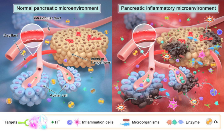
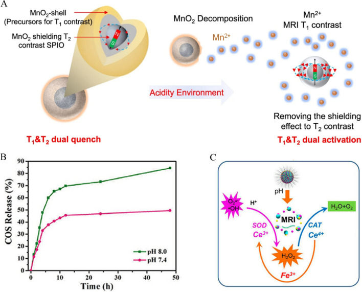
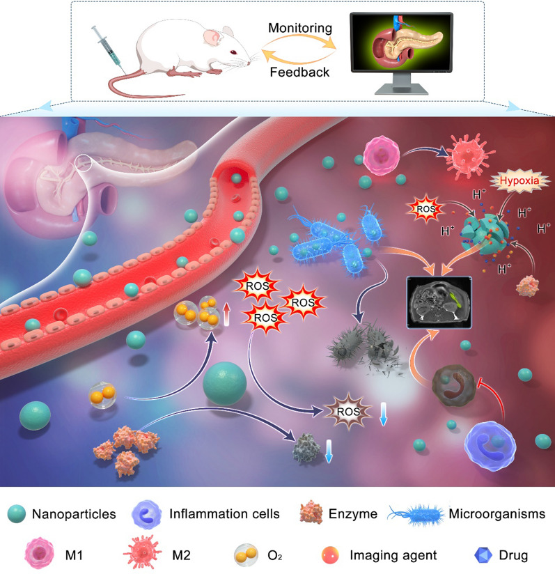
Acute pancreatitis (AP) is a common and life-threatening digestive disorder. However, its diagnosis and treatment are still impeded by our limited understanding of its etiology, pathogenesis, and clinical manifestations, as well as by the available detection methods. Fortunately, the progress of microenvironment-targeted nanoplatforms has shown their remarkable potential to change the status quo. The pancreatic inflammatory microenvironment is typically characterized by low pH, abundant reactive oxygen species (ROS) and enzymes, overproduction of inflammatory cells, and hypoxia, which exacerbate the pathological development of AP but also provide potential targeting sites for nanoagents to achieve early diagnosis and treatment. This review elaborates the various potential targets of the inflammatory microenvironment of AP and summarizes in detail the prospects for the development and application of functional nanomaterials for specific targets. Additionally, it presents the challenges and future trends to develop multifunctional targeted nanomaterials for the early diagnosis and effective treatment of AP, providing a valuable reference for future research.
Keywords: Acute pancreatitis; Diagnosis; Inflammatory microenvironment; Nanotechnology; Targets; Treatment.
© 2023. The Author(s).
Conflict of interest statement
The authors declare that they have no competing interests.
Figures
Similar articles
-
From micro to macro, nanotechnology demystifies acute pancreatitis: a new generation of treatment options emerges.J Nanobiotechnology. 2025 Jan 29;23(1):57. doi: 10.1186/s12951-025-03106-6. J Nanobiotechnology. 2025. PMID: 39881355 Free PMC article. Review.
-
Microenvironment responsive nanomedicine for acute pancreatitis treatment.Colloids Surf B Biointerfaces. 2025 Jul;251:114633. doi: 10.1016/j.colsurfb.2025.114633. Epub 2025 Mar 13. Colloids Surf B Biointerfaces. 2025. PMID: 40112593 Review.
-
Targeting and functional effects of biomaterials-based nanoagents for acute pancreatitis treatment.Front Bioeng Biotechnol. 2023 Jan 10;10:1122619. doi: 10.3389/fbioe.2022.1122619. eCollection 2022. Front Bioeng Biotechnol. 2023. PMID: 36704304 Free PMC article. Review.
-
The apoptosis of peripheral blood lymphocytes promoted by hyperbaric oxygen treatment contributes to attenuate the severity of early stage acute pancreatitis in rats.Apoptosis. 2014 Jan;19(1):58-75. doi: 10.1007/s10495-013-0911-x. Apoptosis. 2014. PMID: 24101212
-
Research Progress of Antioxidant Nanomaterials for Acute Pancreatitis.Molecules. 2022 Oct 25;27(21):7238. doi: 10.3390/molecules27217238. Molecules. 2022. PMID: 36364064 Free PMC article. Review.
Cited by
-
Single-atom nanozymes shines diagnostics of gastrointestinal diseases.J Nanobiotechnology. 2024 May 25;22(1):286. doi: 10.1186/s12951-024-02569-3. J Nanobiotechnology. 2024. PMID: 38796465 Free PMC article. Review.
-
Correction: Microenvironment of pancreatic inflammation: calling for nanotechnology for diagnosis and treatment.J Nanobiotechnology. 2024 Jan 23;22(1):38. doi: 10.1186/s12951-023-02232-3. J Nanobiotechnology. 2024. PMID: 38263033 Free PMC article. No abstract available.
-
An Efficient Electrochemiluminescence Biosensor Based on Ru(bpy)32+@AuNPs@SWCNTs for miRNAs Detection Using a Dual Engine-Triggered DNA Walker.Anal Chem. 2025 May 27;97(20):10841-10848. doi: 10.1021/acs.analchem.5c01244. Epub 2025 May 15. Anal Chem. 2025. PMID: 40371917
-
Nicotinamide adenine dinucleotide phosphate oxidase in pancreatic diseases: Mechanisms and future perspectives.World J Gastroenterol. 2024 Feb 7;30(5):429-439. doi: 10.3748/wjg.v30.i5.429. World J Gastroenterol. 2024. PMID: 38414585 Free PMC article. Review.
-
Studies on Treatment Within the Scope of Medical Biotechnology for Pancreatic Diseases.Mol Biotechnol. 2025 Apr;67(4):1321-1335. doi: 10.1007/s12033-024-01142-5. Epub 2024 Apr 16. Mol Biotechnol. 2025. PMID: 38627328 Review.
References
Publication types
MeSH terms
Substances
Grants and funding
LinkOut - more resources
Full Text Sources
Medical
