. 2023 Nov 23;21(1):846.
doi: 10.1186/s12967-023-04730-2.

Complement factor H attenuates TNF-α-induced inflammation by upregulating EIF3C in rheumatoid arthritis

Affiliations

Complement factor H attenuates TNF-α-induced inflammation by upregulating EIF3C in rheumatoid arthritis

Yimeng Jia et al. J Transl Med. .

Abstract

Objective: To explore the role and underlying mechanism of Complement Factor H (CFH) in the peripheral and joint inflammation of RA patients.

Methods: The levels of CFH in the serum and synovial fluid were determined by ELISA. The pyroptosis of monocytes was determined by western blotting and flow cytometry. The inflammation cytokine release was tested by ELISA. The cell migration and invasion ability of fibroblast-like synoviocytes (FLS) were tested by Wound healing Assay and transwell assay, respectively. The potential target of CFH was identified by RNA sequencing.

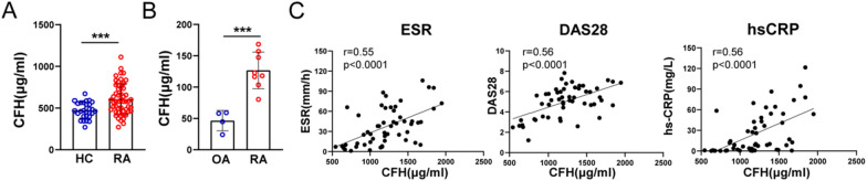
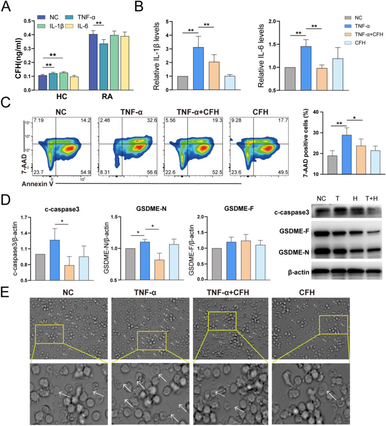
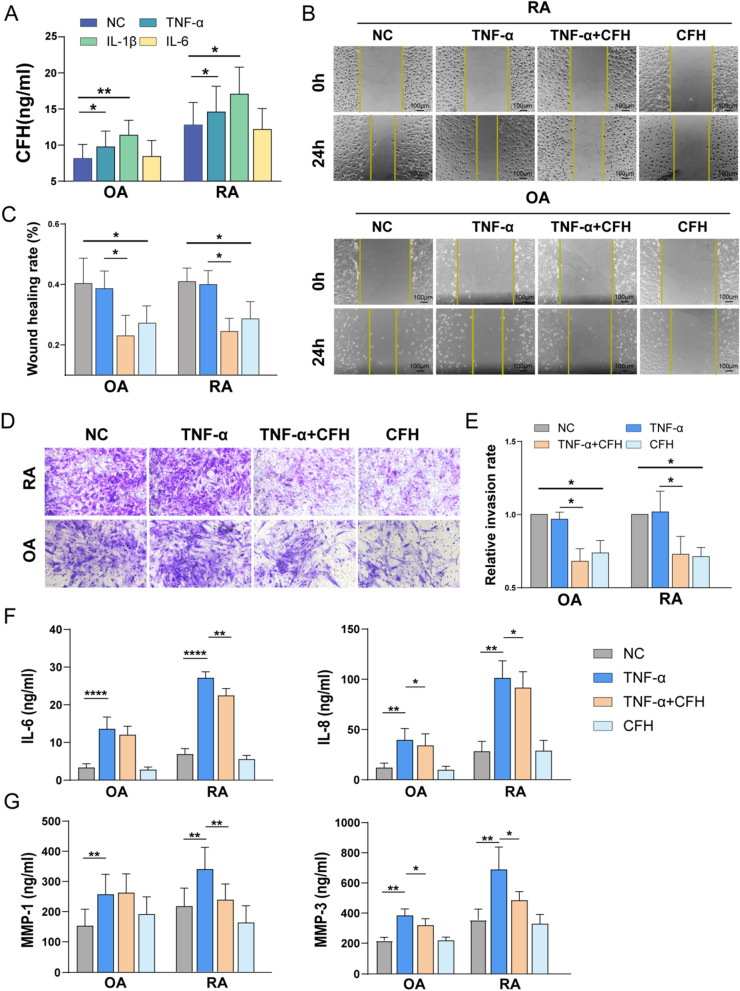
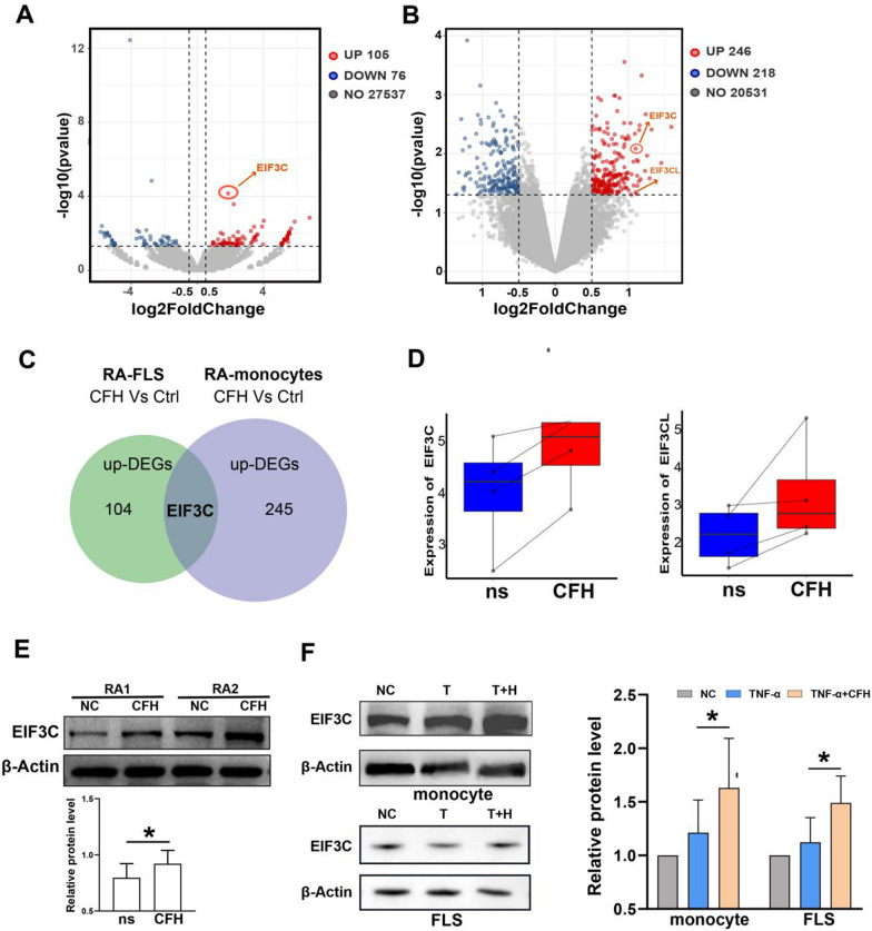
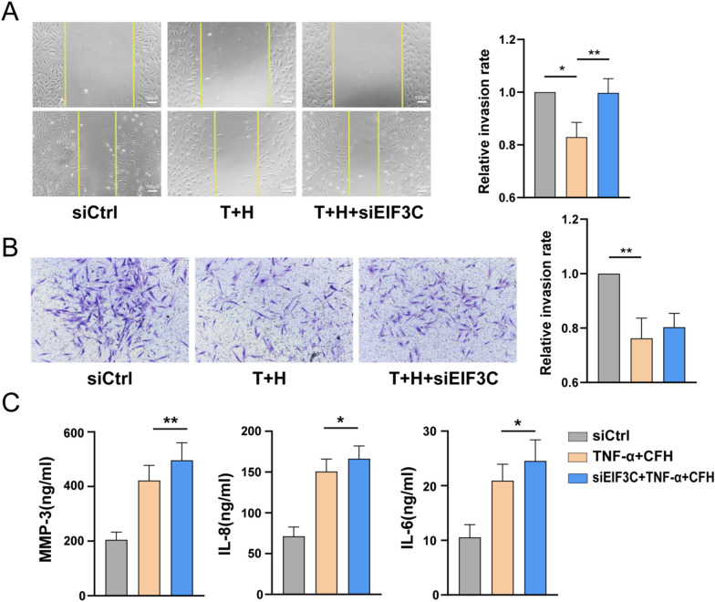
Results: CFH levels were significantly elevated in the serum and synovial fluid from RA and associated with high sensitivity C-reactive protein (hs-CRP), erythrocyte sedimentation rate (ESR), and disease activity score 28 (DAS28). TNF-α could inhibit CFH expression, and CFH combined with TNF-α significantly decreased cell death, cleaved-caspase 3, gasdermin E N-terminal (GSDME-N), and inflammatory cytokines release (IL-1β and IL-6) of RA-derived monocytes. Stimulated with TNF-α increased CFH levels in RA FLS and CFH inhibits the migration, invasion, and TNF-α-induced production of inflammatory mediators, including proinflammatory cytokines (IL-6, IL-8) as well as matrix metalloproteinases (MMPs, MMP1 and MMP3) of RA FLSs. The RNA-seq results showed that CFH treatment induced upregulation of eukaryotic translation initiation factor 3 (EIF3C) in both RA monocytes and FLS. The migration of RA FLSs was promoted and the expressions of IL-6, IL-8, and MMP-3 were enhanced upon EIF3C knockdown under the stimulation of CFH combined with TNF-α.

Conclusion: In conclusion, we have unfolded the anti-inflammatory roles of CFH in the peripheral and joints of RA, which might provide a potential therapeutic target for RA patients.

Keywords: Complement factor H; Eukaryotic translation initiation factor 3 subunit C; Fibroblast-like synoviocytes; Pyroptosis; Rheumatoid arthritis.


Conflict of interest statement

The authors have declared no competing interests.

Figures

Fig. 1
Fig. 1
CFH is elevated in the serum and synovial fluid from RA. A CFH in serum from RA patients and healthy control detected by ELISA. (RA: n = 52; HC: n = 28). B CFH in synovial fluid from RA and OA patients detected by ELISA. (RA: n = 8; OA: n = 4). C Correlation analysis of CFH expression in serum of RA patients with ESR, DAS28, and hs-CRP respectively. Data are expressed as mean ± SEM. ***p < 0.001; RA: rheumatoid arthritis; OA: osteoarthritis; HC: healthy control; hs-CRP: high sensitivity C-reactive protein; ESR: erythrocyte sedimentation rate; DAS28: disease activity score 28
Fig. 2
Fig. 2
CFH attenuates TNF-α-induced pyroptosis and inflammatory cytokines release of RA monocytes. Purified CD14+ monocytes were pretreated with TNF-α(50 ng/ml), TNF-α(50 ng/ml) + CFH (5 μg/ml), or CFH (5 μg/ml) for 24 h (n = 6–9). A The expression of secreted CFH in the culture supernatant of RA patients and healthy control stimulated with TNF-α, IL-1β, and IL-6 detected by ELISA. B The expression of secreted cytokines (IL-1β, IL-6) in the culture supernatant of RA monocytes in each group was detected by ELISA. C Flow cytometric analysis of cells stained with annexin V/7-AAD to determine cell death and percentage of 7-AAD-positive cells. D The protein expression of c-caspase3, GSDME-N, and GSDME-F was measured by using Western blot. E Representative phase-contrast microscopy images of monocytes treated as indicated. Arrows indicate pyroptotic cell bubbles. Data are expressed as mean ± SEM. *p < 0.05; **p < 0.01; NC: negative control; T: TNF-α; T + H: TNF-α + CFH; H: CFH; IL-6: interleukin-6; IL-1β: interleukin-1β; c-caspase3: cleaved caspase-3; GSDME-N: gasdermin E N-terminal; GSDME-F: gasdermin E full length
Fig. 3
Fig. 3
CFH inhibits the migration, invasion, and TNF-α-induced expression of inflammatory mediators of RA FLSs. The FLSs were pretreated with TNF-α(50 ng/ml), TNF-α(50 ng/ml) + CFH (5 μg/ml), or CFH (5 μg/ml) for 24 h (n = 5–10). A The expression of secreted CFH in the culture supernatant of RA-FLS and OA-FLS detected by ELISA. B-C The effect of CFH on cell migration was detected using wound healing assay. The scratching area was photographed at 0 h and 24 h. The scratch assay was presented as the percentage by which the original scratch area decreased at 24 h. Representative images (original magnification, × 200) are shown. D-E FLS invasion ability was measured by Transwell assay, and the invaded cells were photographed. The relative invasion rate was calculated by counting mean invaded cells from 5 randomly selected fields and then normalized to that in the NC group. Representative images (original magnification, × 200) are shown. F–G The expression of secreted cytokines (IL-6, IL-8) and MMPs (MMP-1, MMP-3) in culture supernatant of RA-FLS and OA-FLS in each group were detected by ELISA. Data are expressed as mean ± SEM. *p < 0.05; **p < 0.01; ***p < 0.001; ****p < 0.0001. MMP-1: matrix metalloproteinase-1; MMP-3: matrix metalloproteinase-3
Fig. 4
Fig. 4
EIF3C is a potential target for CFH to play a role in inhibiting FLSs and monocyte function. Monocytes and FLS from RA patients were selected respectively, and cells from each patient were divided into two groups. One group was left untreated, and one group was treated with 5 μg/ml CFH for 6 h. A Volcano plot indicated up-regulated (red dots) and down-regulated (blue dots) genes (P value < 0.05 and |log2FC|> 0.5) by RNA sequencing in CFH-treated versus untreated RA monocytes. EIF3C is indicated. B Volcano plot indicated up-regulated (red dots) and down-regulated (blue dots) genes (P value < 0.05 and |log2FC|> 0.5) by RNA sequencing in CFH-treated versus untreated RA monocytes. EIF3C and EIF3CL are indicated. C We selected the genes upregulated in the CFH-treated monocytes and the genes upregulated in the CFH-treated FLS identified by RNA-sequencing and determined the intersecting proteins. We obtained one gene, namely EIF3C. DEGs: Differentially Expressed Genes. D The boxplot shows the relative expression of EIF3C and EIF3CL. E CFH upregulated the protein expression of EIF3C by using Western blot. F CFH combined with TNF-α upregulated protein expression of EIF3C in both RA FLSs and monocytes by using Western blot. Data are expressed as mean ± SEM (n = 6). *p < 0.05
Fig. 5
Fig. 5
Effect of CFH knockdown on the function of RA FLS. RA FLSs were transfected with siRNAs for EIF3C (siEIF3C) or control siRNA (siCtrl) for 72 h and then stimulated with TNF-α(50 ng/ml) + CFH (5 μg/ml) for 24 h (n = 4–8). A Effect of EIF3C knockdown on the migration of RA FLSs was measured with the wound-healing assay. The relative migration rates were calculated by the percentage by which the original scratch area decreased and then normalized to that in the control group. B Effect of EIF3C knockdown on the invasion of RA FLSs using Transwell assay. The relative invasion rates were calculated by counting invaded cells and then normalized to that in the control group. Representative images (original magnification, × 200) are shown. C Effect of EIF3C knockdown on the inflammatory mediators of RA FLSs detected by ELISA. Data show the mean ± SEM. *p < 0.05; **p < 0.01

References

    1. Conigliaro P, Triggianese P, De Martino E, Fonti GL, Chimenti MS, Sunzini F, et al. Challenges in the treatment of rheumatoid arthritis. Autoimmun Rev. 2019;18(7):706–713. doi: 10.1016/j.autrev.2019.05.007. - DOI - PubMed
    1. Aggarwal A, Bhardwaj A, Alam S, Misra R. Evidence for activation of the alternate complement pathway in patients with juvenile rheumatoid arthritis. Rheumatology. 2000;39(2):189–192. doi: 10.1093/rheumatology/39.2.189. - DOI - PubMed
    1. Wouters D, Voskuyl AE, Molenaar ETH, Dijkmans BAC, Hack CE. Evaluation of classical complement pathway activation in rheumatoid arthritis: measurement of C1q–C4 complexes as novel activation products. Arthritis Rheum. 2006;54(4):1143–1150. doi: 10.1002/art.21729. - DOI - PubMed
    1. Banda NK, Hyatt S, Antonioli AH, White JT, Glogowska M, Takahashi K, et al. Role of C3a receptors, C5a receptors, and complement protein C6 deficiency in collagen antibody-induced arthritis in mice. J Immunol. 2012;188(3):1469–1478. doi: 10.4049/jimmunol.1102310. - DOI - PMC - PubMed
    1. Blom AM, Nandakumar KS, Holmdahl R. C4b-binding protein (C4BP) inhibits development of experimental arthritis in mice. Ann Rheum Dis. 2009;68(1):136–142. doi: 10.1136/ard.2007.085753. - DOI - PubMed

Publication types

MeSH terms